

 PDF
PDF ePub
ePub Citation
 Print
Print


INTRODUCTION
Gastrointestinal cytomegalovirus (CMV) infection in immunocompetent children is very rare. In immunocompromised patients, it can involve the entire alimentary tract from esophagus to rectum [1,2]. In the colon, it can cause colitis-like syndrome including diarrhea, hemotochezia and abdominal pain associated with fever and weight loss [2,3]. Although the association of CMV infection with steroid-refractory or -dependent patients with inflammatory bowel disease (IBD) have been well-defined, it is very rare in steroid-naive patients. Protein losing enteropathy (PLE) as an initial presentation of Crohn's disease (CD) is also very rare [4]. Herein, we reported a pediatric steroid-naive CD patients' presented with disseminated CMV infection and PLE.
CASE REPORT
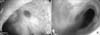
A 15-year-old girl was admitted to our unit with abdominal pain, abdominal distension, lower extremity edema and watery diarrhea (6-10 times/day) for two months. Physical examination revealed mucocutaneous pallor, massive ascites and +2 edema in the lower extremities. She had a weight of 46.5 kg (3-10 percentile) and height of 157 cm (3-10 percentile). Laboratory analysis revealed hemoglobin, 8.1 g/dL; leukocytes, 3,500/mm3 (10% lymphocyte); platelets, 306,000/mm3; total protein, 3.4 g/dL (6.6-8.7 g/dL); albumin, 1.1 g/dL (3.4-5 g/dL); γ globulin, 2.2 g/dL; erythrocyte sedimentation rate, 20 mm/h; C-reactive protein, 10.2 g/dL; and other laboratory analysis including serum electrolytes, liver enzymes and urea and creatinine were normal. Coagulation parameters were mildly increased. Serum immunoglobulin (Ig) G, IgA, IgM and IgE levels were 489 mg/dL (608-1,472), 44.6 mg/dL (>50), 92.9 mg/dL (52-242) and 1.9 IU/mL, respectively. Serum lipid levels were as follows: low-density lipoprotein-cholesterol, <4 mg/dL; high-density lipoprotein-cholesterol, <3 mg/dL; and total cholesterol, 42 mg/dL. Vitamin B12, folic acid and ferritine levels were normal. Urine analysis for the proteinuria including spot and 24 hour collection was negative. The patient had blood, urine, stool culture, stool ova and parasite ×3, and stool Clostridium difficile toxin tested, all of which were negative. Echocardiography was normal. Chest X-ray showed bilateral mild pleural effusion. Based on the clinical (ascites and edema in the lower extremities) and laboratory findings (hypoalbuminemia, leucopenia, low lipid, γ globulin and immunoglobulin levels, negative proteinuria); a diagnosis of PLE was made. Due technical and laboratory inadequacies lymphoscintigraphy using 99mTc-human serum albumin and alpha-1 antitrypsin level in the stool did not studied. Abdominal computed tomography (CT) revealed massive ascites and diffuse thickening (1 cm) in intestinal wall including terminal ileum and whole colonic segments. Additionally, multiple lymph nodes with biggest one in 2.5×3 cm size in the celiac truncus, portal hilus and right parailiac region were seen. Thorax CT showed bronchopneumonia and lymph nodes on the para-tracheal and sub-aortic region with the biggest one in 1 cm size. Upper gastrointestinal endoscopy and colonoscopy was performed for the etiology of PLE. Endoscopy revealed edema and hypertrophy in the gastric mucosa and nodularity and mucosal fissures in the proximal duodenal mucosa. Colonoscopy revealed loss of mucosal vascular pattern, diffuse aphthous and mucosal ulcers with smooth raised edge (like "punched out holes"), mucosal fissures and fragility (Fig. 1). Ulcerated lesions were also seen in the terminal ileum.
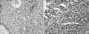
Anti-Saccharomyces cerevisiae antibodies IgA was mildly positive. Purified protein derivative was negative. Celiac serology (tissue transglutaminase [tTG]-IgA and tTG-IgG) and HIV serology was negative. Endoscopic and laboratory diagnosis was compatible with IBD particularly CD. Supportive treatment including antibiotics, albumin, parenteral nutrition and IVIG was initiated for PLE. Mesalazine and oral prednisolone (2 mg/kg/day-2 doses) was prescribed for the colitis. Two days later (after the 4th doses of prednisolone); the patient had massive hematochezia. Hemoglobin levels dropped to 6.6 mg/dl. The patient was transferred to intensive care unit and given 2 units red blood cells Octreotide infusion and fresh frozen plasma (2 times/day) was initiated for the massive bleeding and mild coagulopathy, respectively. Due to prolonged massive bleeding, the patient was also given Factor VII infusion. At the same day, the histopathological analyses of the colonic biopsies revealed severe active colitis and ulceration and CMV inclusions. Immunostaining of colonic biopsies for CMV antigens were positive (Fig. 2).
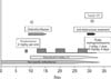
Anti-CMV IgM antibodies, anti-CMV IgG antibodies from the peripheral blood and CMV polymerase chain reaction from peripheral blood, bone marrow, colonic tissue and tracheal sputum was positive. The patient's prednisolone was discontinued, and she was placed on iv ganciclovir (5 mg/kg). Despite the supportive (red blood cell transfusion) and medical treatment (octreotide, fresh frozen plasma and Factor VII), massive bleeding was continued. Pulse methylprednisolone was begun on the 3rd day of ganciclovir and bleeding was mildly subsided. But, the patient needed mechanic ventilation support due to pneumonia. Ganciclovir treatment was continued to 21 days, pulse methylprednisolone was given three courses with the supportive treatment but massive bleeding was resumed and the patient clinical condition was worsened. Factor VII was given for the second time but no respond was achieved. Due to multisystem involvement (pulmonary, intestinal and huge abdominal lymphadenopathy), anti-tuberculous treatment was initiated but the patient died one day later due to massive pulmonary hemorrhage and shock (Fig. 3).
DISCUSSION
In this manuscript, we report an interesting case with CD. She was presenting with diffuse CMV infection and PLE. PLE as a presenting feature of CD is very rare. CMV infection particularly CMV colitis in the steroid-naive patient with IBD is also very rare [1,2,3,4,5].
CMV is a common infection that effects the 40-100% of all adults worldwide. Although it is asymptomatic in 90% of the cases, the classical manifestation of acute symptomatic CMV infection includes mononucleosis-like syndrome. CMV remains latent within the host and can be reactivated later [1,2]. CMV infection may present as interstitial pneumonia, hepatitis, meningoencephalitis, myocarditis, retinitis, colitis and hemolytic anemia in immunocompromised patients [3,4,5]. Gastrointestinal CMV disease in immunocompromised patients can involve the entire alimentary tract from esophagus to rectum. In the colon, it can cause colitis-like syndrome including diarrhea, hemotochezia and abdominal pain associated with fever and weight loss [3].
The association of CMV with IBD has been well-defined in steroid-refractory or -dependent adult patients. Prevalence of CMV in endoscopic biopsies from the patients with steroid-refractory colitis varies from 3% to 66% depends on the methods used for diagnosis [3]. The effect of CMV infection to the clinical course or prognosis is controversial. Kim et al. [2] showed that ulcerative colitis (UC) patients with CMV infection have more severe disease and have longer hospitalization. But, none of the patients with CMV infection developed fulminant colitis, required surgery or died in their study. Similar results were reported from Yoshino et al. [6] and Matsuoka et al. [7]. Contrary to these reports, Berk et al. [8] reported high incidence of toxic megacolon, colectomy requirements and mortality rate in IBD patients with CMV infection. Pfau et al. [9] reviewed the 30 patients with IBD and CMV infection and they found that 56.6% had pancolitis, 56.6% required surgery and 20% of the patients died. These findings are suggested that the CMV infection may cause serious complications and mortality in patients with IBD. There have been little data about CMV infection in pediatric IBD patients. Ghidini et al. [10] reported 6 pediatric IBD patients with CMV infection; most of them had pancolitis and refractory to immunosuppressive agents. They showed that early detection and antiviral treatment was associated with good outcome.
The association of CMV infection with steroid-naive IBD patients is very rare. Inoue et al. [11] reported a steroid-naive IBD patient with CMV colitis and reviewed the 7 adult cases in the literature. Their patient had pancolitis and receiving 5-aminosalicylic acid for two months and admitted with diarrhea. CMV infection was diagnosed by serology and histopathological examination. After antiviral treatment, symptoms did not improved and pulse methylprednisalone was administered. The patients' CMV antigenemia was become negative on the following days but toxic megacolon developed and the patient underwent colectomy. Four of the other seven patients with CMV colitis had pancolitis and all the patients improved after the ganciclovir treatment. To date, we could not found any pediatric steroid-naive IBD patients with CMV infection in the literature. Our patient had some clinical findings suggesting CMV infection such as huge lymph nodes in the abdomen and thorax, and atypical ulcers (with smooth raised edge, like "punched out holes") on colonoscopy. Diagnosis of CMV was made based on the histological and serological examination. Despite the ganciclovir treatment, CMV infection progressed. Although supportive treatment was administered, massive colonic bleeding continued and pulse methylprednisalone was added to the treatment but the clinical condition of the patient worsened and died.
PLE is a rare clinical condition characterized by loss of serum proteins via gastrointestinal tract. The main laboratory findings are reduced serum concentrations of albumin, γ globulins and lipids. The diagnosis is based on the exclusion of other causes of hypoproteinemia, and to demonstrate increase excretion of alpha-1 antitrypsin [4]. Both UC and CD may lead the PLE. Protein loss in these conditions is related to enhanced leakage of protein-rich fluids across the eroded epithelium. The degree of mucosal involvement correlates with the severity of protein loss [12]. CMV associated Menetrier's disease may also cause PLE [13]. Although enlarged and hypertrophic gastric folds were seen in the upper endoscopy, the histopathological finding were inconsonant with Menetrier's disease in our patient.
The association of CMV infection with the new onset IBD suggests the role of CMV in the pathogenesis of IBD. Orvar et al. [14] and van Dorp et al. [15] suggested that CMV infection may initiate an immune response and then autoimmune response in the susceptible host can lead to IBD. But, there are many reports in the literature contrary to this hypothesis [2,3,16]. We thought that severe CD in our case lead the PLE, and hypogammaglobulinemia due to massive and prolonged protein loss via gastrointestinal tract cause susceptible to CMV infection in our case.
It is still debate to test or evaluate the CMV in newly diagnosed IBD patients. CMV colitis is clinically very similar to IBD, and can occur concomitant to UC or CD regardless to the degree of colon involved. On a recent consensus report, evaluation of CMV is recommended on the 3rd day when unresponsive to initial steroid treatment in acute severe colitis in children [17].
In conclusion, we report a unique case of CD. She had PLE and disseminated CMV infection as initial presentation of the disease. Hypogammaglobulinemia associated with PLE in CD may predispose the CMV infection in previously healthy children.
 XML Download