

 PDF
PDF ePub
ePub Citation
 Print
Print


CASE REPORT
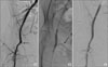
A 50-year-old lady presented to the emergency department at 23.00 on a Saturday night with exsanguinating haemorrhage from a defect in the right groin secondary to tumour invasion and necrosis. She had a previous history of a T1b N1 moderately differentiated squamous cell carcinoma of the vulva. She had undergone radical vulvectomy and bilateral inguinal lymphadenectomy with 3/17 positive lymph nodes in the right groin and extracapsular invasion 10 months earlier. This was followed by adjuvant radiotherapy to the right groin and hemipelvis, 45 Gy in 25 fractions. She was diagnosed with right groin recurrence, four months prior to the urgent admission, and treated with palliative chemotherapy, 3 cycles of Carboplatin and Gemcitabine. An MRI scan a few days before the emergency admission had showed progressive disease but this investigation had been done elsewhere and the result was not available to the admitting team, nor were the patient or her partner aware of the result. Upon presentation to the emergency department she was unstable with blood pressure of 100/80 mmHg and heart rate of 145/minute. She had profuse arterial bleeding from the right groin which was partly controlled by direct pressure. There was no interventional radiology service immediately available at that time. She was taken to theatre where control of the external iliac artery was achieved via an intraperitoneal approach. Exploration of the right groin showed a large cavity with necrotic tissue. The femoral vein was absent, completely destroyed by the recurrent cancer. There was bleeding from the medial aspect of the common femoral artery secondary to destruction by the tumour. The bleeding area was controlled with 4-0 prolene sutures. The cavity was packed and dressed. The patient was taken to intensive care unit for further resuscitation, but a decision was made not to reintervene surgically if there was further haemorrhage. She was transferred to a ward the next day after she became stable. As the femoral artery was still exposed and the tissues were very frail, it was felt that the surgical repair was not adequate and the likelihood of rebleeding high. A right femoral arteriogram with left femoral access was therefore carried out. It showed occlusion of the superficial femoral artery at its origin (Fig. 1A). There was irregularity of the distal common femoral artery, but no extravasation of contrast was demonstrated. Two 8 mm by 20 mm covered stents (Wallgraft™ Endoprosthesis, Boston Scientific, Natick, MA, USA) were deployed across the bifurcation of the common femoral artery into the profunda femoris artery. On balloon expansion of the second stent there was seen to be some extravasation of contrast (Fig. 1B) but a completion angiogram showed the stented segment to be widely patent with no extravasation of contrast (Fig. 1C). After five days of overall stay in hospital she was discharged to a hospice. The oncological decision was for palliative management. The patient died from progressive disease without further bleeding three months later.
DISCUSSION
Recurrent vulvar cancer in the groin particularly after previous lymphadenectomy and radiotherapy is very difficult to manage. It often involves the femoral vessels and it is considered inoperable. The usual treatment is chemoradiotherapy, however surgery in the form of radical resection with vascular grafting and plastic reconstruction or hip disarticulation has been described.4,5 Femoral artery rupture is a rare complication and is usually the result of tumour invasion, radiotherapy and tissue necrosis. Rupture of the femoral artery results in exsanguinating haemorrhage and is usually a terminal event. There are two reports in the literature where the rupture was managed with immediate surgery. One patient had a successful graft bypass from the external iliac artery to the superficial femoral artery by tunnelling the graft laterally to the open wound.1 No follow-up was documented. The other patient had an initial repair of the bleeding superficial femoral artery with 4-0 prolene and subsequently, as the leg became ischaemic, a graft bypass from the external iliac artery to the distal superficial femoral artery.2 The graft was tunnelled through the obturator foramen. Her leg was well at five months follow-up. In another report a patient with common femoral artery erosion and severe haemorrhage was successfully managed with percutaneous endovascular stent-graft placement and subsequent graft bypass a month later. She only survived for three months.3 Endovascular stent graft placement has also been described in two patients with external iliac artery erosion and haemorrhage from a recurrent uterine carcinoma.6,7
In our case the patient presented with exsanguinating haemorrhage at night when the interventional radiology service was not immediately available. In hindsight, she would not have survived the delay of organising an angiogram even if the service had been available. At the time of surgery it was felt that a graft bypass was not suitable for her as the entire groin was very indurated and oedematous from the local recurrence and the previous radiotherapy, and there was also a large defect with necrotic tissue. The bleeding vessel was therefore repaired locally during open surgery and endovascular placement of a covered stent graft was subsequently carried out the following day. This was successful and she went on to live another three months without further bleeding. In conclusion, the management of these patients should be individualisedand when intervention is considered appropriate, the option of combined open surgery and endovascular repair should be kept in mind.
 XML Download