

 PDF
PDF ePub
ePub Citation
 Print
Print


Introduction
According to the third edition of the Japanese gastric cancer treatment guidelines, the treatment for upper-third advanced gastric cancer is total gastrectomy (TG) with lymph node (LN) dissection.1 For early gastric cancer (EGC), gastric resection can be modified as proximal gastrectomy (PG), but a consensus regarding the extent of resection has yet to be reached. Survival rates do not significantly differ between TG and PG, as has been reported by many studies.2 Compared with TG, PG is less invasive, requires less surgical time, provides a relatively more physiological anastomosis, and mitigates vitamin B12 deficiency with less nutritional loss given a portion of the stomach is preserved. Despite these advantages, PG has not been widely used because of associated complications such as reflux esophagitis and dysphagia, as well as its unproven long-term outcomes.34 Some efforts have been made to reduce reflux esophagitis and preserve the lower esophageal sphincter (LES).56 However, previous studies had significant limitations, including a small number of enrolled patients. In laparoscopy-assisted PG (LAPG), anastomosis is performed using extracorporeal methods, which allows for a longer portion of the intra-abdominal esophagus to be preserved than with open methods. This study aimed to evaluate functional and oncological outcomes of PG in comparison with TG for upper-third EGC. Additionally, subgroup analysis was performed to evaluate factors affecting the incidence of postoperative reflux esophagitis after PG.
Materials and Methods
1. Patient selection
The medical records of upper-third EGC patients who underwent PG or TG were retrospectively reviewed. The PG grou p was subdivided into conventional open PG (cPG) and modified laparoscopy-assisted PG (mLAPG). We aimed to preserve a longer portion of the intra-abdominal esophagus in patients who had undergone mLAPG than in those who had undergone cPG. We reviewed 157 patients who had undergone cPG between 2002 and 2012 and 35 patients who had undergone mLAPG between 2006 and 2012. For the control group, we reviewed the medical records of 157 consecutive patients who had undergone TG between 2007 and 2011. EGC was defined as a preoperative T1 cancer. The indication for TG and cPG was upper-third EGC. For patients who had undergone mLAPG, we intended to preserve the intra-abdominal esophagus as much as possible. Therefore, the indication for mLAPG was upper-third EGC 2 cm or further away from the gastroesophageal junction (GEJ). Surgical morbidity, recurrence, long-term nutritional status, and the incidence of reflux esophagitis were compared. The study was approved by the Institutional Review Board of Seoul National University Hospital.
2. Operative procedures
The surgical methods used for cPG and mLAPG are represented in Fig. 1. For mLAPG, the dissection of the regional LN and mobilization of the stomach were performed by laparoscopy, but anastomosis (direct esophagogastrostomy) was performed extracorporeally. With preservation of the spleen, LNs 1, 2, 3a, 4sa, 4sb, and part of 10 were dissected. In particular, LN 4sb was dissected with preservation of the right gastroepiploic artery. In addition, the LNs 7, 8a, and 9, and LNs along the splenic artery (nos. 11p and 11d) were dissected to complete the D1+ dissection. After thorough dissection of the distal esophagus and surrounding LNs, a 4- to 6-cm-long midline incision was made on the epigastrium. After extraction of the entire stomach through the minilaparotomy site, distal resection was performed at the mid stomach and angle using three GIA 60 (United States Surgical Corp., Norwalk, CT, USA). An anvil clamp was then applied at the level of the GEJ, which was above or below the abdominal wall depending on the obesity of the patient, with preservation of the abdominal esophagus as much as possible. Proximal resection was performed at a margin of more than 1 cm from t he tumor. After gastrotomy on the anterior side of the antrum, the shaft of an end-to-end anastomosis stapler (United States Surgical Corp.) was inserted and esophagogastrostomy anastomosis was performed at the staple line or slightly posterior with a 25-mm anvil. Special attention was taken to preserve the hepatic branch of the vagus nerve if it was visible, and pyloroplasty was not performed. Preoperative clipping and intraoperative frozen biopsy were conducted to secure the distal margin in all of the PG cases. In contrast, all procedures in cPG were performed via the open method. The range of LN dissection and the method of anastomosis (direct esophagogastrostomy) were similar to those used in mLAPG. A proximal resection was made at the distal esophagus, which was 1 to 2 cm proximal to the GEJ. The vagus nerve was routinely sacrificed and pyloroplasty was selectively performed depending on the surgeon's preference.
TG was performed via the open method, and Roux-en-Y esophagojejunostomy was performed for every case. With preservation of the spleen, LNs 1, 2, 3, 4sa, 4sb, 4d, part of 10, and LNs along the greater and lesser curvatures of the stomach (the right pericardial LNs, left pericardial LNs, LNs along the lesser curvature, LNs along the short gastric vessels, and LNs along the great curvature) were dissected. In addition, the LNs along the left gastric artery (No. 7), common hepatic artery (No. 8a), celiac artery (No. 9), suprapyloric area (No. 5), infrapyloric area (No. 6), LNs along the splenic artery (No. 11p and selectively No. 11d), and selectively LN No. 12 were dissected to complete the D1+ dissection. Proximal resection was made at the distal esophagus with sacrifice of the vagus nerve.
3. Outcomes
For short-term outcomes, we analyzed surgical complications that developed within 30 days after surgery. Long-term outcomes were assessed according to patient survival and recurrence. Postoperative gastrointestinal symptoms were considered present when the Visick score reflecting the symptomatic level and Los Angeles (LA) classification based on endoscopic observation confirmed the presence of reflux esophagitis. LA classification was graded as A, B, C, and D according to the extent of mucosal breaks. Grade A is defined as one or more mucosal breaks confined to the mucosal folds and each fold is no longer than 5 mm; grade B is defined as at least one mucosal break longer than 5 mm confined to the mucosal folds but not continuous between the tops of two mucosal folds; grade C is defined as at least one mucosal break continuous between the tops of two or more mucosal folds but not circumferential; and grade D is defined as a circumferential mucosal break.7
To evaluate nutritional status, we measured the level of postoperative vitamin B12, hemoglobin (Hb), and serum albumin. Severe anemia was determined on the basis of Hb level <10 mg/dl and hypoalbuminemia was defined as an albumin level <3.5g/dl. The TG group was routinely supplemented with vitamin B12 every two or three months after the first postoperative year, and the cPG and mLAPG groups were selectively supplemented only in cases when a vitamin B12 level less than 200 pg/ml was detected after routine blood screening. The follow-up periods were 36.3±27.6 and 46.0±19.7 months for PG and TG, respectively, and the mean follow-up period was 49.0 months (range, 1~143 months).
4. Statistical analysis
PASW ver. 18.0 (IBM Co., Armonk, NY, USA) was used for statistical analyses. Categorical variables were compared using the chi-square test, and continuous data were compared using the Student's t-test or the Mann-Whitney U-test. Data were presented as mean±standard deviation values. Recurrence-free survival curves from the time of primary surgical treatment to the final follow-up assessment were calculated (in months) using the Kaplan-Meier method. For multivariate analysis, we included variables that were P<0.20 in the univariate analysis. A P-value of 0.05 was considered significant.
Results
1. Demographic characteristics
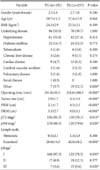
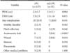
Table 1 presents the demographic characteristics of the patients in this study. The sex ratios were 2.1:1.0, and 2.7:1.0 in the PG and TG groups, without significant differences (P=0.346). The mean ages were 59.7±11.2 and 57.4±11.9 years without any significant differences (P=0.061). The mean body mass indexes and proportions of underlying diseases were not significantly different. In the PG group, the operating time was shorter than in the TG group (181.0±50.5 vs. 216.8±106.1 minutes; P<0.001). The number of retrieved LNs from the PG group was significantly smaller than that from the TG group (28.8±14.5 vs. 42.0±18.2; P<0.001). However, the number of metastatic LNs was not significantly different between the two groups (0.5±2.1 vs. 1.3±4.9; P=0.306). Stage I was 87.5% in the PG group and 78.3% in the TG group (P=0.030).
2. Postoperative morbidity
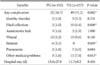
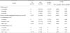
Hospital stay durations were similar across the groups (13.6±27.8 and 11.7±8.2 days for PG vs. TG; P=0.404). For short-term outcomes, postoperative complication rates were significantly lower for the PG versus the TG group (16.7% vs. 31.2%; P=0.002). Complications included wound problems, fluid collection, motility disorder, anastomotic leak, stenosis, pneumonia, and other medical problems. In particular, stenosis developed in 3.1% and 0% of the patients in the PG and TG groups, with significant differences. However, fluid collection and other medical problems developed in a significantly lower proportion of the PG group (P=0.008 and P=0.013) (Table 2).
3. Recurrence and survival
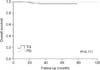
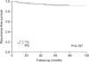
The five-year overall survival rates were 99.3% and 96.3% for the PG and TG groups, without any significant differences (P=0.111) (Fig. 2). The recurrence-free survival rates did not significantly differ between the groups (P=0.787) (Fig. 3). During the follow-up periods, eight and nine patients in the PG and TG groups, respectively, experienced disease recurrence. In particular, local recurrence including cancer on the remnant stomach was observed in 6 and 0 patients in the PG and TG groups, respectively.
4. Nutritional status
In the PG group, decreased vitamin B12 levels (<200 pg/ml) were observed in 64 patients (33.3%). Severe anemia, defined as Hb<10 g/dl, was detected in 13 patients (6.8%) and 41 patients (26.1%), and hypoalbuminemia (serum albumin <3.5 g/dl) was detected in 12 patients (6.3%) and 34 patients (21.7%) in the PG and TG groups, respectively, with significant differences (P<0.001) (Table 3).
5. Subgroup analysis of proximal gastrectomy
There was no significant difference in proximal resection margin (PRM) or distal resection margin (DRM) between the cPG and mLAPG groups. In addition, there was no significant difference in the rate of overall postoperative complications except reflux esophagitis (Table 4).
6. Reflux esophagitis
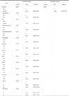
The proportion of patients with Visick scores higher than 3 were 0%, 0%, and 0% for TG; 19.1%, 18.5%, and 9.6% for cPG; and 11.4%, 5.7%, and 2.9% for mLAPG at 3 to 6 months, 12 months, and 24 months, respectively. The incidence of reflux symptoms decreased gradually with time and finally there was no statistical difference between the TG and mLAPG groups after 24 months (P=0.131). In contrast, the reflux symptoms associated with the cPG group remained.
The incidence of reflux esophagitis by LA classification was higher for PG than TG (37.4% vs. 3.7%; P<0.001). Overall reflux esophagitis by LA classification was significantly lower for mLAPG than for cPG (14.3% vs. 42.0%; P=0.002). Reflux classified as LA classification A or B did not differ significantly between the cPG and mLAPG groups (P=0.482 at 6 months; P=0.611 at 12 months; and P=0.771 after 1 year). There was a lower tendency for reflux grade C or D to appear subsequent to mLAPG, but there was no statistically significant difference. For cPG, 11 patients (7.1%) experienced severe reflux esophagitis defined as LA classification C or D, but no cases were noted in the mLAPG group at 1 year( Table 5).
Univariate and multivariate analyses indicated that the operative methods of PG were related to the incidence of reflux esophagitis. mLAPG was the only independent factor associated with a lowering of the incidence of reflux esophagitis compared to cPG (P<0.001) (Table 6).
Discussion
The choices regarding operative technique, extent of resection, and reconstruction method for upper-third EGC remain controversial. An et al.8 indicated that PG is associated with a high risk of esophageal reflux, which substantially affects patients' quality of life. Meanwhile, some authors have reported good radicality and safety in patients who undergo PG.2910 In this study, we aimed to comparatively analyze the functional and oncological outcomes of PG and TG. We reported data with special reference to postoperative reflux esophagitis, given it is the most common complication after PG. Thus, we evaluated factors affecting postoperative reflux esophagitis after PG by subgroup analysis (cPG vs. mLAPG). We attempted to preserve the LES by resection of the PRM at the GEJ. Pyloroplasty was not performed in mLAPG because the pyloric sphincter was considered to be able to reduce biliary or pancreatic reflux, as reported by Imada et al.11 and Park et al.12
Recently, esophagojejunostomy with double tract reconstruction or jejunal interposition after PG has shown acceptable incidence rates of anastomotic stricture and reflux esophagitis.13 However, laparoscopic jejunal interposition has not yet gained acceptance owing to its complicated technique, including the formation of a pedicled jejunal flap, the formation of three anastomoses, and longer surgical time.1415 For double tract reconstruction, the long term benefit must be followed up because food passage through the distal stomach/duodenum may not be consistent.
PG required less surgical time (181.0±50.5 minutes) than TG (216.8±106.1 minutes), as reported in previous studies.16 Regarding the LN dissection range, TG showed a greater number of examined LNs, but the difference in the number of metastatic LNs was not statistically significant. The short-term complications, except for reflux within the first postoperative month, were significantly lower in the PG group versus the TG group. In particular, the rates of fluid collection and medical problems excepting pneumonia were significantly lower. However, the occurrence rate of stenosis was significantly higher in the PG group (3.1%) compared with the TG group (0%). In our study, stenosis was effectively managed by balloon dilatations in most patients, which concurs with other reports.1718 The stenosis can develop because of postoperative complications, such as anastomotic leakage, infection, poor vascularity, or fistula formation in the upper part of the gastric tube.1920 Pierie et al.17 concluded that poor vascularization of the gastric tube and anastomotic leakage are associated with anastomotic stricture development because three of the four arteries are sacrificed during cervical esophagogastrostomy. According to the pattern of complications, it is possible that inflammation caused by reflux and poor vascularity are important risk factors. Meanwhile, the length of hospital stay was not different between the two groups. For TG patients, the incidence of wound complication (9.6%) and other medical problems (7.0%) including phlebitis, cystitis, voiding difficulty, atrial fibrillation, gout, and zoster was relatively higher than that of patients who underwent PG. However, those complications did not affect beginning meals, so there were only immaterial alterations to hospital stays.
PG in the upper third of the stomach is believed to be appropriate in terms of radicality and safety.1014 Similarly, in the present study, the overall survival and recurrence-free survival did not significantly differ between the PG and TG groups. However, eight and nine patients in the PG and TG groups, respectively, experienced disease recurrence during the follow-up period. There was no difference in the overall recurrence rate. However, local recurrence including cancer on the remnant stomach was observed in 6 and 0 patients in the PG and TG groups, respectively (4 of 157 patients in the cPG and 2 of 35 patients in the mLAPG group). This is a cause for concern, but the recurrences may have resulted from initial experiences. Therefore, future studies must include a larger number of cases. Three patients in the mLAPG group succumbed to disease recurrence: the first case (pT2N1M0, 1/34) was a systemic recurrence that involved liver metastasis. The second case of recurrence was observed in a male patient who underwent mLAPG (pT1bN0M0, 0/22); the proximal and DRMs were 2 cm and 3.4 cm. After 4 months, recurrence in the anastomosis site was detected after a routine upper gastrointestinal series. This was an early experience, but the margin was adequate. The third case occurred in a male patient who underwent mLAPG after endoscopic submucosal dissection. On pathological examination, no residual tumor was found. After 2 years, recurrence in the anastomosis site was detected on endoscopy, which we considered to be caused by signet ring cell type cancer.
Of the PG patients, 66.7% did not require supplementation with vitamin B12, and only 7.3% required periodic supplementation. The higher level of Hb in the PG group clearly indicated that PG was superior to TG in reducing the incidence of anemia. The major cause of hypoalbuminemia seemed to be a decrease in nutritional intake2122; therefore, hypoalbuminemia was more severe in the TG group than in the PG group.
At our institution, TG was usually performed using open methods during the study period. However, in PG cases, the incidence of LAPG cases has increased recently, so that PG was modified to mLAPG. Moreover, patients who underwent mLAPG had better reflux symptoms than patients who underwent cPG. Therefore, we evaluated factors affecting the incidence of postoperative reflux esophagitis after PG by subgroup analysis (cPG vs. mLAPG). Subgroup analysis showed no significant difference in PRM or DRM between the cPG and mLAPG groups. In addition, there was no significant difference in overall postoperative complications, except for reflux esophagitis (Table 4). Therefore, it is important to make efforts to decrease the incidence of reflux esophagitis after PG. A major finding of this study is that the lower incidence of overall reflux esophagitis in mLAPG was lower, and that mLAPG was found to be a significant risk factor for reflux esophagitis after PG.
Symptoms of post-TG esophagitis include bile reflux, pain caused by the alkaline reflux in the digestive tract induced by bile reflux, and stenosis. Meanwhile reflux symptoms after PG may be caused by bile, acid, or both.23 Reflux after PG has been a matter of concern,816 and the PG group in our study had a higher incidence of reflux symptoms than the TG group. However, in mLAPG, the incidence rates gradually decreased with time and there was no statistically significant difference between TG and mLAPG after 24 months (P=0.131). This finding could be attributed to symptom control through lifestyle modification and medication, as well as adaptation. In contrast, reflux symptoms were found to be persistent in cPG. In particular, a comparison between the cPG and mLAPG groups based on LA classification showed that the mLAPG group had a lower tendency toward severe reflux esophagitis of grade C or D. Overall, reflux esophagitis by LA classification was significantly lower in the mLAPG group compared with the cPG group (14.3% vs. 42.0%; P=0.002). Univariate and multivariate analyses showed that mLAPG was the only significant negative risk factor for reflux esophagitis after PG (P<0.001). When using mLAPG, we aimed to preserve the intra-abdominal esophagus as much as possible by resecting at the GEJ to save the LES. However, we could not measure the length of the preserved esophagus. Instead, we investigated the mean length of resected esophagus according to pathologic specimen pictures (0.47±0.09 cm for cPG and 0 cm for mLAPG; P<0.001). Thus, mLAPG may be more beneficial than cPG for lower reflux esophagitis. Pyloroplasty was also selectively performed according to surgeon's preference, especially in the cPG group. All three operators performed pyloroplasty whenever indicated, mainly in the early period of the study rather than the more recent period. When analyzing the incidence of reflux esophagitis for the pyloroplasty and non-pyloroplasty groups, no significant difference was found between the two groups. In addition, several studies of fundoplication after PG have been reported.242526 However, the results were not confirmative or promising. Moreover, fundoplication after PG is not technically easy, even when using the laparoscopy-assisted method. Therefore, we did not perform anti-reflux procedures in this study.
Our study has several limitations. First, selection bias may have come into play. The number of PG and TG cases was different; in addition, data regarding both older and recent PG cases were collected, but only recent data from TG cases were collected. Moreover, the length of the preserved esophagus could not be measured. This was a retrospective study and based on the operation record. For all procedures, we dissected the enitre intra-abdominal esophagus from the crus to the GEJ, but the length of the esophagus was different in every case. Accordingly, the length of the preserved esophagus could not be measured, which made it impossible to determine the incidence of reflux esophagitis in relation to the length of the preserved section. The relatively small number of mLAPG cases was analyzed owing to the limited experience with this technique. Finally, we did not evaluate the incidence of dysphagia, which is another common complication. Further studies investigating the issue of dysphagia are needed.
In conclusion, PG had an advantage in terms of postoperative morbidity and nutrition, and yielded a comparable prognosis to TG. The incidence of reflux esophagitis in PG can be lowered by preserving the intra-abdominal esophagus.


 XML Download