

 PDF
PDF ePub
ePub Citation
 Print
Print


INTRODUCTION
Strongyloides stercoralis has both free-living and parasitic life cycles with reproduction occurring in the latter by parthenogenesis (1). Infections are acquired when larvae penetrate the skin and migrate to the small intestine to mature, and internal autoinfective cycle allows the parasite to persistently survive within the human being for years (1). S. stercoralis has a cosmopolitan distribution in tropical and subtropical regions (2), and S. fuelleborni occurs in African primates where identical infection can be shared with humans (3).
S. stercoralis cause opportunistic infection in immunocompromised patient and is being increasingly detected due to increasing numbers of immunosuppressed patients in Korea. A total of 40 cases have been reported in Korea (4). Parasitological diagnosis is usually made by identifying worms in feces, or sometimes in body fluids or tissue samples (1). Most of the worms identified in clinical specimens were rhabditoid larvae, while the adults could only be seen in vitro. We report a case in which free-living S. stercoralis adults were recovered from human stool for the first time.
CASE REPORT
A 76-year-old male, living in Cheonan-si, Chungcheongnam-do, Republic of Korea, came to the emergency room of Dankook University Hospital on August 17th, 2007. He complained of dyspnea and peripheral edema, and also had abdominal pain, which developed 10 days ago. His past medical history showed that he was suffering from alcohol addiction and had undergone gastrectomy 30 years ago due to gastric cancer. In 2004, he was diagnosed with liver cirrhosis, and multifocal hepatoma was discovered one year later. Thereafter, he has been treated with transcatheter arterial chemoembolization (TACE) on twelve occasions at National Cancer Center. He appeared chronically ill and was very thin. He was intermittently suffering from abdominal pain, which was aggravated 10 days prior to visiting emergency room. Physical examination revealed tenderness over the umbilicus region. Pitting edema was present on both feet. Hemoglobin level was 9.9 g/dL and platelet level was 94,000/mm3. Serum GOT and GPT levels were 114 IU/L and 67 IU/L, respectively, while total bilirubin level was 2.23 mg/dl. White blood cell count was within normal range, while eosinophil count was increased to 22%.
Stool was obtained on the morning of 18th of August, 2007 and examined two days later. Numerous adult and larval worms were detected with the former being predominant. In total, 140 worms (103 adults) were recovered; the ratio of male to female was 1:1.3. Since the patient was suffering from constipation, second stool sample was available not until after six days. Observation of second stool specimen revealed no adult worms in microscopic examination but showed rhabditiform larvae. He was treated with albendazole, and transferred to the National Cancer Center.
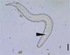
The morphological characteristics of the worms were as follows. The larvae were 412.2 µm (347.5-460.0) in length, and 26.3 µm (22.5-35.0) in width. The buccal cavity was rectangular. The esophagus, which occupied nearly one-fifth of the body, was 75.4 µm (62.5-87.5) and had central narrowing with club-shaped end, indicating typical rhabditoid larvae of S. stercoralis. Beneath the esophagus, the intestine was connected to the opened anus. The tail contained a pointed end (Fig. 1).
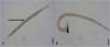
Female worm was 913.1 µm (795.0-1045.0) in length and 56.6 µm (42.5-67.5) in width. Esophagus consisted of three part: the anteriorly cylindrical procorpus, the narrow isthmus, and the round, posterior bulb, measuring 123.5 µm (110.0-152.5) in length. Short vulva opened at the middle of the body, and two-horned uterus was seen. Most of the body was occupied by intrauterine eggs. The average number of eggs was 15.5 (7-26) per worm, and the average size of eggs was 41.8 (40.0-42.5) × 26.9 µm (25.0-30.0). Tail portion was pointed, and the anus opened laterally (Fig. 2A).
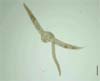
The male worm was stout, measuring 654.7 (617.5-685.0) µm in length and 38.3 (30.0-42.5) µm in width, being shorter than the female, and had a curved end. Esophagus was rhabditiform, measuring 112.9 (110-122.5) µm in length and had similar appearance compared to that of the female. The testis occupied the posterior body, with two ventrally protruded spicules. The spicules were parallel to each other, and nearly of equal size measuring 39.0 (37.5-40.0) µm in length (Fig. 2B). Mating adults were also found (Fig. 3). After the collection of second stool sample, the patient was treated with albendazole, and transferred to National Cancer Center for treatment of hepatoma. On the basis of morphological and structural characteristics of nematodes, adults and rhabditiform larvae were identified as S. stercoralis.
DISCUSSION
The average size of identified female worms in this case report was 913.0 µm in length and 56.6 µm in its greatest transverse diameter, and the male worms measured 654.7 µm in length and 38.3 µm in width on average. Although the size of the worms seemed to be slightly smaller then usual, they approximately met the criteria of the size of free-living adults of Strongyloides stercoralis (5): 1.0 to 1.7 mm by 50 to 75 µm for free-living female worms and 0.7 to 1.0 mm by 40 to 50 µm for male worms. Morphological characteristics such as the short vulva in female worms and two spicules in males also supported that they belonged to the free-living adults of S. stercoralis.
Free-living forms of S. stercoralis resembled Rhabditis sp., requiring careful examination for differential diagnosis. While the infection by adult worms of S. stercoralis from a human being had not yet been reported, there have been several reports on human infection by Rhabditis species (6, 7). Hence, the free-living adults of S. stercoralis could be misidentified as those of Rhabditis sp. In this study, species identification was mainly decided based on the length of buccal cavity. It was very short, nearly indistinguishable, in these worms, but Rhabditis sp. had relatively long buccal cavity (5).
In any case, recovery of the free-living adults from human stool was a very peculiar phenomenon. The stool of this patient was obtained on Saturday morning, and transferred to the department of Laboratory Medicine two days later. It could be possible that some of the rhabditiform larvae developed into free-living adults during the two-day delay, but this finding could not explain why no previous cases of adult S. stercoralis have not been reported from other patients, given the habitual delay between stool obtaining and examination. In vitro culture of S. stercoralis, there have been several reports on obtaining free-living adults. Lok observed that a small proportion of the first stage larva (L1) develop directly to the third stage larva (L3) within 24-48 hrs of deposition, and the time needed for growing into free-living adults was 72 hrs at 20℃ and 48 hrs at 22℃ (8). Nolan et al. suggested the importance of temperature by observing that the larvae developed to free-living adulthood in a day at temperatures below 34℃ (9). Hence, it was supposed that the fecal environment of this patient was optimum for the growth of S. stercoralis, but this hypothesis was not enough to explain the absence of S. stercoralis adult worms in other fecal samples. Further investigation is required to elucidate this phenomenon.
 XML Download