

 PDF
PDF ePub
ePub Citation
 Print
Print


INTRODUCTION
Hemangioblastomas (HBs) are benign vascular neoplasms that originate in the central nervous system (CNS) and comprise about 1.5-2.5% of intracranial tumors. They are usually in the cerebellum (76%) and uncommon in the cerebral hemispheres (9%), spinal cord (7%), and brainstem (5%) (1). A single tumor may be sporadic, but multiple tumors are almost always associated with Von Hippel-Lindau disease, an autosomal dominant disorder characterized by various lesions, including renal cell carcinoma, pheochromocytoma and visceral cysts.
Cerebellar HBs are traditionally classified into two morphologic types: cystic and solid (1). Cystic-HBs are associated with peritumoral edema (2), but solid HBs are not.
Here, we report a case of solid cerebellar HB with massive peritumoral edema.
CASE REPORT
An 83-year-old female visited our hospital due to a sudden headache. The headache was dull, continuous, and concentrated in the left temporo-occipital area. She did not complain of nausea.
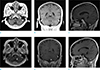
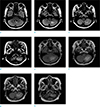
Five years ago, she had been admitted to our hospital with a similar headache. Brain magnetic resonance imaging (MRI) (Figs. 1,2) at the first admission showed a homogeneous enhancing mass (ovoid shape, 7.2 × 5.9 mm in size) with surrounding edema in the left lower cerebellar hemisphere. However, she refused management and did not attend the hospital thereafter.
The patient had no history of hypertension or diabetes mellitus, and no relevant family history was found. Her vital signs were within the normal ranges. A neurological examination was normal. Her blood tests were normal.
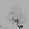
Follow-up brain MRI (Figs. 1,2) 5 years later showed an increased size of the homogeneous enhancing mass (9.0 × 7.4 mm) with aggravated peritumoral edema in the left lower cerebellar hemisphere. Cerebral angiography (Fig. 3) showed a highly vascularized mass in the cerebellum, which was compatible with a solid-type HB.
Chest and abdominal computed tomography were normal. The patient underwent gamma knife surgery. After the treatment, her symptoms improved.
DISCUSSION
This report describes a patient with solid cerebellar HB with massive peritumoral edema. Although peritumoral edema of HB is associated with cystic lesions of HB (2), it is interesting that the solid-type HB may also contribute to formation of peritumoral edema.
Cerebellar HBs are divided into four subtypes according to histological (3) and MRI (4) findings: simple cyst without a macroscopic nodule (5% of posterior fossa HBs), cyst with a mural nodule (60%), solid tumors (26%), and solid tumors with small internal cysts (9%). In this case, the cerebellar HB was solid. However, histologically, there is no distinct differentiation between the mainly cystic and solid tumor subtypes (5). Both subtypes present with a biphasic differentiation composed of large vacuolated stromal cells and a dense capillary network.
Generally, HB tends to enlarge extremely slowly. In particular, solid lesions remain stable in size and therefore asymptomatic for many years (1). The most common symptom of cerebellar HB is headache. Vomiting, ataxia, vertigo, diplopia and nystagmus are also common symptoms. The majority of symptomatic tumors are associated with cysts (6), and the progressive growth of the cystic component of the tumor results in mass effects.
Peritumoral edema is a precursor to formation of HB-associated cysts, which could progress into peritumoral cysts in the cerebellum (2). Peritumoral cyst formation in CNS is initiated by increased vascular permeability of the tumor. As the plasma ultrafiltrate enters the interstitial space of the tumor, interstitial pressure increases and plasma extravasates with convective distribution into the surrounding tissue. If the delivery of plasma from the tumor exceeds the capacity to absorb the extravasated fluid of the surrounding tissue, edema and subsequent cyst formation occur (2).
However, this case showed a solid HB with peritumoral edema and aggravated edema at the 5-year follow up. Although similar imaging cases that showed peritumoral edema in solid HB have been reported (57), they did not emphasize its importance. By cerebral angiography, solid lesions often demonstrate a complex arteriovenous malformation with a clear vascular blush, multiple feeding vessels and draining veins (5). It could be hypothesized that peritumoral edema of solid HB has a similar mechanism to that of peritumoral cysts. Based on the pathogenesis of HB, which depends on increased tumor vascular permeability, extravasation of fluid may contribute to formation of both edema and cyst. Indeed, chemotherapeutic agents that directly or indirectly decrease vascular permeability, may reduce both edema and cyst formation (8).
This case suggests that solid cerebellar HB might also be accompanied by peritumoral edema; clinicians should be aware of these imaging characteristics of solid HB.
 XML Download