

 PDF
PDF ePub
ePub Citation
 Print
Print


INTRODUCTION
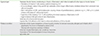
Atopic dermatitis (AD) is a chronic, relapsing and remitting inflammatory skin condition that usually develops in early childhood. Data derived from the International study of asthma and allergies in childhood (ISAAC) has shown a worldwide prevalence ranging from 2% to 20% with a tendency for higher prevalence in affluent European and Australasian settings, and rising eczema burden in most developing country settings and in younger children [1, 2, 3]. Diagnosis depends on clinical assessment and the National Institute for Health and Care Excellence (NICE) guideline recommends the use of the validated criteria for AD (Table 1), which was derived from the Hanifin and Rajka diagnostic criteria [4, 5]. Most cases of childhood AD are mild with infrequent exacerbations and minimal impact to quality of life. However, more severe AD is correlated with poorer overall health, impaired sleep and increased healthcare utilization [6, 7]. There also appears to be an association between severe AD and multiple comorbid chronic health disorders, both atopic and nonatopic, such as asthma, hay fever, food allergies, recurrent ear infections, visual problems and impaired dental hygiene [6, 7].
Much remains to be understood about the complex pathophysiology of AD, however, it is widely accepted that the skin commensal microorganism Staphylococcus aureus (SA) has an important role in the disease process [8]. The skin of patients with atopic eczema has a tendency to be colonized by SA [8, 9, 10, 11, 12]. This bacteria has been isolated from up to 90% of lesional skin [13, 14, 15] and has also been found in clinically uninfected areas, as well as in several reservoirs such as the anterior nares, the axillae and the perianal region [16, 17] in patients with AD. It has been reported that self-contamination from these reservoir sites or colonized skin lesions can be as high as 73% [18, 19, 20]. In comparison, SA can be isolated from the skin of 5-30% of unaffected individuals [17, 21, 22, 23]. Colonization with SA within the first year of life has been linked to AD prevalence in the first and second years of life and poses a 4.29-fold risk of moderate to severe AD in early childhood, although this link has been disputed [24, 25]. Staphylococcal colonization also persists despite eradication strategies with studies showing recurrence in almost 100% of patients with eczema after antistaphylococcal treatment, with pathogenic strains being reisolated after months of adequate treatment [17, 26]. Family members also often serve as a source of rapid recolonization [27]. The clinical significance of colonization is that there may be a correlation between SA colonization with severity and exacerbation of disease [11, 12, 22].
STAPHYLOCOCCAL COLONIZATION IN ATOPIC ECZEMA
Extensive research has shown that the propensity for SA colonization is complex and multifactorial [12]. It has been suggested that the underlying allergic skin inflammation of AD may contribute to increased colonization of SA as skin inflammation injures the skin barrier and exposes extracellular matrix adhesins which facilitate SA adherence [12]. The role of inflammation and bacterial adherence is based on studies showing significantly reduced colonization on the skin following topical anti-inflammatory drugs alone, such as corticosteroids or calcineurin inhibitors [28, 29, 30, 31]. There is also an understanding that individuals with skin disease may be genetically predisposed to microscopic structural changes in the skin barrier, including increased synthesis of extracellular matrix adhesins, fibronectin and fibrinogen for SA [32, 33], reduced skin lipid content, an alkaline skin surface pH [34, 35, 36] and reduced production of endogenous antimicrobial peptides due to defective innate immune responses [10, 12]. Collectively, this skin barrier dysfunction enhances SA colonization. It also allows the entry of SA superantigens (SSAgs), as well as allergens and irritants, thus contributing to exacerbation of skin disease [10].
SSAgs are exotoxins produced by SA which play a key role in the chronic inflammatory nature of AD. They have the ability to trigger an enhanced inflammatory response through the stimulation of a variety of T-cell clones and cytokine secretions [12]. Over 70% of SA strains isolated from the skin of AD patients produce superantigens such as alpha-toxin, toxic shock syndrome toxin-1 and staphylococcal enterotoxins and their role has been established in several immunohistological studies based on SA strains isolated from the lesional skin of patients with AD [26, 37, 38, 39]. Increased levels of antistaphylococcal superantigen specific IgE and IgM antibodies, in addition to cytokines such as interleukin (IL) 4 and interferon-γ, have also been quantified in the sera of most patients with AD compared to normal controls and have been shown to decrease with treatment [11, 40]. There is also a positive correlation between colonization with superantigen producing strains of SA and clinical severity of AD [41, 42]. SSAgs can also induce corticosteroid resistance, which may increase the severity of skin disease [43, 44]. This has led to the hypothesis that eradication of SA may lead to a steroid sparring effect [11], which is an incentive to management as long term topical corticosteroid use may lead to adverse effects such as local irritation, skin atrophy and skin depigmentation [5].
There is also an emerging role on the impact of environmental proteases in the pathogenesis of AD. SA, in addition to environmental allergens such as cockroaches, dust mites, pollen and fungi, also produce proteases which may enhance the passage of allergens across the skin barrier, or act as allergens themselves, and contribute to the inflammatory response [45]. Extracellular proteases produced by SA, such as serine-, cysteine- and metalloproteases [46, 47], have been found in in vivo mice studies to compromise the epidermal permeability barrier [48]. These bacterial proteases are also insensitive to and have the ability to inactivate some human plasma proteases inhibitors and interact with host defense mechanisms and tissue components in a complex manner that further augments the infection process and enhances SA survival [46, 47].
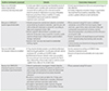
SA infection is therefore related to the pathogenesis of eczema (Table 2). Consequently, there is a theoretical basis for the eradication of SA in patients with eczema in order to prevent aggravation of skin disease.
THE RATIONALE FOR ANTISEPTIC USE IN ATOPIC ECZEMA
There has been wide interest in the use of antistaphylococcal agents as ancillary therapy for the management of AD. Treatment strategies can be divided into the management of clinically infected skin disease and the prophylactic treatment of individuals who are at risk of colonization or infection, and ranges from the use of systemic and topical antibiotics as well as antiseptic agents [49, 50]. Antiseptics represent an alternative to topical antibiotics in patients with AD. The main advantages of antiseptics over antibiotics are that they have low potential of inducing bacterial resistance, rarely cause delayed-type hypersensitivities or allergic reactions and come in a variety of preparations to suit individual needs. Antiseptics include triclosan, potassium permanganate, sodium hypochlorite or bleach and chlorhexidine gluconate [49]. It is widely accepted that disease flares caused by secondary infection with SA requires and responds to treatment with antibiotics and antiseptic agents [11, 51, 52]. However, good scientific data on the subject are rare. Clinically infected or 'impetiginised' skin disease can be characterized by increased erythema, superficial pustular eruption or purulent exudation with crusting and can spread easily [5]. Antiseptic baths can be particularly useful in reducing crusting and consequently, bacterial load [49]. Antibiotic treatment alone has no impact on allergic skin inflammation although a number of studies showed that topical or systemic antibiotics were able to reduce colonization density, resulting in partial improvement of skin lesions [17, 53, 54]. The NICE guideline recommends a one to two weeks course of flucloxicillin as first-line treatment to treat widespread infection with SA in children with atopic eczema [5]. Topical antibiotics, including preparations combined with topical corticosteroids, can be used for no longer than two weeks in patients with clinical infection in localized areas. However, the benefit of antibiotic use in AD is still disputed with a Cochrane review presenting studies showing no significant difference in global outcome for clinically infected eczema when oral antibiotics were compared with placebo or when topical steroid antibiotic combinations were compared with steroid alone [50]. Given the contradictory results available, it would be prudent to be restrictive in prescribing antibiotics, mainly because of an increasing resistance problem worldwide [55, 56, 57, 58].
For children with AD and are prone to recurrent infections, the short term use of antiseptics, such as triclosan or chlorhexidine, can also be used as adjunct therapy to reduce bacterial load [5]. One percent to 2% triclosan or 0.5% to 1% chlorhexidine added to emollient may be applied on a daily basis on the whole body or on affected areas. Alternatively, twice weekly antiseptic bathes have been recommended [49]. However, it appears that the recommendations for the topical use of antibiotics and antiseptics are not substantiated by good quality clinical data [49, 50].
Antiseptics in clinically infected eczema
In a Cochrane review released in 2010, Bath-Hextall et al. [50] and Birnie et al. [50, 59] performed a systemic review of 26 randomized controlled trials (RCTs), involving 1,229 participants, of interventions to reduce SA in eczema to determine whether they were superior than standard therapy in clinically infected or clinically uninfected atopic eczema. The review found one RCT assessing the effect of an antibacterial bath additive on clinically infected AD. This study by Huang et al. [51] involved 31 children with clinically infected moderate to severe AD who were pretreated with oral cephalexin then treated with either intranasal mupirocin ointment and bleach baths (treatment arm) or intranasal petrolatum ointment and plain water baths (placebo arm) for 3 months. Whilst the RCT concluded that there was reduced clinical severity of clinically infected AD in the treatment arm based on eczema area and severity index (EASI) scores (7.9 points, p = 0.017 at 1 month; 12.1 points, p = 0.004 at 3 months), the study was criticized as it was unclear if any adjustment was made for the differing baseline EASI scores between the two group of patients who were treated for more than one month (26.9 in the treatment group and 17.7 in the placebo group) [50, 51]. Of further clinical relevance is that eradication of SA was not achieved despite pretreatment with daily cephalexin for 2 weeks followed by 3 months of twice daily intranasal mupirocin treatment for 5 consecutive days each month and twice weekly bleach baths [51]. With regards to the use of topical antiseptic agents, the Cochrane review found one poorly reported study assessing the benefit of povidone-iodine in 15 children and adults with clinically infected mild to moderate AD but was unable to perform a group analysis [50].
Topical antiseptics in clinically uninfected eczema
The role of antistaphylococcal agents used preventively in the setting of uninfected skin in AD against exacerbation is less clear and remains a controversial area in the management of AD, particularly with the fears of the emergence of antibiotic-resistant strains of SA [56, 60]. Therefore, antiseptics used in this setting may be a suitable alternative as they have less potential to induce resistance [49]. The Cochrane review reported a lack of good quality large RCTs and found that there is no good evidence to support the use of antistaphylococcal agents in uninfected eczema [50]. A review by Schnopp et al. [49] presented 15 studies which assessed the efficacy of a range of antiseptic treatments including triclosan, chlorhexidine, didecyldimethylammoniumchloride, povidone iodine, bleach, silk coated textiles and special silk fabric. However, differing endpoints, inclusion criteria and treatment protocol in the studies made it challenging to determine the benefit of antibacterial therapy in AE. A key finding was the importance of anti-inflammatory therapy and its role in reducing bacterial colonization.
AN UPDATED REVIEW OF STUDIES ASSESSING ANTISEPTIC USE IN ATOPIC ECZEMA
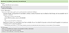
Given the growing potential of antiseptic use in AE, we reviewed studies assessing the efficacy of antiseptics in AD in the last five years (to March 2014). PubMed was searched for studies in English containing bleach, triclosan, chlorhexidine, sodium hypochlorite, povidone iodine or potassium permanganate and eczema or AD. The efficacy of fabrics treated to provide antimicrobial activity was not assessed in this review. The results were hand searched for relevant studies, with 4 studies found, Table 3, including the bleach bath study by Huang et al. [51] which has already been reviewed [49, 50].
Sodium hypochlorite in AD management
It appears that there is growing interest in the role of sodium hypochlorite in the management of AD. Its antistaphylococcal activity, including against methicillin-resistant SA, has been established in in vitro and in vivo studies, with concentrations as low as 0.005% being shown to be safe and effective [61, 62, 63, 64]. Its antimicrobial effect is probably due its ability to cause irreversible aggregation of essential bacterial proteins [65]. Traditional dilute bleach baths have concentrations that range from 50 parts per million (ppm; 0.005%; equivalent to 0.25 cup bleach in half a tub of water) to 90 ppm (0.009%; equivalent to half a cup of bleach in half a tub of water) [66]. Furthermore, sodium hypochlorite is better tolerated, is easily accessible and cheaper compared to other antiseptic agents [49].
In a pilot feasibility study, Ryan et al. [66] assessed the efficacy of a 0.0061% sodium hypochlorite containing cleansing body wash in 18 children with clinically uninfected moderate to severe AD who also had lesional cultures positive for SA at baseline. Patients used the wash three days a week for 12 weeks and were allowed to continue with their individualized topical and systemic treatment regimen. Clinical response was measured using an investigator global assessment (IGA) score and the percentage of body surface area (BSA) affected. Acceptability and tolerability of the product was also assessed retrospectively using a questionnaire completed by parents [66]. The use of sodium hypochlorite gel cleanser was reported to result in statistically significant mean reductions in IGA scores at all follow-up time points-1.0 at 2 weeks (p = 0.01, n = 6), 0.8 at 1 month (p = 0.005, n = 11), 0.8 at 2 months (p = 0.01, n = 12), and 0.9 at 3 months (p = 0.002, n =13). The overall mean reduction in IGA score from baseline to final measurement was 0.9 (p = 0.001, n = 18). The mean reduction from baseline of BSA affected was 10.7% at 2 weeks (p = 0.22, n = 6), 10.9% at 1 month (p < 0.001, n = 11), 14.6% at 2 months (p = 0.001, n = 12), and 16.8% at3 months (p = 0.01, n = 13). The overall mean reduction from baseline to final measurement in all patients was 14.8% (p = 0.005, n =18). A statistically significant reduction in bacterial count was found at 1 month only (p = 0.01, n = 10), despite following a decreasing trend. There were no reports of patients developing infective exacerbations requiring oral antibiotic treatment during the course of the study. The use of sodium hypochlorite gel cleanser was also highly accepted and tolerated by patients and their parents with high scores recorded on the patient satisfaction questionnaire. Patients also unanimously preferred this product over traditional dilute sodium hypochlorite bleach baths [66]. The authors acknowledged that the limitations of the study were that it was a small non blinded study that lacked a placebo arm and had many of the retrospective patients in the initial phase of the study lost to follow up [66]. A larger randomized controlled study assessing the clinical efficacy of sodium hypochlorite containing body wash is thus needed to confirm the encouraging results of this pilot study and should include the impact on the usage of the patient's topical and systemic regimen and whether it has any benefit in clinically infected AD.
Wong et al. [67] performed a 2-month prospective randomized, double-blinded, placebo-controlled study assessing the efficacy of 0.005% sodium hypochlorite baths in 36 patients aged between 2 and 30 years old with uninfected moderate to severe AD. Patients were given either 100mL of 5% sodium hypochlorite (bleach treatment arm) or distilled water (placebo arm) and instructed to add this into 100 L of water. They were instructed to soak neck down in these baths for 10 minutes, twice a week for two months. Patients then rinsed off with normal tap water and were allowed to maintain their individualized regimen of topical anti-inflammatory and emollient therapy. Aqueous cream as soap was provided to standardize their bathing regimen. Follow-up was performed at baseline, week 2, week 4, and week 8 (end) of treatment [67]. Response to treatment was assessed using the EASI score, a validated composite score of BSA from four regions (head and neck, upper limbs, trunk and lower limbs) and a physician's assessment of erythema, edema/induration/papulation, excoriation and lichenification on a 4-point scale. Patients' assessment of overall response and intensity of itch was assessed using visual analog scale and bacteriological assessment was performed by calculating SA density from a swab of the worst affected sites. Safety based on clinical adverse events reported by patients or observed by the physician was also assessed [67]. Overall, the study reported that the use of diluted bleach baths resulted in clinically improved AD from 1 month and was tolerated by patients. The study reported a significant reduction in EASI scores between the treatment and placebo groups at 1 and 2 months (p < 0.001) suggesting clinical improvement was achieved and sustained after 1 month of treatment. There was also significant improvement in EASI scores and various clinical parameters corresponding to the upper limbs, trunk and lower limbs (areas of the body submerged in the baths) at 2 months in the treatment group. Whilst the head and neck of patients in the treatment group showed improvement in the above outcomes, this was not statistically significant and may have been due to partial submersion of these areas. The improvement in EASI scores in the treatment group also corresponded to a significant reduction in percentage BSA affected at 2 months (p = 0.002). These improvements appeared to be evident across all age groups [67]. The use of bleach baths was also associated with a reduction in density of SA over time, although this was not statistically significant. There was also no statistical difference in colony counts between the treatment and placebo groups at either 1 or 2 months. These findings were similarly found in the bleach bath study by Huang et al. [51] suggesting that clinically improvement in AD does not require complete eradication of SA [67]. There were no statistically significant differences in patient's overall assessment of response between treatment and placebo groups at either 1 or 2 months. Of patients in the treatment group, 27.8% experienced side effects which included burning/stinging and mild dry skin. A similar proportion of patients in the placebo group also experienced similar side effects. No patients withdrew from the study because of intolerance to the baths [67]. Limitations of this study were that it was a small study with short treatment and follow-up and since patients were responsible for administering their own treatment, the study was not performed in a controlled environment. Furthermore, this study was not a true double blinded study as patients would have been able to differentiate between bleach and placebo on the basis of odour. This was recognized by Huang et al. [51] in their own bleach bath study. Future studies also need to further assess the relationship between degree of SA colonization and clinical severity of AD since it appears complete eradication of SA colonization may not be necessary for disease improvement. Whilst dilute sodium hypochlorite is bactericidal in vitro [62], persistent colonization despite treatment in patients may reflect recolonization from sites not exposed to the antiseptic, such as the nares, or from members of the household who may be carriers. Improvement in disease may also reflect a shift in skin flora, as opposed to being solely related to reduction in SA colonization, as suggested in a separate study assessing the temporal shifts in the skin microbiome associated with disease flares and treatment in children with AD [68]. Based on ribosomal RNA bacterial gene sequencing performed on DNA obtained directly from serial skin sampling of children with AD, it was found SA was greater during disease flares and correlated with worsened disease severity and that AD treatments diversify skin bacteria preceding improvements in disease activity [68].
Triclosan in AD management
One study presented in this review was a double blinded RCT assessing the efficacy of emollient containing 1% triclosan in 60 patients with mild to moderate uninfected AD [69]. It was reported that emollient containing 1% triclosan significantly reduced AD severity at day 14 compared to vehicle control, as determined by mean SCORAD change from baseline (-8.86 vs.-4.75, p < 0.05), and was found to be safe and highly acceptable to patients after a study period of 27 days. Adverse events were reported in 15 patients however only 4 were considered related to treatment and included transient stinging pain after application of 1% triclosan containing emollient. This resolved spontaneously even with continued use of the cream. There were no subjects who withdrew from the study due to adverse events [69]. There was also improvement of severity at day 27 in the treatment group, however this was not significant (-11.46, 95% CI, 15.2-8.1 for treatment group vs. -9.71, 95% CI, 13.06-5.99 for control group; p > 0.05) [69]. A low potency corticosteroid was allowed to be used concurrently during the treatment period and interestingly, the authors reported significantly lower amount of topical steroid application by patients in the treatment group compared to the control (22 g and 44.2 g, respectively; p < 0.05), thus implying a steroid sparring and synergistic effect of 1% triclosan containing emollient [69]. It has been suggested that the reduction of SA colonization associated with antiseptic use produces this steroid sparring effect by reducing local SA enterotoxin induced corticosteroid resistance [43, 44]. Since this study was performed in patients with clinically uninfected eczema, no bacteriological analysis was performed [69]. Therefore, this putative effect requires further investigation as there is also a paucity of long term, good quality studies assessing the relationship between the reduction in SA colonization with antimicrobials, its impact on amount of corticosteroid used, whether it has any implications to corticosteroid potency required and how it affects clinical outcome.
CONCLUSIONS
As the skin of patients with AD has intrinsic properties that increase susceptibility to SA colonization, it is unlikely that the use of antistaphylococcal therapy alone, with the aim of eradicating primary infection, will be of long-term benefit, particularly with the high rates of recolonization. Optimal management requires addressing the key factors known to cause exacerbation of disease, primarily targeting improvement in skin barrier function and reducing skin inflammation. Consequently, the mainstay of AD management, which constitutes irritant and allergen avoidance, emollients to enable the repair and protection of the skin barrier with proper hydration and topical anti-inflammatory agents such as corticosteroids or calcineurin inhibitors, will remain important therapeutic options (Table 4) [70, 71]. The use of emollients and topical corticosteroids alone are able to reduce microbial colonization thus reducing the risk of secondary infection and the need for specific antimicrobial therapy [8, 72]. There is also an emerging rationale for a 'proactive' approach to the management of AD to achieve long-term remission between flares. This involves continued intermittent application of low dose topical anti-inflammatory agent over previously eczematous but now normal looking skin, together with daily application of emollient [73]. Again, this is based on the recognition that normal appearing skin is not immunologically normal and is often colonized by SA [8, 73, 74]. However, the proactive approach of topical anti-inflammatory agents is considered 'offlabel' use and the adverse effects of their long-term use cannot be ignored. It may also be difficult to achieve patient compliance thus hindering the reported benefits of this 'proactive' approach.
The studies available on the role of antiseptic agents in the management of AD suggest some benefit for the inclusion of antiseptic use. However, there are many limitations to these studies which therefore warrant further investigation on the impact of antiseptic use in AD. The approach of adding antiseptic agents to emollients and body washes is a convenient measure of introducing an additional agent into the AD management regimen. Future studies need to be large scale double blinded RCTs and should assess long-term efficacy, safety, tolerability and cost effectiveness. In particular, these studies should assess whether any flares or infected exacerbations occur with continued antiseptic use. The reported steroid sparring effect associated with antiseptic use should also be further investigated.
 XML Download