

 PDF
PDF ePub
ePub Citation
 Print
Print


INTRODUCTION
Pituitary adenoma accounts for 10-20% of all intracranial tumors, and is a highly angiogenic tumor that secretes vascular endothelial growth factor (VEGF) [12]. Angiogenesis or neovascularization plays an important role in tumor growth, which is determined by angiogenesis stimulatory factor and its inhibitors, VEGF and endostatin. VEGF is secreted by the normal pituitary glands and all types of adenomas may be involved in pituitary tissue growth [345]. Endostatin is one of a number of endogenously generated anti-angiogenic protein fragments that have anti-tumoral activity in a murine model [6].
Surgical resection is the initial treatment for pituitary adenoma; however, delayed recurrence after complete resection can occur in 24-80% of cases [7]. Stereotactic radiosurgery (SRS) has been used for pituitary adenoma in cases when the tumor has recurred or was not completely removed. Although pituitary adenoma control rates after SRS are high, analysis of angiogenic factors after Gamma Knife radiosurgery (GKS) may be needed to understand the anti-tumoral and anti-angiogenic effect of GKS [2].
Angiogenesis plays an important role in the progression of pituitary adenomas; however, no study has evaluated plasma endostatin and VEGF levels in patients with pituitary adenoma before and after radiosurgery. In this study, we evaluated plasma endostatin and VEGF levels in patients with pituitary adenoma before and after SRS.
MATERIALS AND METHODS
Patient population
Five patients with pituitary adenoma, all of whom provided written informed consent, were enrolled in this prospective protocol that was reviewed and approved by the Institutional Review Board of our hospital (No. KNUH 2012-07-021-001). All patients underwent SRS following surgical resection. SRS was considered for patients with recurrent or residual pituitary adenoma after an initial transsphenoidal resection. Three patients experienced three operations and two patients underwent one surgery before SRS. All pituitary adenomas were pathologically proven to be non-functioning pituitary adenomas. Peripheral blood samples donated from 10 volunteers wi-thout known malignancy or pregnancy were used as controls.
Sample collection and enzyme-linked immunosorbent assays for endostatin and VEGF
Peripheral venous whole blood was obtained before radiosurgery (baseline sample) and 1 week and 1 month after SRS. Blood samples from study participants were collected in ethylenediaminetetraacetic acid tubes and centrifuged at 3,200 rpm for 10 minutes at 4℃. The supernatant including the plasma fraction was transferred to a microtube and frozen im-mediately at -80℃ until analysis. The analysis was performed with commercially available enzyme-linked immunosorbent assays kits (Quantikine Human VEGF Immunoassay, R&D Systems, Minneapolis, MN, USA), according to the manufacturer's instructions. All patient samples collected were assayed simultaneously.
Radiosurgical technique and volume measurement
SRS was carried out using a Gamma Knife Model C (Elekta AB, Stockholm, Sweden). T1-, T2-, and enhanced T1-weighted magnetic resonance imaging (MRI) with a slice thickness of 2 mm was used for three-dimensional reconstructions and treatment planning. The MRIs were transferred to a workstation for post-processing and analysis. The Gamma Plan system was used to determine the GKS for all patients. Multiple small isocenters were used to deliver a highly conformal dose to the tumor. Tumor volume ranged from 1.1 to 4.9 mL (mean, 2.3 mL). Mean marginal dose was 13.6 Gy (range, 12-15 Gy). The 50% isodose line was used as the margin in all patients. Maximum radiation dose was 24-30 Gy (mean, 27.2 Gy). The size of each tumor before and 6 months and 1 and 2 years after GKS was measured retrospectively using the Gamma Plan workstation and a picture archiving communication system. Tumor progression was defined as an increase in tumor volume of at least 10%. Tumor regression was defined as at least a 10% decrease in tumor volume. Tumors that were ±10% of their original volume were defined as stable [7].
Statistical analysis
All analyses were performed using SPSS 18.0 for Windows (SPSS Inc., Chicago, IL, USA). Because the data were not distributed normally, they were analyzed using the Wilcoxon's rank-sum test, the Mann-Whitney U-test. Correlations were assessed using the nonparametric Spearman's rank correlation analysis. p-values<0.05 were considered significant.
RESULTS
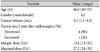
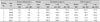
Of the five consecutive patients enrolled, three were men and two were women (mean age, 46.6 years; range, 40-55 years). Six men and four women with a mean age of 47.6 years (range 30-55 years) were included as controls. Blood samples were drawn from five pituitary adenoma patients before SRS and 1 week after SRS, and 1 month after SRS. The patient and tumor characteristics as well as the radiosurgical parameters, are summarized in Table 1. Four tumors had decreased in size and one tumor had increased in size at the 2 year MR follow-up (Table 2). No radiosurgical complications were noted.
Plasma endostatin and VEGF concentrations in controls
Mean plasma endostatin and VEGF concentrations in controls were 86.6 ng/mL and 29.3 pg/mL, respectively. There was no significant differences in mean plasma endostatin as well as VEGF levels between men and women. There was no correlation between mean plasma concentrations of endostatin and VEGF levels and age.
Plasma endostatin and VEGF concentrations in patients
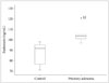
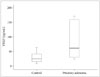
Mean baseline plasma endostatin level was 105.3 ng/mL (range, 97.0-120.2 ng/mL) in patients with pituitary adenoma, which was significantly higher than that in the control group (mean, 86.6 ng/mL; range, 71.3-98.2 ng/mL) (p=0.001) (Fig. 1). There was no significant difference in plasma endostatin levels between men and women (p=0.638). No correlation was found between endostatin level and age (p=0.873) or tumor size (p=0.391). Mean plasma VEGF level was 89.5 pg/mL (range, 24.1-171.8 pg/mL) at baseline in patients with pituitary adenoma and 29.3 pg/mL (range, 9.2-64.3 pg/mL) in the control group (p=0.050) (Fig. 2). Plasma VEGF levels in patients with pituitary adenoma were higher than those in the control group; however, no significant difference was observed between the two groups. No correlation was found in plasma VEGF levels between men and women (p=0.863). No correlation was observed between VEGF level and age (p=0.285) or tumor size (p=0.391).
Plasma endostatin and VEGF concentrations after radiosurgery
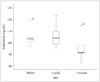
The effect of SRS for pituitary adenomas was evaluated by sequential measurement of plasma endostatin and VEGF concentrations before SRS and 1 week and 1 month after SRS. Mean plasma endostatin level changed to 106.6 ng/mL 1 week after SRS and then dropped to 95.9 ng/mL after 1 month (Fig. 5). There was a trend toward decreased plasma endostatin concentrations 1 month after SRS compared to baseline levels (p=0.195). Plasma endostain concentration 1 month after SRS was still elevated over control levels, even though it was not significantly different from control levels (p=0.140).
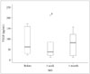
Mean plasma VEGF level following SRS decreased to 74.1 pg/mL at 1 week and 79.0 pg/mL at 1 month (Fig. 6). Mean plasma VEGF level 1 month after SRS decreased compared to pretreatment level; however, there was no significant difference from baseline levels (p=0.812). Plasma VEGF level 1 month after SRS was still significantly increased over controls (p=0.033).
Four patients with a decrease in tumor size experienced a constant decrease in endostatin and VEGF concentrations. One patient with an enlarged pituitary adenoma after SRS had an elevated VEGF level and a slightly decreased endostatin level (Fig. 7). This patient underwent surgical resection for a recurrent tumor 2 years after SRS. No patient had radiation necrosis following SRS.
DISCUSSION
Angiogenesis is essential for the development and progression of brain tumors. Angiogenesis is thought to be induced by an imbalance between pro- and anti-angiogenic factors. VEGF is the most potent angiogenic factor known so far and endostatin is a potent endogenous angiogenesis inhibitor [389]. Endostatin and VEGF have recently been implicated in the pathophysiology of pituitary adenoma. VEGF is a tyrosine kinase that plays an important role in angiogenesis and modulation of vascular permeability. VEGF-A binds with high specificity to VEGF receptor-1 and VEGF receptor-2 on vascular endothelial cells. These receptors modulate downstream signaling pathways affecting various cellular processes [10]. Increased plasma levels of VEGF are observed in patients with pituitary adenomas, compared to those in controls were detected [4]. Endostatin is a cleaved fragment of collagen XVIII, and inhibits migration of endothelial cells and induces endothelial cell apoptosis [6]. Plasma endostatin concentrations are significantly higher in patients with pituitary adenoma compared to those in controls [3].
Patients with pituitary adenoma usually undergo surgical resection; however, complete resection is often difficult. SRS is an effective and well-tolerated management option for recurrent or residual pituitary adenoma [2]. Recurrent or residual tumors are related to significant morbidity over the lifetime of a patient. Monitoring tumor response with biomarkers may be useful to improve long-term treatment outcomes of patients who undergo SRS for pituitary adenoma.
We evaluated plasma endostatin and VEGF levels in patients with pituitary adenoma before/after SRS and in a control group. We sought to understand angiogenic mechanisms regulating pituitary adenomas and the anti-angiogenic effect of SRS by studying changes in plasma endostain and VEGF levels in patients with pituitary adenoma undergoing SRS. A better understanding of the role of abnormal angiogenesis in patients with pituitary adenoma may lead to improved patient management and novel therapeutic options.
Plasma endostain and VEGF levels in patients with pituitary adenoma
Plasma endostain and VEGF levels were significantly higher in patients with pituitary adenoma at baseline in comparison to the controls in this study, which is consistent with those of previous studies [34]. Although several studies have found that abnormal local expression of endostain and VEGF plays a role in angiogenesis in pituitary adenomas, the cause of increased endostain and VEGF still remains unclear [13456]. Feldman et al. [1112] found that circulating endostatin and VEGF levels increase in patients with clear cell renal cancer and colorectal cancer with liver metastasis. They suggested that the correlation between circulating endostatin and VEGF may be associated with secretion of proteases that cleave endostatin from collagen XVII, both from tumor and endothelial cells. VEGF upregulates the release of proteases. Elevated plasma endostain levels may be a defense mechanism to protect the host from angiogenesis by VEGF [13]. Angiogenesis inhibitors are effectively suppress growth of experimental pituitary adenoma [14].
Plasma VEGF is detectable in the peripheral blood of normal healthy volunteers [9]. The origin and biologic role of VEGF in healthy subjects are unknown; however, this finding suggests a role for endothelial mitogens in the maintenance of physiologic endothelial integrity [8]. In healthy controls, a significant negative correlation between circulating endostatin and VEGF levels was found [3]. It results from balance mechanism between angiogenic and anti-angiogenic factors. Our results showed that VEGF and endostatin levels in the peripheral blood in patients with pituitary adenoma increased at the same time. Gruszka et al. [3] suggested that simultaneous elevation of endostatin and VEGF may attenuate the pro-angiogenic action of VEGF and could be responsible for the ra-ther weak neovascularization of pituitary adenomas. Pituitary adenomas are mostly benign and have lower vascular density than that of non-tumorous glands. Low microvascular density or inhibited angiogenesis may be a partial explanation for the relatively low growth potential observed in pituitary tumors [3515].
Change in plasma endostatin and VEGF levels
The plasma endostatin and VEGF levels in the patients with pituitary adenoma dropped 1 month after SRS but were still elevated over control levels, confirming abnormal angiogenesis in pituitary pathophysiology. This finding suggests that the natural history of a pituitary adenoma is related to disruption of endostatin and VEGF expression. Our result suggests that Gamma Knife-irradiated pituitary adenoma tissue has significantly less angiogenic activity than that of a previously untreated pituitary adenoma. Our results support the anti-angiogenic effects of SRS, which may be important for treating pituitary adenomas.
Unlike four patients, one patient experienced increased endostain and VEGF levels after SRS. That patient experienced a tumor recurrence 2 years after SRS. Systemic expression of endostatin and VEGF may be related to the development, progression in pituitary adenoma patients. O'Sullivan et al. [16] reported that the 5- and 10-year actuarial rates of recurrence or growth of a residual adenoma after resection are 24.4% and 51.5%, respectively. Monitoring peripheral blood biomarkers in patients with pituitary adenoma may be valuable for those with highly angiogenic tumors, which tend to recur. Serial plasma endostatin and VEGF measurements may be a useful to monitor the treatment efficacy of SRS for angiogenic tumor cells in the future.
The main limitations of our study were the small number of patients and the short follow-up period. Sufficient plasma endostatin and VEGF data in a control group and abnormal levels in patients with pituitary adenoma are needed to use them as biomarkers and a diagnostic tool to monitor the tumor response after SRS. A controlled systematic prospective study with a longer follow-up is necessary to investigate the anti-angiogenic effect of SRS in patients with pituitary adenoma.
In conclusions, plasma endostatin and VEGF levels in patients with pituitary adenoma are elevated over controls at baseline and decrease 1 month after SRS. This was a preliminary study on the potential association between plasma endostatin and VEGF levels and pituitary adenoma. Our results provide evidence for further work related to determining whether plasma endostatin and VEGF levels could be useful to monitor the tumor response after SRS.

 XML Download