Journal List > Brain Tumor Res Treat > v.3(2) > 1059672

Kim, Moon, Kim, Lim, Lee, Jang, Jung, Kim, and Jung: Role of Craniofacial Resection for Malignant Tumors Involving the Anterior Skull Base: Surgical Experience in a Single Institution

Abstract

Background

Craniofacial resection (CFR) has been regarded as a standard treatment for various tumors involving the anterior skull base. The purpose of this study was to evaluate the results of CFR for the patients with anterior skull base malignancies in our hospital.

Methods

We retrospectively analyzed 17 patients with anterior skull base malignancies treated with CFR between 2001 and 2012. Mean follow-up duration was 41 months (range, 2-103 months).

Results

Intracranial involvement was found in 11 patients (65%) and orbital extension in 6 patients (35%). Classical bifrontal craniotomy was combined with endoscopic endonasal approach in 14 patients and external approach in 3 patients. Vascularized flap was used for reconstruction of the anterior fossa floor in 16 patients (94%). The most common pathological type was squamous cell carcinoma (6 patients). Gross total resection was achieved in all cases. Postoperative complications developed in 4 patients (24%) and included local wound problem and brain abscess. One patient with liver cirrhosis died from unexpected varix bleeding after the operation. Although postoperative treatment, such as radiotherapy or chemotherapy, was performed in 14 patients, local recurrence was seen in 6 patients. The mean overall survival time after the operation was 69.0 months (95% confidence interval: 47.5-90.5 months) with a 1-, 2-, and 5-year survival rate of 82.3%, 76.5%, and 64.7%, respectively. Postoperative radiotherapy was found to be the powerful prognostic factor for favorable survival.

Conclusion

Considering the higher local control rate and acceptable complication or mortality rate, CFR with adjuvant radiotherapy is a gold standard treatment option for malignant tumors involving anterior skull base, especially with extensive intracranial involvement.

INTRODUCTION

Craniofacial resection (CFR) for anterior skull-base tumors has been used for about 30 years as a general surgical procedure since its original description [1]. Classical CFR consists of transfacial/transnasal and transcranial approaches. Recently, the endonasal approach using the endoscope, substitutive for the open transfacial approach, has been used as a universal surgical route. Furthermore, due to the development of the endoscope technique and instruments, a pure endoscopic approach, without a transcranial approach, has been attempted for tumor removal in the anterior skull base [2].
Although the pure endoscopic approach has been comprehensively compared with the classical traditional combined approach, there are many differences between the two groups of patients, and many constraints on the accurate comparison of the surgical results [3]. One study used purely endoscopic approach for the lesions without definitive invasion into skull base [4]. The study was limited by patient selection in being able to compare the surgical results of the classic CFR with the endoscopic techniques. Also, surgical outcomes by transcranial approach have recently improved as the result of advancement in microsurgical instruments and techniques.
In the light of these advancements, there is a need to re-evaluate the indications of CFR with endoscopic approach. The purpose of this study is to evaluate the surgical results for CFR in our hospital with the review of the recently published data.

MATERIALS AND METHODS

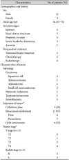
This study fulfilled all the requirements for patient anonymity and was approved by the Institutional Review Board (MP 2015-010). From 2001 to 2012, 17 patients underwent CFR for anterior skull-base malignancies in our hospital. The benign pathologic case was excluded. The medical records were evaluated for patient demographics, clinical features, staging, preoperative treatment, operative procedure, extent of tumor extension, postoperative complication, follow-up, recurrence rate, and survival rate. Mean follow-up duration was 41 months (range, 2-103 months).
Based on the preoperative computed tomography scan and/or magnetic resonance imaging and intraoperative findings, the exact location of tumor and its extension into orbit or intracranial structures were evaluated. The tumors were classified according to both the Kadish and Biller staging systems. The Kadish staging system describes three stages to determine the location and extension of olfactory neuroblastoma [4]: Stage A: tumor confined to the nasal cavity; Stage B: tumor confined to the nasal cavity and one or more paranasal sinus; and Stage C: tumor extending beyond the nasal cavity or paranasal sinuses, and includes involvement of the orbit, base of skull, or intracranial extension. The Biller staging system has four stages to determine the location and extension of remaining other tumors [5]. T1 stage is defined as a tumor involving the nasal cavity and adjacent paranasal sinuses (excluding sphenoid), with or without erosion of the bone of the anterior cranial fossa. T2 stage is defined as a tumor extending into the periorbital tissue or protruding into the anterior cranial fossa. T3 stage is defined as a tumor involving the brain that is resectable with margins. T4 stage is defined as a non-resectable tumor. All specimens were examined in our pathology department.
Overall survival (OS) was calculated from the date of surgery until death, or until the date of the last follow-up visit for patients who were still alive. Relapse-free survival (RFS) was also calculated as the time from the date of surgery to the date of recurrence or death. Survival rate was analyzed by the Kaplan-Meier method and compared with the log-rank test. For the multivariate analysis, independent prognostic factors were determined using the Cox's proportional hazards model. The statistical analysis was performed using SPSS version 20.0 software program for Windows (SPSS Inc., Chicago, IL, USA). The level of significance was set at p<0.05.

RESULTS

Patient demographics and clinical symptoms at the time of initial diagnosis are listed in Table 1. The most common symptoms were epistaxis and nasal obstruction. Two patients with paranasal malignancies underwent radiochemotherapy before CFR. The median age was 56 years (range, 34-74 years) and male to female ratio was 12 to 5. The most common pathological type was squamous cell carcinoma (6 patients). Tumors invaded only in the cribriform plate in 5 patients, extended through dura in 4 patients, and involved brain parenchyma in 7 patients. Orbit involvement was noted in 6 patients. All olfactory neuroblastomas were classified as stage C of the Kadish system. Majority of the remaining tumors were classified as T3 or T4 of the Biller classification system.
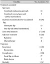
Classical bifrontal craniotomy was combined with endoscopic endonasal approach in 14 patients. External approach was used in 3 patients with severe orbital involvement for orbital exenterating (Table 2). Reconstruction of the anterior fossa floor was performed using vascularized flap in 16 patients (94%). Apart from 3 cases requiring free flap after orbital exenterating, the remaining 13 cases underwent nasoseptal flap. Gross total resection was achieved in all cases. Four patients (20%) experienced postoperative complications including local wound problem [3 patients; 2 with cerebrospinal fluid (CSF) leakage] and brain abscess (1 patient). One patient with liver cirrhosis died from unexpected varix bleeding. Although postoperative treatment such as radiotherapy or chemotherapy was performed in 14 patients, local recurrence was seen in 6 patients (35%).
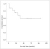
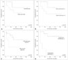
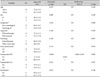
The mean OS time after CFR of anterior skull base tumors was 69.0 months [95% confidence interval (CI): 47.5-90.5 months, the median survival time was not reached]. One-, 2-, and 5-year survival rate was 82.3%, 76.5%, and 64.7%, respectively (Fig. 1). The results of analyses of the variables that could be correlated with OS are shown in Fig. 2 and Table 3. On univariate and multivariate analysis, postoperative radiotherapy was significantly related with survival time.
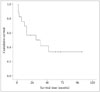
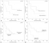
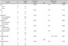
In a view of RFS, the mean RFS time after the operation was 47.1 months (95% CI: 25.9-68.4 months, the median survival time was 30.0 months). One-, 2-, and 5-year RFS rate was 64.7%, 47.1%, and 30.7%, respectively (Fig. 3). On univariate and multivariate analysis, postoperative radiotherapy was also significantly related with RFS time (Fig. 4, Table 4). Considering the small number of this study, however, pathologic type showed somewhat relationship with OS (p=0.057) and RFS (p=0.053) after the operation.

DISCUSSION

Anterior CFR has remained the gold standard for the management of tumors involving the anterior skull base. The addition of postoperative radiation with or without chemotherapy has shown a favorable effect on treatment result of advanced malignancies of the paranasal sinus extending to the anterior skull base [6]. The continuous improvement in endoscopic surgical instrumentation led to the advance of endoscopic-assisted CFR for sinonasal malignancies [7]. In recent years, there has been increasing popularity of purely endoscopic resection of malignant tumors involving the anterior skull base [289]. Resection should be radical, regardless of approach modalities for the lesion, because patients with local relapse after previous treatment have a worse prognosis [2].
Because of the relative rarity of anterior CFR, there are few centers that treated adequate numbers of patients for meaningful analysis of outcomes [10]. In an effort to overcome these difficulties, an international collaborative study on craniofacial surgery for malignant skull base tumors collected data from 1,307 patients in 2003. With a median follow-up of 25 months, the 5-year overall, disease-specific, and recurrence-free survival rate was 54%, 60%, and 53%, respectively [11]. Tumor-related variables, such as the histological variation, intracranial tumor extent, status of surgical margin, and postoperative complication, influence outcomes [1112]. Many studies of CFR have reported postoperative complications as high as 40%, and postoperative mortality rate has remained about 5% [111213]. According to the literatures, complications after CFR includes CSF leakage, frontal pneumocephalus, local wound infection, meningitis and disturbance of central nervous systems. The CSF leakage, wound infection, meningitis occurred more frequently than other complications. The CSF leakage was recovered with conservative treatment but wound infection and meningitis were fatal requiring invasive antibiotics or reoperation [91014]. These surgical limitations have prompted the search for more safe approaches to the anterior skull base [9]. Endonasal techniques are thought to offer several advantages. When compared to the traditional anterior CFR, the use of pure endonasal endoscopic resection is associated with a decreased blood loss, benefit of a desirable cosmetic outcome and faster recovery [9]. Comparing the results of traditional CFR with pure endoscopic resection continues to be difficult because most published reports on exclusive endoscopic approach resection include small case series of patients with a short follow-up [10]. In addition, Hanna et al. [2] treated with exclusive endoscopic approach only patients with earlier disease stage with limited or no skull base involvement. As the interest of endoscopic resection of tumors involving the anterior skull base increases, concurrent improvements in traditional techniques are occurring [3]. The traditional anterior CFR and the endoscopic resection might not be two opposing techniques, but two useful methods to achieve the goal of a safe and radical resection when they were properly applied [10].
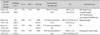
In order to better understand the role of CFR, we assessed our surgical outcomes in the context of recently published series [31014]. Previously reported 5-year OS rate, gross total resection rate, complication rate are 46-72%, 98-100%, and 9.7-47%, respectively (Table 5). Histology, extent of intracranial involvement, increasing age, incomplete tumor removal, and staging were associated with poor survival outcome [101516]. The status of the surgical margins is an important predictor for overall, disease-specific, and recurrence-free survival [14]. The histological finding of the primary tumor and extent of intracranial involvement is also a significant predictor of overall, disease-specific, and recurrence-free survival [1217]. Increased age was reported as a risk factor for poor survival [14]. In our study, postoperative radiotherapy was significant predictor for longer OS and RFS. Neuroblastoma type seemed to be related with prolonged OS and RFS, compared to other pathologies.
In conclusion, this study was basically a retrospective investigation of a relatively small number of patients, possibly leading to selection bias. Although there are limitations in number of cases and follow-up duration, our study shows that CFR followed by adjuvant radiotherapy can remain the primary option for malignant tumors involving anterior skull base, especially with intracranial extension.

Figures and Tables

Fig. 1

Overall survival in 1,917 patients (except 1 mortality case) after CFR for malignant tumors involving anterior skull base tumors. Note that the mean survival time was 69.0 months (95% CI: 47.5-90.5 months, the median survival time was not reached) and 1-, 2-, and 5-year survival rates were 82.3%, 76.5%, and 64.7%, respectively. CFR, craniofacial resection; CI, confidence interval.

btrt-3-81-g001
Fig. 2

Kaplan-Meier analyses of overall survival for 17 patients according to different predictors (overall comparison was estimated using a log-rank test). A: Pathology. B: Brain involvement. C: Combined approach methods. D: Postoperative adjuvant radiotherapy.

btrt-3-81-g002
Fig. 3

Relapse-free survival in 17 patients after CFR for malignant tumors involving anterior skull base. Note that the mean survival time was 47.1 months (95% CI: 25.9-68.4 months, the median survival time was 30.0 months) and 1-, 2-, and 5-year relapse-free survival rates were 64.7%, 47.1%, and 30.7%, respectively. CFR, craniofacial resection; CI, confidence interval.

btrt-3-81-g003
Fig. 4

Kaplan-Meier analyses of relapse-free survival for 17 patients according to different predictors (overall comparison was estimated using a log-rank test). A: Pathology. B: Brain involvement. C: Combined approach methods. D: Postoperative adjuvant radiotherapy.

btrt-3-81-g004
Table 1

Summary of clinical & radio-pathological characteristics in patients who underwent craniofacial resection

btrt-3-81-i001
Characteristics No. of patients (%)
Demographics and history
 Sex
  Male 12
  Female 5
 Mean age (yr) 56 (34-74)
 Symptom/signs
  Epistaxis 6
  Nasal obstruction/mass 5
  Proptosis, eye pain 3
  Severe headache, drowsiness 3
  Anosmia 1
 Preoperative treatment
  Transnasal biopsy/resection 5
  Chemotherapy 2
  Radiotherapy 0
Characteristics of tumor
 Pathology
  Carcinoma
   Squamous cell 6
   Adenocarcinoma 1
   Adenoidcystic 1
   Small cell neuroendocrine 2
  Metastatic melanoma 1
  Teratocarcinosarcoma 1
  Neuroblastoma 5
 Extension of tumor*
  Cribriform plate 5 (29)
  Intracranial involvement 11 (65)
   Dura 4
   Parenchyme 7
  Orbit involvement 6 (35)
 Tumor stage
  T stage (n=12)
   T1 0
   T2 1
   T3 5
   T4 6
  Kadish stage (n=5)
   B 0
   C 5

*confirmed by radiological and intra-operative findings, T stage for eleven cases except olfactory neuroblastomas, Kadish stage for only olfactory neuroblastoma cases

Table 2

Summary of treatment and outcomes in patients who underwent craniofacial resection

btrt-3-81-i002
Variables No. of patients (%)
Treatment associated
 Approach
  Combined endoscopic approach 14
  Combined external approach (orbital exenteration) 3
 Skull base reconstruction by vascularized flap 16 (94)
  Nasoseptal flap 13
  Free flap (all orbital exenteration) 3
 Gross total resection 17 (100)
 Adjuvant treatment 14 (82)
  Radiation 13
  Chemotherapy 2
Outcomes
 Recurrence 6 (35)
  Reoperation 2
 Complication 4 (24)
  Local flap problems 3*
  Brain abscess 1
 Mortality 1 (5)

*all free flap case, 2 with CSF leak, unpredicted varix bleeding from known liver cirrhosis 1 month after operation. CSF, cerebrospinal fluid

Table 3

Univariate and multivariate analysis for overall survival predictors

btrt-3-81-i003
Variables No Mean±SD (mo) Univariate Multivariate
p-value HR 95% CI p-value
Age 0.515 ND 0.166
 <60 yr 10 73.6±13.5
 ≥60 yr 7 49.7±13.8
Sex 0.898 ND 0.339
 M 12 68.3±13.5
 F 5 53.0±15.5
Symptoms* 0.584 ND 0.980
 Non-neurological 12 65.2±12.7
 Neurological 5 55.6±12.0
Approach 0.140 ND 0.599
 With endoscopic 14 75.3±11.4
 With external 3 27.7±17.0
Pathology 0.338 ND 0.057
 Neuroblastoma 5 84.8±15.8
 Other cancer type 12 46.5±9.3
Brain involvement 0.154 ND 0.974
 No 10 70.0±9.5
 Yes 7 56.6±14.7
Postoperative RT <0.001 0.004-0.369 0.005
 No 4 11.5±6.6 0.039
 Yes 13 88.5±9.3 1
Postoperative Cx 0.510 ND 0.297
 No 13 73.5±12.1
 Yes 4 42.5±14.1
Recurrence 0.676 ND 0.148
 No 11 66.6±14.3
 Yes 6 61.7±11.0

*non-neurological symptom; mainly related with nasal symptom including epistaxis, nasal obstruction or mass, neurological sign; mental changes, headache, nausea/vomit, cranial nerve signs, orbital pain, seizure. CI, confidence interval; Cx, complication; HR, harzard ratio; ND, non-detected; RT, radiotherapy; SD, standard deviation

Table 4

Univariate and multivariate analysis for relapse-free survival predictors

btrt-3-81-i004
Variables No Mean±SD (mo) Univariate Multivariate
p-value HR 95% CI p-value
Age 0.505 ND 0.218
 <60 yr 10 37
 ≥60 yr 7 15
Sex 0.453 ND 0.173
 M 12 37
 F 5 15
Symptoms* 0.160 ND 0.295
 Non-neurological 12 ND
 Neurological 5 15
Approach 0.637 ND 0.894
 With endoscopic 14 37
 With external 3 12
Pathology 0.193 ND 0.053
 Neuroblastoma 5 50
 Other cancer type 12 15
Brain involvement 0.298 ND 0.893
 No 10 50
 Yes 7 15
Postoperative RT 0.005 1.417-23.972 0.015
 No 4 2 1
 Yes 13 50 5.827
Postoperative Cx 0.740 ND 0.183
 No 13 37
 Yes 4 30

*non-neurological symptom; mainly related with nasal symptom including epistaxis, nasal obstruction or mass, neurological sign; mental changes, headache, nausea/vomit, cranial nerve signs, orbital pain, seizure. CI, confidence interval; Cx, complication; HR, harzard ratio; ND, non-detected; RT, radiotherapy

Table 5

Summary of recent studies assessing craniofacial resection for malignant tumor involving anterior skull base

btrt-3-81-i005
Author (yr, No. of case) Histology (m/c) IC (+) OB (+) GTR rate Cx rate (m/c)/ mortality rate OS Survival factors
Cantu et al. (2012, 366) AD (49%) 25% 30% 98% 30% (CSF leak)/3.6% 46% (5 yr)/34% (10 yr) Histological type, surgical margin, INT classification, postsurgical radiotherapy
Mine et al. (2011, 30) SQ (38%) 66% NA 100% (87%)* 47% (local infection)/3.2% 80% (2 yr)/72% (5 yr)/63% (10 yr) Surgical margin
Raza et al. (2012, 41) OFN (29%) 72% 54% 100% (85%)* 9.7% (pneumocephalus)/0% NA NA
Present series (2014, 17) SQ (35%) 65% 35% 100% 24% (wound problem)/6% 882% (1 yr)/77% (2 yr)/65% (5 yr) Postsurgical radiotherapy

*% in microscopic examination, histological type was possibly related with overall survival without statistical significance. AD, adenocarcinoma; Cx, complications; GTR, gross total resection; IC, intracranial involvement; INT, Istituto Nazionale Tumori; m/c, most common type; NA, not available; No., number; OB, orbit involvement; OFN, olfactory neuroblastoma; OS, overall survival rate; SQ, squamous carcinoma

Acknowledgments

This study was supported by a grant of Chonnam National University Hospital Research Institute of Clinical Medicine.

Notes

Conflicts of Interest The authors have no financial conflicts of interest.

References

1. Ketcham AS, Wilkins RH, Vanburen JM, Smith RR. A combined intracranial facial approach to the paranasal sinuses. Am J Surg. 1963; 106:698–703.
crossref
2. Hanna E, DeMonte F, Ibrahim S, Roberts D, Levine N, Kupferman M. Endoscopic resection of sinonasal cancers with and without craniotomy: oncologic results. Arch Otolaryngol Head Neck Surg. 2009; 135:1219–1224.
crossref
3. Raza SM, Garzon-Muvdi T, Gallia GL, Tamargo RJ. Craniofacial resection of midline anterior skull base malignancies: a reassessment of outcomes in the modern era. World Neurosurg. 2012; 78:128–136.
crossref
4. Kadish S, Goodman M, Wang CC. Olfactory neuroblastoma. A clinical analysis of 17 cases. Cancer. 1976; 37:1571–1576.
crossref
5. Biller HF, Lawson W, Sachdev VP, Som P. Esthesioneuroblastoma: surgical treatment without radiation. Laryngoscope. 1990; 100:1199–1201.
6. Harrison LB, Pfister DG, Kraus D, et al. Management of unresectable malignant tumors at the skull base using concomitant chemotherapy and radiotherapy with accelerated fractionation. Skull Base Surg. 1994; 4:127–131.
crossref
7. Richtsmeier WJ, Briggs RJ, Koch WM, et al. Complications and early outcome of anterior craniofacial resection. Arch Otolaryngol Head Neck Surg. 1992; 118:913–917.
crossref
8. Dave SP, Bared A, Casiano RR. Surgical outcomes and safety of transnasal endoscopic resection for anterior skull tumors. Otolaryngol Head Neck Surg. 2007; 136:920–927.
crossref
9. Eloy JA, Vivero RJ, Hoang K, et al. Comparison of transnasal endoscopic and open craniofacial resection for malignant tumors of the anterior skull base. Laryngoscope. 2009; 119:834–840.
crossref
10. Cantu G, Solero CL, Miceli R, et al. Anterior craniofacial resection for malignant paranasal tumors: a monoinstitutional experience of 366 cases. Head Neck. 2012; 34:78–87.
crossref
11. Patel SG, Singh B, Polluri A, et al. Craniofacial surgery for malignant skull base tumors: report of an international collaborative study. Cancer. 2003; 98:1179–1187.
crossref
12. Ganly I, Patel SG, Singh B, et al. Craniofacial resection for malignant paranasal sinus tumors: Report of an International Collaborative Study. Head Neck. 2005; 27:575–584.
crossref
13. Kraus DH, Shah JP, Arbit E, Galicich JH, Strong EW. Complications of craniofacial resection for tumors involving the anterior skull base. Head Neck. 1994; 16:307–312.
crossref
14. Mine S, Saeki N, Horiguchi K, Hanazawa T, Okamoto Y. Craniofacial Resection for Sinonasal Malignant Tumors: Statistical Analysis of Surgical Outcome over 17 Years at a Single Institution. Skull Base. 2011; 21:243–248.
crossref
15. Chen AM, Daly ME, El-Sayed I, et al. Patterns of failure after combined-modality approaches incorporating radiotherapy for sinonasal undifferentiated carcinoma of the head and neck. Int J Radiat Oncol Biol Phys. 2008; 70:338–343.
crossref
16. Bridger GP, Kwok B, Baldwin M, Williams JR, Smee RI. Craniofacial resection for paranasal sinus cancers. Head Neck. 2000; 22:772–780.
crossref
17. Ganly I, Patel S, Matsuo J, et al. Postoperative complications of salvage total laryngectomy. Cancer. 2005; 103:2073–2081.
crossref
TOOLS
Similar articles