

 PDF
PDF ePub
ePub Citation
 Print
Print


INTRODUCTION
Aneurysmal bone cysts (ABCs) are benign bone tumors that most commonly occur in people younger than 30 years. The cysts are most often found in the metaphyses of long bones [1]. The involvement of the skull base, particularly the clivus, is an uncommon location for ABCs [2]. It is an expansile, multi-loculated destructive bone lesion. ABC is clinically a benign condition that does not always require surgical treatment. Chordomas, on the other hand, are low-grade malignant tumors that arise from the remnants of the notochord. Radiological differential diagnosis of these tumors is clinically important. Localization is the main confusing part for radiologists in the differential diagnosis of these lesions. Care must be taken for the presence or absence of sclerotic rim, fluid levels and internal septations.
Herein, we present the magnetic resonance (MR) imaging features of a sphenoid bone and clivus ABC misdiagnosed as a skull base chordoma, which is the first case in the English nomenclature according to our knowledge.
CASE REPORT
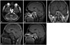
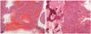
A 21-year-old male presented to a state hospital with headache and decreased visual acuity. A history of trauma was denied. On physical examination decreased visual acuity and nar-rowing of visual field of the eyes were noted. Routine laboratory tests were within the normal range. At the outer radiology department, cranial MR imaging was performed. There was a space occupying lesion in the skull base. According to the MR imaging findings, he underwent excision of the skull base mass, which was misdiagnosed as chordoma. Because of the pathologic discrepancy, the pre-operative cranial MR imaging was consulted to our radiology department. In the pre-operative contrast enhanced MR imaging a lobulated and well-demarcated expansile clival and sphenoidal lesion was seen. There were multiple fluid-fluid cavities showing the presence of degraded blood. T1-weighted images (T1WI) demonstrated small high signal foci that was heterogeneous and iso to high signal in T2-weighted images (T2WI). Also T2WI revealed surrounding hypointense sclerosis. There was heterogeneous enhancement on contrast enhanced T1WI (Fig. 1). ABC was considered according to imaging findings, and also the histopathology showed multiple blood filled cystic cavities separated by thin fibrous septa and many multinucleated giant cells. Histological evaluation confirmed that the lesion was an ABC (Fig. 2). Pathologically, ABCs usually have multiloculated blood-filled spaces like our case, which is characteristic. After surgery, the patient had a headache for two weeks which disappeared completely with medical treatment.
DISCUSSION
ABCs are relatively uncommon, well differentiated benign tumors that present most frequently before the age of 20 years [34]. The knee joint and vertebrae are the most frequently affected sites. They are commonly located in the metaphysis of the long bones. However, the involvement of the skull base is rarely reported [2]. Chordomas are relatively rare low-grade malignant tumors that arise from primitive notochord remnants of the axial skeleton. The majority occur in the sacrum or in the clivus. Chordomas are slow-growing tumors, can invade locally, but rarely metastasize. In advanced disease, metastases to the lung, bone, soft tissue, lymph node, and skin occurs. They may occur around 60 years of age; whereas, presentation with skull base tumors are seen at a younger age and has been reported in children and adolescents. Chordomas account for 1% of intracranial tumors and 4% of all primary skeletal tumors. Intracranial chordomas constitute one-third of all chordomas and usually occur in the vicinity of the clivus [567].
In the diagnosis of ABC, several imaging modalities can be used. Plain films may reveal a complex cystic lesion with a thin shell of periosteal bone. Computed tomography (CT) images disclose erosion, thinning of the bone cortex and demonstrate heterogeneous mass with multiple fluid levels. MR imaging also shows these fluid levels, with heterogeneous signal intensity on both T1WI and T2WI and frequently multiple internal septations, as a well-defined hypointense sclerosis [8]. Fluid levels within ABCs are indicative of hemorrhage with sedimentation and are better demonstrated with MR imaging. On T1WI, they may have increased signal intensity due to methemoglobin in either the dependent or nondependent component. Contrast enhanced MR imaging reveals internal septas within lesion. MR imaging demonstrates low signal intensity rim around the lesion due to thickened, intact periosteal membrane [9]. Conversely chordoma's MR imaging findings do not include hypointense peripheral sclerotic rim. MR imaging is the single best modality for radiologic evaluation of intracranial chordomas [10]. It is considerably superior to CT in the description of lesion extent because it provides excellent tissue contrast and perfect anatomic detail [11]. Skull base chordomas are generally isointense or hypointense on T1WI relative to muscles. The high signal intensity on T1WI indicates hemorrhagic foci and collection of dense content within the lesion. Because of the fluid content, chordomas have a high signal intensity on T2WI [9]. After contrast injection, the majority of intracranial chordomas display moderate to marked enhancement, occasionally ring, arc and peripheral enhancement have been defined on MR imaging [12].
Clinical symptoms of skull base and sphenoid bone ABCs include headache, ptosis, strabismus exophthalmus, double vision, swelling, visual loss, and nasal obstruction for ABCs of the sphenoid bone due to pressure and mass effect and includes headache, sinus congestion, and cranial nerve palsies [13]. Treatment aims at complete surgical excision through a transcranial-transbasal or transfacial approach. In pathological evaluation description of the lesion as a blood-filled sponge is characteristic for the diagnosis of an ABC. Chordomas are often contained within a pseudocapsule and these lesions have clear cells containing intracytoplasmic vacuoles [9]. Clinical manifestation of chordoma is often subtle because it is a slow-growing lesion [9]. The clinical features of chordoma will depend on the site and line of spread of the tumor. The major complaints consist of headache, cranial nerve palsies, nasal obstruction with discharge and dysphagia due to intracranial spread and compression of the tumour [14]. In our case, the localization of the lesion suggests chordoma. The MR imaging findings of chordoma do not show hypointense peripheral sclerotic rim nor fluid levels like an ABC. A reasonable differential diagnosis for skull base chordoma and ABC can be made on the basis of radiologic findings, especially MR imaging.
In conclusion, MR imaging has an important role in the differential diagnosis of skull base lesions. Radiation therapy is used as an adjunct treatment in chordomas, despite not being an option in ABC treatment. Therefore, skull base ABCs should be differentiated from chordomas in accordance with hypointense peripheral sclerotic rim, fluid levels and multiple internal septations.
 XML Download