

 PDF
PDF ePub
ePub Citation
 Print
Print


Abstract
Objective
Seizures are common consequence of traumatic brain injury and have been reported in clinical series as an incidence of 15% to 22%. Among them, nonconvulsive seizures (NCS) are often unrecognized during the early period of neurosurgical hospitalization because their clinical presentations can be misunderstood as consequent symptoms of clinical course, and the diagnosis can be confirmed only by the electroencephalographic (EEG) recording.
Methods
We retrospectively reviewed our clinical database of traumatic brain injury (TBI) patients admitted between March 2008 and September 2012. Twenty one patients with suspicious symptoms of NCS, such as decrease of consciousness, aphasia or irritability, were included. Routine wake and sleep EEG or bedside continuous EEG monitoring were done in all patients.
Results
Ten out of twenty-one patients showed abnormal activities on EEG. Ictal discharges were documented on four patients. Based on clinical symptoms and EEG findings, these four patients were diagnosed as NCS. Two out of four NCS patients showed EEG findings of nonconvulsive status epilepticus (NCSE). Another six patients with abnormal EEG activities were considered as ‘suspicious NCS' because only interictal activities were recorded on EEG but increasing dose or adding on antiepileptics relieved their symptoms. All NCS/NCSE were successfully controlled by appropriate antiepileptic therapy.
Conclusion
Our result showed that NCS was diagnosed in about 20% of patients with suspicious symptoms. There's a possibility that actual NCS might have happened more. Because untreated NCS/NCSE might cause worse clinical outcome, careful observation and urgent EEG recordings should be considered in a patient with suspicious NCS symptoms.
References
1. Aldenkamp A, Arends J. The relative influence of epileptic EEG discharges, short nonconvulsive seizures, and type of epilepsy on cognitive function. Epilepsia. 45:54–63. 2004.

2. Annegers JF, Coan SP. The risks of epilepsy after traumatic brain injury. Seizure. 9:453–457. 2000.
3. Chen CW, Kuo JR, Lin HJ, Yeh CH, Wong BS, Kao CH, et al. Early postoperative seizures after burrhole drainage for chronic subdural hematoma: correlation with brain CT findings. J Clin Neurosci. 11:706–709. 2004.
4. Drislane FW. Presentation, evaluation, and treatment of nonconvulsive status epilepticus. Epilepsy Behav. 1:301–314. 2000.
5. Friedman D, Claassen J, Hirsch LJ. Continuous electroencephalogram monitoring in the intensive care unit. Anesth Analg. 109:506–523. 2009.
6. Jirsch J, Hirsch LJ. Nonconvulsive seizures: developing a rational approach to the diagnosis and management in the critically ill population. Clin Neurophysiol. 118:1660–1670. 2007.
7. Jordan KG. Nonconvulsive status epilepticus in acute brain injury. J Clin Neurophysiol. 16:332–340. ; discussion 353,. 1999.
8. Kennedy JD, Gerard EE. Continuous EEG monitoring in the intensive care unit. Curr Neurol Neurosci Rep. 12:419–428. 2012.
9. Liesemer K, Bratton SL, Zebrack CM, Brockmeyer D, Statler KD. Early posttraumatic seizures in moderate to severe pediatric traumatic brain injury: rates, risk factors, and clinical features. J Neurotrauma. 28:755–762. 2011.
10. Ronne-Engstrom E, Winkler T. Continuous EEG monitoring in patients with traumatic brain injury reveals a high incidence of epileptiform activity. Acta Neurol Scand. 114:47–53. 2006.
11. Vespa PM, Nuwer MR, Nenov V, Ronne-Engstrom E, Hovda DA, Bergsneider M, et al. Increased incidence and impact of nonconvulsive and convulsive seizures after traumatic brain injury as detected by continuous electroencephalographic monitoring. J Neurosurg. 91:750–760. 1999.
12. Westmoreland BF. Periodic lateralized epileptiform discharges after evacuation of subdural hematomas. J Clin Neurophysiol. 18:20–24. 2001.
FIGURE 1.
A case of nonconvulsive status epilepticus in 80-year-old woman. She showed repeated episodes of facial twitching and nystagmus. A: Computed tomography demonstrating acute subdural hematoma on right fronto-temporo-parietal area with hemorrhagic contusion. B: Typical EEG findings of nonconvulsive seizure. Ictal activities are seen on right fronto-temporal (F8-T4, T4–6) and right fronto-parietal (F4-C4, C4-P4) area. Note that during the ictal period, there is no artifact from muscle contraction in other surface electrodes, which is normally accompanied by convulsive seizures. EEG: electroencephalographic.
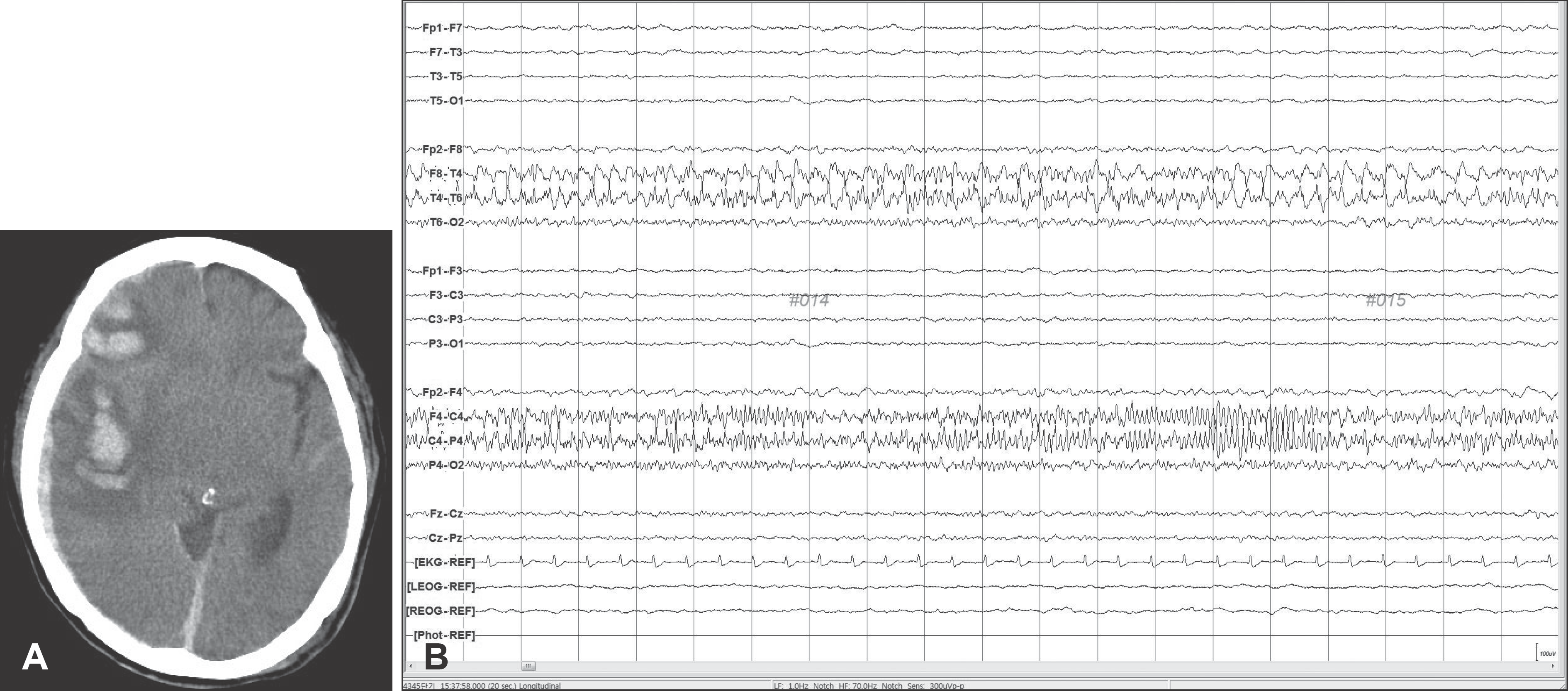
TABLE 1.
Demographics of the study population
| Total | 21 |
|---|---|
| Sex | |
| Male: Female | 13: 8 |
| Total | 21 |
| Age (years) | 65.7 (10–87) |
| GCS score | 12.7 (4–15)0 |
| Type of injury | |
| ASDH | 06 |
| CSDH | 10 |
| EDH | 02 |
| HCONT | 02 |
| Skull fracture∗ | 01 |
| Symptoms | |
| Aphasia | 04 |
| Twitching | 04 |
| Confusion | 04 |
| Lethargy | 01 |
| Tremor | 02 |
| Decreased consciousness | 06 |
TABLE 2.
The clinical characteristics in patients of NCS and ‘suspicious NCS' group
| Case no. | Sex | Age | Type of injury | Initial GCS∗ score | Surgery | Day of† seizure | EEG finding | Symptoms | |
|---|---|---|---|---|---|---|---|---|---|
| NCS | 1 | M | 68 | HCONT | 07 (2/1/4) | None | 1 | Cont gen ictal spikes | Decreased consciousness |
| 2 | M | 60 | ASDH | 15 (4/5/6) | None | 4 | Cont gen delta slowing | Decreased consciousness | |
| 3 | F | 81 | ASDH | 13 (3/4/6) | Decomp & H/R | 14 | Focal ictal spikes | Facial twitching, Nystagmus | |
| 4 | F | 83 | ASDH | 14 (3/5/6) | None | 11 | Genictal spikes | Decreased consciousness | |
| Suspicious NCS | 5 | M | 72 | CSDH | 15 (4/5/6) | Burr hole drainage | 13 | TIRDA | Aphasia |
| 6 | F | 64 | CSDH | 15 (4/5/6) | Craniotomy & H/R | 15 | FIRDA | Aphasia | |
| 7 | F | 75 | CSDH | 15 (4/5/6) | Burr hole drainage | 9 | Focal interictal spikes | Lethargy | |
| 8 | M | 36 | ASDH | 04 (1/1/2) | Decomp & H/R | 8 | PLEDs | Facial twitching | |
| 9 | M | 72 | CSDH | 08 (2/1/5) | Burr hole drainage | 11 | Focal interictal spikes | Decreased consciousness | |
| 10 | M | 71 | ASDH | 11 (3/3/5) | None | 3 | PLEDs | Decreased consciousness |
† ‘postinjury day 0' indicates the day of injury. EEG was taken at the ‘day of seizure' in all patients. EEG: electroencephalographic, NCS:nonconvulsive seizures, GCS: Glasgow Coma Scale, HCONT: hemorrhagic contusion, ASDH: acute subdural hemorrhage, CSDH: chronic subdural hemorrhage, Decomp: decompressive craniectomy, H/R: hematoma removal, Cont: continuous, Gen: generalized, TIRDA: temporal intermittent rhythmic delta activity, FIRDA: frontal intermittent rhythmicdelta activity, PLEDs: periodic lateralized epileptiform discharges
 XML Download