

 PDF
PDF ePub
ePub Citation
 Print
Print


Abstract
Superficial siderosis (SS) in central nervous system is a rare, slowly progressive disease and usually misdiagnosed or diagnosed too late when the patient is chronically devastated. A 55-year-old man with deafness and gait disturbance for ten years was referred from otorhinologist for evaluation of brain. Magnetic resonance image (MRI) showed symmetric hypointense rim partially delineated the bilateral hemisphere on gradient-recalled-echo T2-weighted image, and it was diagnosed as hemosiderin deposition in subarachnoid and subpial meningeal layer. The correct diagnosis of cerebral superficial siderosis can be achieved by careful neurological examination and MRI because computed tomography findings and symptoms are ambiguous. Serial follow-up of imaging study and education for patient are necessary to prevent progression of SS.
References
1. Fearnley JM, Stevens JM, Rudge P. Superficial siderosis of the central nervous system. Brain 118 (Pt 4):1051–1066. 1995.

2. Hathaway B, Hirsch B, Branstetter B. Successful cochlear implantation in a patient with superficial siderosis. Am J Otolaryngol. 27:255–258. 2006.
3. Koeppen AH, Michael SC, Li D, Chen Z, Cusack MJ, Gibson WM, et al. The pathology of superficial siderosis of the central nervous system. Acta Neuropathol. 116:371–382. 2008.
4. Kondziella D, Zetterberg H. Hyperphosphorylation of tau protein in superficial CNS siderosis. J Neurol Sci. 273:130–132. 2008.
5. Kumar N. Superficial siderosis: associations and therapeutic implications. Arch Neurol. 64:491–496. 2007.
6. Kumar N, Cohen-Gadol AA, Wright RA, Miller GM, Piepgras DG, Ahlskog JE. Superficial siderosis. Neurology. 66:1144–1152. 2006.
7. Levy M, Turtzo C, Llinas RH. Superficial siderosis: a case report and review of the literature. Nat Clin Pract Neurol. 3:54–58. ; quiz 59,. 2007.
8. Scheid R, Frisch S, Schroeter ML. Superficial siderosis of the central nervous system – treatment with steroids? J Clin Pharm Ther. 34:603–605. 2009.
9. Sydlowski SA, Cevette MJ, Shallop J, Barrs DM. Cochlear implant patients with superficial siderosis. J Am Acad Audiol. 20:348–352. 2009.
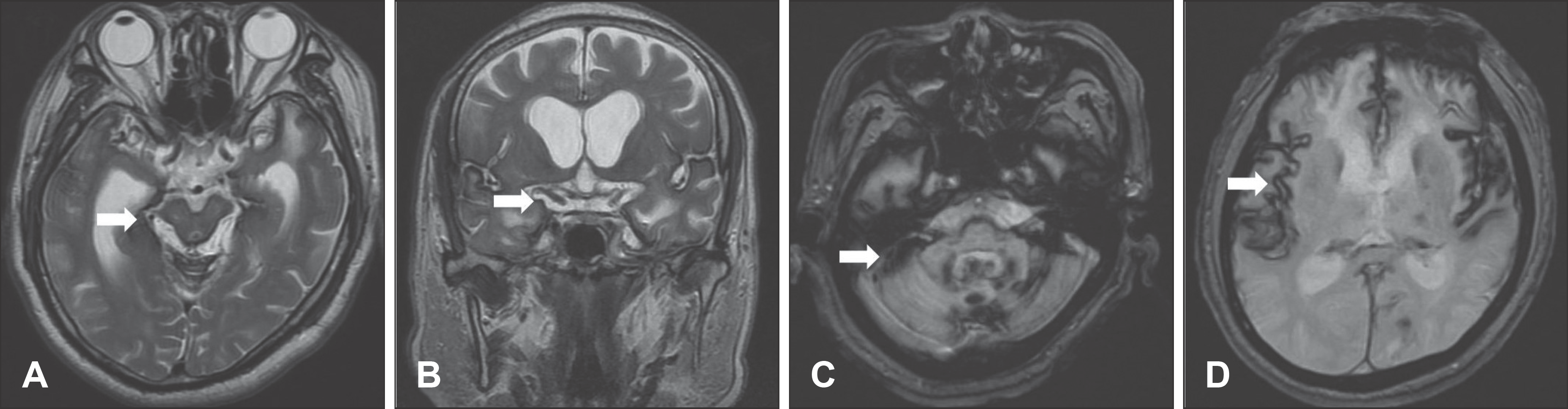
FIGURE 1.
A, B: Axial and Coronal T2-weighted MR images show a variably thick hypointense rim outlined the brain stem and various cisterns. C, D: Axial gradient-recalled-echo T2-weighted images show a symmetric well-defined hypointense rim that delineated the cerebellum, some of the lower cranial nerves, bilateral sylvian fissures and other leptomeningeal layers.
 XML Download