

 PDF
PDF ePub
ePub Citation
 Print
Print


Introduction
Since the development of various shunt systems, shunt operation has been regarded as one of the foremost surgical treatment modality for patients with symptomatic hydrocephalus. The Codman-Hakim programmable valve (Codman, Johnson & Johnson, Raynham, MA, USA) was developed by Drs. Salomon and Hakim during the late 1980s, and it has been one of most popular shunt systems used in the clinical practice for the treatment of hydrocephalus.7) It allows for a precise control of the cerebrospinal fluid (CSF) drainage by noninvasive adjustments using magnetic impulses into one of 18 different pressure settings within a range of 30 to 200 mmH2O.2367)
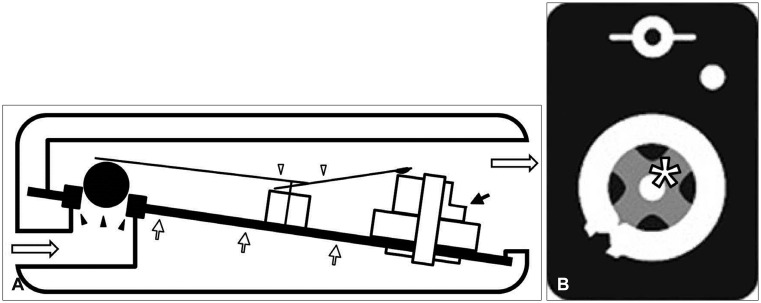
Similar to other shunt systems, this system consists of three parts: a ventricular catheter, a valve, and a distal catheter. The valve has a unique and delicate feature. The stainless steel flat spring has two ends. The proximal end presses the synthetic ruby valve ball landed on a matching valve seat through which CSF is passed distally. The other end is supported by the spiral staircase cam (pressure control cam) which controls the intensity of the flat spring (Figure 1A). The bottom side of the cam has a protrusion of "X" in the center and this part is fixed to a corresponding X-shaped slit on the titanium base plate by the pressure of the flat spring (Figure 1B).6)
Even with these precise and exquisite microtechnics of this system, malfunctions have been also reported which lead to underdrainage or overdrainage of the CSF.246) According to these reports, obstruction of the ventricular catheter by tissue materials or hematoma and catheter disconnections are relatively common, while the malfunction of the valve itself is rare.2) A direct trauma or an exposure to magnetic fields may cause a mechanical malfunction of the programmable valve.456) Herein, we report on a rare case of shunt overdrainage caused by displacement of the pressure control cam after pressure adjustment.
Case Report
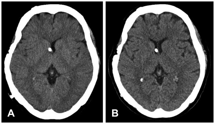
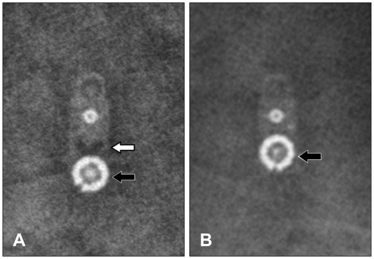
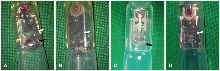
Eight years ago, a 57-year-old female underwent a ventriculoperitoneal shunt operation with a Codman-Hakim programmable valve for hydrocephalus after subarachnoid hemorrhage. Her symptoms of hydrocephalus had been improving since the operation, but she complained of slowly aggravating headache and nausea for recent years. Her brain computed tomography (CT) showed a slit-ventricle (Figure 2A), and the skull X-ray revealed the valve pressure at 10 mmH2O. The valve pressure was adjusted to 12 mmH2O at the outpatient department. A few days later, she presented with even worsening headache and nausea. She had no history of direct trauma on the valve or an exposure to magnetic fields. The skull X-ray showed that the pressure control cam was detached from the base plate and displaced distally (Figure 3). She underwent revision of the shunt system to exchange the valve. A microscopic inspection of the valve revealed the detachment of the cam from the base plate leaving the X-shaped slit empty (Figure 4). The displacement of the cam relaxed the flat spring pressure on the valve ball, and it led to overdrainge of the CSF. After the shunt revision, her symptoms dramatically resolved and her brain CT showed recovery from the slit-ventricle (Figure 2B).
Discussion
Patients undergone shunt operation have a narrow therapeutic window between overdrainage and underdrainage.3) Since the required amount of the CSF drainage inconsistently varies depending on patients’ condition, life style, and status of physical activity, more than half of these patients require postoperative adjustments of valve pressure to maintain an ideal shunt function.17)
Because the Codman-Hakim programmable valve allows noninvasive fine tuning of the valve pressure using magnetic impulses and gives clinicians a broad choice of pressure settings within a range of 30 to 200 mmH2O, this device has been used popularly in the clinical practice for the treatment of hydrocephalus.2367)
This valve is composed of complex systems, and each component has a delicate function. The flat spring presses the valve ball as well as the pressure control cam on the base plate, and the set of these complex parts controls the drainage of the CSF in an appropriate pressure.6) Unintended or accidental alterations in any of these components may cause valve malfunction. In order to recognize the presence of valve malfunction, clinicians require a working knowledge of the valve structure and an ability to interpret the valve setting on an X-ray.3)
Okazaki et al.4) reported a case of shunt malfunction resulting from a direct trauma to the valve. A crack was found at the top of valve housing, and the cam was detached from the base plate. According to the product manual and other reports, it is mentioned that the use of magnetic resonance (MR) systems up to 3 Tesla will not damage the valve itself but may change the setting of the valve, and it is recommended to confirm the valve setting after an MR procedure.56) However, Watanabe et al.6) reported a rare case of shunt malfunction caused by detachment of the cam after 3 Tesla MR imaging (MRI). Therefore, a direct trauma to the valve or an exposure to strong magnetic fields may cause a mechanical malfunction of the programmable valve.
In the present study, the patient had no definite history of head trauma or MRI. Skull X-ray taken prior to the valve adjustment was unremarkable. Since pressure adjustment normally does not lead to the cam displacement, we assume that there has been a minor problem with durability of the valve causing a reduction of the flat spring pressure during the eight years of postoperative periods and a magnetic impulse to adjust the valve pressure finally attributed to the detachment of the cam leading to overdrainge of the CSF.
Conclusion
Although the Codman-Hakim programmable valve is generally very useful in terms of noninvasive pressure adjustment and fine tuning of valve pressure, a rare complication such as displacement of the pressure control cam may occur. An accurate knowledge on the valve structure as well as an ability to interpret the valve status on an X-ray are essential to confirm maintenance of this elaborate device and recognize the presence of valve malfunction.
 XML Download