

 PDF
PDF ePub
ePub Citation
 Print
Print


I. INTRODUCTION
Bonding to dentin has been one of the most important and challenging issues in restorative dentistry since the introduction of adhesive technology nearly 50 years ago1). The process of bonding to dentin generally begins with acid etching, which is necessary to remove the smear layer and to create a 1 - 5 µm-deep demineralized zone in the dentin surface. In the next step, hydrophilic primers are applied, which diffuse across the demineralized layer to displace water and stabilize the collagen network. Finally, the adhesive resins are applied to the primed dentin and polymerized. The hybrid layer or resin-dentin interdiffusion zone is formed based upon this micromechanical interlocking, which is considered the most important mechanism for dentin bonding2).
Dentin can be regarded as a biological composite of a collagen matrix which is highly filled with nanometer-sized apatite crystal. After extracting the mineral from the collagen fibrils, the voids can be filled with a resin, thus forming a new composite made up of resin matrix filled with a fibrous collagen3).
In order to obtain consistent hybrid layers, the three-dimensional structure of a collagen network should be exposed by acid etching and the interfibrillar space of the collagen network should be maintained by being floated in water during infiltration of resin monomers. Collapse of collagen network reduces the porosity of the demineralized dentin, inhibits resin penetration through the demineralized layer4) and forms a barrier between the demineralized layer and the underlying intact or unreacted dentin surface. Because optimal interfacial bonding can be achieved by resin diffusion through the conditioned dentin, it is essential to maintain collagen network without collapse5).
The lack of complete diffusion throughout the demineralized dentin with adhesive systems that inhibit collagen collapse through "wet" bonding can be attributed to a variety of factors, including dissimilar solvents and differences in the hydrophobicity of the adhesive. For example, under moist bonding conditions, the channels between demineralized dentin collagen fibrils are filled with water. The only mechanism available for monomer infiltration is diffusion of monomers into whatever solvent is in the spaces of the substrate and along the collagen fibers3).
This dentin bond, if still intact after resin composite placement and polymerization, can degrade over time, leading to clinical restorative failures. Biodegradation of the collagen matrix and/or synthetic resin components of this hybrid layer is possibly due to incomplete penetration/infiltration of resin into the dentin substrate, heterogeneous distribution of monomers through the interdiffusion zone, suboptimal polymerization and hydrolysis6).
In order to improve the durability of dentin adhesives, it is necessary that complete removal of water from the exposed collagen network without deteriorating the integrity of its three-dimensional structure and substitution of hydrophilic monomers in the adhesives with more hydrophobic monomers which can resist hydrolysis. Most primers in currently available systems usually contain hydrophilic monomers, such as 2-hydroxylethyl methacrylate (HEMA), as surface-active agents to enhance the wettability of the hydrophobic adhesive resins. HEMA can polymerize in the presence of water to form 'microporous' hydrogel (polyHEMA) with pore sizes ranging from 10 to 100 nm7). The loss of interfibrillar resin may have been caused by slow hydrolysis of polyHEMA8). Like HEMA, alcohols at higher concentration also significantly stabilized collagen9) and high vapor pressure of these solvents have been used to compete with and displace water and to facilitate the permeation of monomers10).
In order to increase the durability of dentin-adhesive interfaces, by priming the acid-etched dentin surfaces with organic solvents, we tried to remove water from the exposed collagen network without collapse. We also tried to penetrate the experimental adhesives containing relatively hydrophobic monomers into the collagen network floating in the solvent.
The hypothesis tested was that priming the collagen network with organic solvents might displace water from it without collapse and thereby represent less nanoleakage.
The purpose of this study was (1) to compare nanoleakage patterns of a conventional 3-step etch and rinse adhesive system and two experimental hydrophobic adhesive systems and (2) to investigate the change of the nanoleakage patterns after load cycling.
II. MATERIAL AND METHODS
Preparing experimental adhesives
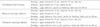
Scotchbond Multi-Purpose (3M ESPE, St. Paul, MN, USA) was used as control. Two experimental adhesives were prepared by dissolving a mixture (1 : 1) of two hydrophobic monomers, Bis-GMA (Bisphenol-A-glycidyl dimethacrylate, Shin-Nakamura chemical Co., Inc.) and TEGDMA (Triethyeneglycol dimethacrylate, Shin-Nakamura chemical Co., Inc.), into an equal amount of an organic solvent (50 wt%), one of ethanol and methanol. Camphoroquinone (CQ) and Ethyl 4-dimethylaminobenzoate (4E) were each added to the mixture by 1.0 wt% of resin monomers (Table 1).
Bonding Procedures
Thirty freshly extracted human molars stored in 0.5 % chloaramine T solution were used. Occlusal enamel and coronal dentin were removed using a low-speed saw (Isomet, Buehler Ltd., Lake Bluff, IL, USA) under running water. The exposed dentin surfaces were wet ground with 500 grit silicone carbide paper (Rotopol-V, Struers Ltd, Glasgow, UK) for 60 seconds. The three dentin adhesive systems were used in this study (Table 1). Scotchbond Multi-Purpose (MP) and two experimental adhesive systems, ethanol containing adhesive (EA) and methanol containing adhesive (MA), were used. The polished dentin surface was etched with a 35 % phosphoric acid etching gel (3M ESPE, St. Paul, MN, USA) for 15 seconds and rinsed with distilled water for 15 seconds. Ten teeth were used for each adhesive. In the MP group, dentin surfaces were treated following the manufacturer's instruction. In the EA and MA groups, immediately after rinsing, the teeth were primed with ethanol or methanol for 15 seconds and then the experimental adhesives containing the same solvent as the priming solvent were applied on the solvent-primed dentin with two successive coats and agitated for 10 seconds. The remaining solvent was dried with gentle air blow. The adhesive was cured for 20 seconds using a dental light-curing unit (Hilux™ Ultra+, Benlioglu Dental Inc., Ankara, Turkey, power density: > 500 mW/cm2). Z-250 hybrid composite resin (A2 shade, 3M ESPE, St. Paul, MN, USA) was built-up in two increments on the adhesive-treated surfaces to 4.0 mm thickness (Table 2).
Load Cycling
Five teeth of each dentin adhesive group were subjected to mechanical load cycling. The cyclic mechanical loading device used was a hydraulic dynamic fatigue testing machine (858 Bionix II, MTS, USA). The tooth was mounted in acrylic resin block and 100N was loaded on the middle of each tooth by a steel tip attached to the machine for 1,000,000 cycles in water at room temperature.
Nanoleakage evaluation
The teeth were sectioned occluso-gingivally perpendicular to the bonded interface into approximately 2.0 mm thick slabs using a diamond saw (Isomet, Buehler Ltd., Lake Bluff, IL, USA) under water cooling. Two slabs were obtained from each tooth. And then the slabs were coated twice with nail varnish except within 1 mm of the bonded interfaces. The slabs were placed in 50 % (w/v) silver nitrate solution in total darkness for 24 hours, rinsed in running water for 5 minutes, and immersed into photo-developing solution under fluorescent light for 8 hours in order to reduce the silver ions to metallic silver. After removal from the developing solution, the teeth were placed in running water for 5 minutes11). Specimens were polished with incre-asingly fine diamond pastes (6, 3 and 1 µm particles, Buehler Ltd., Lake Bluf, IL, USA). The specimens were cleaned ultra-sonically, air dried, mounted on aluminum stubs, placed in a desiccator for 24 hours, and coated with thin gold. Ten specimens for each group were examined under the scanning electron microscope (SEM, S-4700 Hitachi, Japan) in backscattering electron mode.
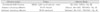
The image analysis method was modified from a protocol previously described by Tay et al12). All photographs were analyzed using image analysis software (Scion Image Beta 4.03, Scion Corp., Frederick, MD, USA)13). The amount of silver nitrate within the hybrid layer was measured with gray value in three regions (3.0 µm × 3.0 µm) of the bonded interface (left, center and right of each specimen). In SEM image, silver nitrate was observed as white particle and the silver nitrate uptake was expressed as average gray value ranging from 0 (black) to 255 (white). Data were statistically analyzed by two-way ANOVA and post-hoc testing of multiple comparisons was done with the Scheffe's test.
III. RESULTS
All specimens in the six groups showed nanoleakage at the adhesive/dentin interface. Typical leakage patterns at the resin-dentin interfaces for each dentin adhesive system are illustrated in Figures 1 - 7.
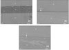
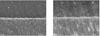
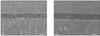
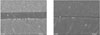
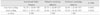
The Scotchbond Multi-Purpose system produced a very distinct hybrid layer which was approximately 3 µm thick. Distinct resin tags were also observed as shown in Figures 2 and 3. Silver particles were deposited along the lower half of the hybrid layer that were different, however, from those of the experimental adhesive systems. The leakage pattern revealed a thin but very dense deposition of silver at the bottom of the hybrid layer. There was also distinct deposition of silver between the resin tag and tubule walls. The average gray scale was 91.78 in the non-load cycling group and 110.11 in the load cycling group (Table 3). The Scotchbond Multi-Purpose system produced an approximately 300 µm thick adhesive layer (Figure 1a).
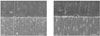
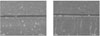
The leakage patterns for the ethanol containing adhesive were shown in Figures 4 and 5. The hybrid layer was approximately 3 µm thick. There were diffuse silver deposits within the hybrid layer, and dentinal tubules rarely took up silver. The average gray scale was 20.70 in the non-load cycling group and 24.20 in the load cycling group (Table 3). This system produced an approximately 50 µm thick adhesive layer (Figure 1b).
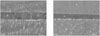
The leakage patterns for the methanol containing adhesive are shown in Figures 6 and 7. The hybrid layer was approximately 3 µm thick. Silver particles were evenly deposited within the hybrid layer, but of relatively low density. The tubule walls rarely took up silver particles. The average gray scale was 32.88 in the non-load cycling group and 32.27 in the load cycling group (Table 3). This system produced an approximately 30 µm thick adhesive layer (Figure 1c).
The Scotchbond Multi-Purpose groups (non-load cycling and load cycling) had significantly higher silver content than other groups (p < 0.0001) (Table 3). SEM images showed that samples subjected to load cycling had leakage patterns similar to non-load cycled samples for all dentin bonding systems.
IV. DISCUSSION
The durability of the bond between adhesive resins and dentin is of critical importance for the longevity of bonded restorations14). This study was designed to evaluate whether new experimental adhesive systems, which contain less hydrophilic domains, might resist hydrolytic de-gradation. Sano et al.15,16) hypothesized that nanoleakage revealed the location of defects at the resin-dentin interface, and could be the pathway for degradation of resin/dentin bonds over time.
All adhesives presented a certain degree of nanoleakage with differences among densities and patterns of silver deposition, depending on their composition. This may be a consequence of incomplete penetration of the adhesive resin into the demineralized dentin, leaving the collagen not enveloped by resin, or this may result from incomplete polymerization of resin monomer16). The nanoleakage pathway may be located within the hybrid layer, within demineralized dentin or within the hydrophilic domain which exists in adhesive resin17). Although nanoleakage was shown to occur throughout the hybrid layer and/or adhesive resin, the clinical significance of nanoleakage was unclear. The spaces are too small to allow bacterial penetration, but they were large enough for enzymes or bacterial product to enter.
The original interpretation of nanoleakage was that silver occupied nanometer-sized spaces around naked collagen fibrils, where resin failed to infiltrate, or where residual water had not been displaced by adhesive resin18,19). But Tay and Pashley12) demonstrated that water could pass from dentin, around resin tags, to form water-filled channels that project from the hybrid layer into the overlying adhesive. When these water-filled channels are stained with silver, those often look like microscopic trees. They called them "water trees"and suggested that those might act as potential sites for hydrolytic degradation of resin/dentin bonds. Thus far, all marketed products permitted some amount of nanoleakage and water-tree formation.
In this study, there were much less silver depositions in the experimental adhesive groups. Ideally, HEMA, which is a primary component in many commercial dentin adhesive systems, conditions the collagen to remain expanded during adhesive infiltration20,21). However, results from a recent study indicate that HEMA can dramatically reduce the evaporation of water22). The addition of HEMA reduces the mole fraction of water and therefore reduces the partial pressure of water according to Dalton's law of partial pressures. As the partial pressure of water drops, it becomes more and more difficult to remove residual water from the demineralized dentin. Hydrophobic monomers, such as Bis-GMA, would resist diffusing into these sites where there is residual water5). Because of high vapor pressure of alcohol solvents that were used as priming material in the experimental groups, the solvents might have competed with water and effectively replaced it. Therefore, the collagen network was expected to shrink much less and not to collapse, and then solvents might carry the monomers effectively into the spaces and facilitate the development of a hybrid layer10). Furthermore, the alcohols and monomers used in the experimental adhesives had hydroxyl groups. The hydrogen-bonding between the hydroxyl groups might facilitate their penetration.
In the experimental adhesive groups, the adhesive layer was thinner than that of the Scotchbond Multi-Purpose group. In substance, the viscosity of both experimental adhesives was relatively low and it was very difficult to apply adhesive evenly. Therefore, this must be improved for clinical use.
Teeth are continually subjected to stresses during chewing, swallowing, and parafunctional habits. Vertical loading introduced by a food bolus between opposing teeth can be evenly distributed over the entire occlusal surface. Mechanical load cycling has been studied due to its potential capability of simulating mastication23,24). Anderson25) recorded axial loads from 70 to 150 N during chewing and swallowing. A load of 100 N was used in this study, as it was considered to be within the normal functional range.
In this study, the load cycling did not influence the leakage pattern of each dentin adhesive system. For micro-leakage evaluation, discrepant results related to the effect of load cycling have been reported26-29). Other studies, however, found no effect of load cycling on nanoleakage, which is in accordance with the finding of this study23,30). After hybridization of the adhesive and demineralized dentin via micromechanical interlocking, the bond between the adhesive and dentin obtained from the dentin bonding agents used, is believed to be strong enough to resist a moderate amount of occlusal force for some time. Although porosity existed at the hybrid layer, it did not increase after the loading; that is, there was no greater penetration of silver nitrate. However, the forces and movements during mastication are highly complex and factors such as age, gender, bruxism, and bite habits have a significant influence on the forces measured25). Also, the testing machine used could only produce axial cyclic loads, while the movements of mastication in the oral environment are three-dimensional in pattern31). Thus, the test conditions simulate but do not duplicate clinical conditions, and this must be considered when interpreting the findings. This study was performed for the quantitative analysis of silver distribution by using SEM Images. However, it might be speculated that TEM can give an informative image for quantitative analysis.
V. CONCLUSION
Durability of long-term dentin adhesion can be influenced by hydrolytic degradation. This study was aimed to evaluate the nanoleakage patterns of two experimental adhesives containing relatively hydrophobic monomers before and after load cycling. From this study, the following results were drawn.
The dentin adhesive systems used in this study did not achieve perfect sealing at the adhesive/dentin interface. Silver particles were observed in all the groups at the hybrid layer.
However, silver particles were more sparsely distributed in the ethanol containing adhesive group and the methanol containing group than in the Scotchbond Multi-Purpose group (p < .0001).
There were no changes in nanoleakage patterns after load cycling.
 XML Download