

 PDF
PDF ePub
ePub Citation
 Print
Print


INTRODUCTION
Dental implant is widely used in the field of reconstructive dentistry as a predictable treatment modality.12 Implant surfaces and design are continuously being improved; the ideal dental implant system should have a contour similar to that of a natural tooth, be esthetic, and demonstrate strength and long-term durability.345 In addition, the maintenance cost should be low, and the patient should agree that the dental implant is a satisfactory replacement of a missing natural tooth.6
Despite their widespread use, biological and mechanical failures of implant-supported fixed dental prostheses (ISFDPs) and implant-supported single crowns (ISSCs) are still frequent.78 The high survival rate of ISSCs is similar to that of ISFDPs, but mechanical, biological, and esthetic complications occur more frequently with ISSCs.7 Esthetic complications (e.g., soft-tissue recessions, unfavorable color, and visible crown margins) occur frequently in the anterior region, while mechanical complications (e.g., screw loosening, screw fracture, and fractures of the veneer material) are more frequent in the posterior region.9
Various dental implant systems have been studied and developed with the aim of improving the mechanical, biological, and esthetic properties and overcoming the above-mentioned disadvantages.10 The lateral-screw-retained implant prosthesis (LSP) was designed with a lateral screw access hole replacing an occlusal screw access hole. The implementation of physiologically shaped occlusal surfaces increases the esthetic value of the implant prosthesis and essentially removes any possibility of unfavorable occlusion interference.6 Removal of the occlusal screw holes can also prevent mechanical complications, in particular screw loosening and loss of the access-hole resin and sealing materials. 111213 Despite these merits, the experimental and clinical studies of the LSP have been insufficient, and the previous treatments have been empirical only.
The modified LSP was designed to improve on the advantages of the LSP by making it easy to separate the implant prosthesis. The purpose of this structural design is to reduce the rates of mechanical and biological complications and to make the prosthesis easier to retrieve. Easy retrievability facilitates maintenance care, which allows mechanical and biological complications to be treated.
The aim of this study was to determine the mechanical and biological complication rates of ISSCs inserted in the posterior region using the modified LSP, with a focus on the cases that could be followed up after an average of 4 years of loading. The effects of the following clinical factors were also considered: gender, age, position in the jaw, placement location, functional duration, clinical crown-toimplant length ratio (C/I ratio), crown height space (CHS), and the use of a submerged or nonsubmerged placement procedure.
MATERIALS AND METHODS
This retrospective study evaluated the clinical feasibility of using the modified LSP in the posterior region. The study was approved by the Institutional Review Board of National Health Insurance Service (NHIS) Ilsan Hospital (approval no. #2014-074) and was carried out in the Department of Periodontology and Prosthodontics, NHIS Ilsan Hospital.
The patients who have been concluded prosthodontic treatments with modified LSPs were reviewed from January 2009 to January 2012. The following inclusion criteria were used: (1) aged 20 to 80 years, (2) good systemic health (including controlled medical or dental diseases), and (3) placement of ISSCs in the posterior region. The following exclusion criteria were applied: (1) severe systemic disease, (2) advanced or untreated periodontal disease, (3) heavy smoking habit (> 20 cigarettes/day), and (4) severe parafunctional activity (heavy clenching or bruxism).
Internal-connection-type implants processed with a resorbable blasting material (RBM) and sandblasted, large-grit, acid-etched surface were placed by a single periodontist. Either one- or two-stage surgery was performed depending on the bone quality and quantity, and all of the procedures, including using a prepared surgical stent, followed the manufacturer's recommended protocol. The final prosthesis (porcelain-fused-to-metal or all-ceramic crown) was attached by a single prosthodontist at least 3 months after the implant fixture had been placed in the ideal prosthesis position. Occlusion was adjusted at the centric relation and eccentric relations to obtain the optimal occlusal contact. After the final setting of the ISSCs with the modified LSP, maintenance care–with a focus on oral hygiene-was provided every 6 months, and an intraoral radiograph was obtained every 12 months using the parallel-cone technique.
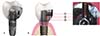
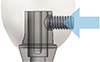
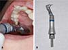
The modified LSP (YK Implant Prosthetic System, Dipstek, Seoul, Korea) consists of a lateral screw, an abutment with a lateral hole, an anti-loosening abutment screw, and a crown with a negative screw housing (Fig. 1). The modified LSP has a negative screw housing located within the crown, which provides mechanical locking by pushing against the walls of the abutment. The lateral screw penetrates the abutment and makes direct contact with the abutment screw (Fig. 2). The implant abutment/crown is manually tightened to a torque of 5 - 10 Ncm using a lateral screwdriver with a contra-angle attachment (Fig. 3).
Mechanical complications (i.e., lateral screw loosening [LSL], abutment screw loosening [ASL], lateral screw fracture [LSF], and ceramic fracture [CF]) were identified by examining the patients' treatment records, clinical photographs, and periapical and panoramic radiographs. Biological complications were diagnosed by assessing various clinical and radiographic parameters (probing depth, bleeding on probing, suppuration, mobility, and periapical radiographic bone loss) with the aid of a UNC periodontal probe (Hu-freidy, Chicago, IL, USA). The presence of only reversible inflammatory reactions (e.g., easy bleeding on probing, gingival swelling, or redness) was diagnosed as peri-implant mucositis (PM), while it was diagnosed as peri-implantitis (PI) if these symptoms were accompanied by bone loss (as detected with a periodontal probe and in the periapical radiograph), probing depth > 5 mm, and/or mobility.1415 The clinical C/I ratio was measured as the length of the clinical crown (with the fulcrum located at the crestal bone) divided by the length of the implant, while CHS was measured as the length from the crestal bone to the occlusal plane.1617 To correct the distortion errors, such as magnification, the clinical C/I ratio, and CHS, were calibrated using the inter-thread distance as a reference on a PACS workstation (Centricity GE Healthcare, Waukesha, WI, USA).
Statistical analyses were performed with SPSS Statistics software (19 version, SPSS, Chicago, IL, USA), using Student's t (two-tailed with independent samples), Fisher's exact, and chi-square tests to identify the relationships between clinical factors and complication rates. Standard deviation values and 95% confidence intervals were calculated, and the cutoff for statistical significance was set at P < .05.
RESULTS
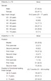
Seventy patients (37 male and 33 female patients) with a mean age of 54.9 years (range: 25 - 77 years) met the inclusion criteria. All of the 73 implants investigated from these patients were the internal-connection type (types: Implantium [n = 60], GS2 [n = 1], GS3 [n = 5], SS2 [n = 7], Dentium and Osstem, Seoul, Korea). Fifty-six implants were placed using a one-stage nonsubmerged procedure without any advanced surgery, while 17 implants were placed using a two-stage submerged procedure.
The implants were distributed in the posterior region as follows: maxillary first premolar, n = 2 (2.7%); second premolar, n = 6 (8.2%); first molar, n = 16 (21.9%); second molar, n = 2 (2.7%); mandibular first premolar, n = 1 (1.4%); first molar, n = 31 (42.5%); and second molar, n = 15 (20.5%). In total, 26 implants were placed in the maxilla (35.6%) and 47 in the mandible (64.4%). The mean loading period was 43.7 months (range: 31 - 56 months; Table 1).
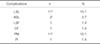
Mechanical complications were present in 14 (19.8%) of the 73 investigated posterior ISSCs inserted with the modified LSP. LSL was the most common complication (n = 11, 15.1%), followed by ASL (n = 2, 2.7%), LSF (n = 1, 1.4%), and CF (n = 1, 1.4%). LSL and ASL occurred simultaneously in an implant. Biological complications occurred in 11 (15.1%) of the investigated posterior ISSCs. PM (n = 11, 15.1%) and PI (n = 1, 1.4%) were also present, and PM occurred twice in two of the implants (Table 2).
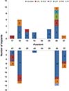
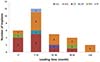
The frequency distributions of the implants according to their positions in the upper and lower jaw are shown in Fig. 4. Complications occurred in 1 (16.7%) of the 6 implants placed in the maxillary second premolar (LSL, n = 1), 8 (50%) of the 16 implants placed in the maxillary first molar (LSL, n = 2; ASL, n = 2; LSF, n = 1; PM, n = 3), 2 (100%) of the 2 implants placed in the maxillary second molar (LSL, n = 1; PM, n = 1), 8 (25.8%) of the 31 implants placed in the mandibular first molar (LSL, n = 4; PM, n = 3; PI, n = 1), and 8 (53.3%) of the 15 implants placed in the mandibular second molar (LSL, n = 3; CF, n = 1; PM, n = 4).
Based on the incidence of mechanical complications relative to the duration of functional loading, 11 (73.3%) of 15 implants exhibited mechanical complications during the first 12 months after undergoing functional loading. The incidence of LSL during the first 12 months was particularly noticeable (n = 9, 60%). ASL occurred after LSL in one case before any repair took place. During the first 12 months, six implants showed PM, with an even distribution of the complications across the duration of functional loading. During the mean follow-up period of 43.7 months, there were no further mechanical complications after 24 months of functional loading (Fig. 5).
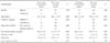
Analyses using the Fisher's exact, chi-square, and the t-tests indicated that the incidence of mechanical complications was significantly related to gender (P = .024). The other clinical factors of age, position in the jaw, placement location (premolars were excluded due to the small size of the sample), functional duration, clinical C/I ratio, CHS, and the use of a submerged or nonsubmerged placement procedure were not significantly associated with the mechanical complication rates. Biological complications showed no statistically significant association with any of the clinical factors (Table 3).
DISCUSSION
The present study found that the modified LSP was similar to the conventional implant prosthetic system. A systematic review found that the cumulative 5-year complication rates of CF and screw fracture were 7.8% and 1.3%, respectively.8 Kreissl et al.18 reported a similar rate of CF (5.7%) over an observation period of 5 years. In the present study, the mean of 4-year cumulative incidence rate was 1.3% for both CF and LSF (CF, n = 1; LSF, n = 1).
Many studies have indicated that screw loosening is a major mechanical complication of ISSCs.819 The occurrence rates of ASL have been reported to range from 2.4% to 37.7%, and a maximum occurrence rate of 22.2% has been reported for occlusal screw loosening (OSL).891011 Among the cases studied in the present study, 11 (15.1%) showed LSL and 2 (2.7%) showed ASL, including a case in which ASL accompanied LSL. The occurrence rate of LSL was similar to or lower than that of OSL, while the occurrence rate of ASL was significantly lower.1920 Screw loosening results in non-ideal occlusion and is the main cause of mechanical complications such as screw fracture of ISSCs placed in the posterior region.21 When mild LSL is detected during routine follow-up, a screwdriver with a contra-angle attachment can be used to tighten the lateral screw, with the occlusal contact also being re-evaluated and adjusted. This intervention results in the modified LSP being effective in reducing the occurrence of serious mechanical complications, such as screw or ceramic fracture. Mechanical complications are most likely to take place within 2 years after functional loading (in contrast to biological complications), 22 and therefore our data are considered to be meaningful although longer, more-detailed prospective and larger controlled studies may be needed.
Structural defects in the implant prosthesis cause clinical problems such as food impaction and cement retention that make it difficult to maintain oral hygiene, and this problem in turn causes biological complications, lowering the survival and success rates of the implants.23 A recent study found that the presence of excess cement in cementretained implant prostheses was one of the main causes of peri-implant disease.24 Wilson reported that excess cement caused peri-implant disease in 81% of cases of a cementretained implant prosthesis.25 Among biological complications, PM reportedly occurred in 80% of patients and 50% of implants, while PI occurred in 20 - 56% of patients and 10 - 43% of implants.26 The present study found that biological complications occurred in 11 implants (15.1%) during recall checks, and no patient complained of discomfort. Nonsurgical mechanical debridement was carried out for the 10 implants (13.7%) with PM,27 while regenerative surgical therapy was carried out for the PI that occurred in 1 implant (1.4%). After the treatments, the implant prosthesis was regularly separated to allow meticulous scaling and professional care during periodic recall checks.1528
The modified LSP allows easy separation of the prosthesis and provides an environment for easy access when PM or PI occurs, which facilitates effective peri-implant treatment. When cleaning and caring for the peri-implant mucosa, a wound is formed in the connective tissue while the screw-cement-retained prosthesis is disconnected and subsequently reconnected, causing marginal bone resorption and recession.29 However, in the modified LSP, recession of the mucosa can be prevented by disconnecting only the crown–and not the abutment–when treating any inflammation present in the marginal gingiva and connective tissue, thereby enhancing gingival health.30
An analysis of correlations among observed clinical variables in ISSCs with the modified LSP revealed that gender was the only variable that was significantly related to the occurrence of mechanical complications (P = .024); this may be because the biting force and occlusal contact area are greater in male subjects.3132 It has been reported that an unfavorable C/I ratio (anatomical and/or clinical C/I ratio of ≥ 2) does not affect the rate of biomechanical complications associated with implants, while an unfavorable CHS value (≥ 15 mm) does affect the rate of prosthesis complications. 33343536 The present study found that the clinical C/I ratio and CHS were not significantly associated with mechanical and biological complication rates. Since both values fell within the favorable range, they were not considered to affect the incidence of complications. The possibility of mechanical complications increases with the horizontal distance between the most distally positioned ISSCs and the mesially adjacent natural tooth.37 The present study investigated the 35 most distally positioned ISSCs, and the rate of mechanical complications was found to not vary significantly with horizontal distance (P = .099).
The present study found that the incidence of mechanical and biological complications in the posterior region of modified LSP was similar to that of the conventional implant prosthetic system. Despite the considerable differences between the modified LSP and conventional implant system in terms of mechanical and physical aspects, the fact that we did not include a finite element method (FEM) analysis can be considered a limitation of this study. A previous study that performed FEM analysis between cemented-retained dental implants and screw-retained dental implants showed that screw-retained implants had low dissipation of overload energy and mechanical stress, which is likely to have resulted in weak-linked components.3839 Therefore, an appropriately designed FEM using a modified LSP should be carried out precisely to investigate the dissipation and distribution of mechanical tension and stress among fixture, lateral screw, and prosthetic components.
A long-term study of prosthesis survival and complication rates of ISSCs found that 66% of patients experienced at least one complication.40 The present study focused on the treatment outcomes of ISSCs inserted with the modified LSP in the posterior region, and in particular the rate of mechanical and biological complications. ISSCs with the modified LSP were found to be clinically acceptable and effective in preventing and treating these complications. Although the modified LSP structurally compensates for the considerable drawbacks of the conventional prosthetic system, structural constraints associated with using an additional negative screw housing cause overcontouring and restrict the design of the implant prosthesis. In addition, there have been no reports of accumulated long-term success rates, so future studies should perform long-term observations with a larger number of cases, including the anterior region and ISFDPs.
CONCLUSION
Within the limitations of this study, the mechanical and biological complication rates for ISSCs inserted with the modified LSP were found to be similar to those of conventional implant prosthetic systems. In addition, the modified LSP is amenable to maintenance care, which facilitates the prevention and treatment of mechanical and biological complications.
 XML Download