

 PDF
PDF ePub
ePub Citation
 Print
Print


INTRODUCTION
Most commonly, contractures arise where adequate burn care and delivery have not occurred and scar management has not been instigated in a vigorous manner. Repair by regeneration can no longer occur when the depth of injury extends beneath the reticular dermis, and healing by secondary intention.1 The resultant wound contraction can lead to contractures over flexor surfaces and its surroundings. The incidences with contractions in oral region can limit proper prosthodontic treatments. For example, limitation of opening jaws may cause difficulties in accessing the dentitions and tissues of the oral cavity, and in using full size of removable denture for the patients. Telescopic implant-supported removable partial prosthesis with milled abutments has been used for the patient with burn contracture to provide enough retention and stability for the prosthesis.
CASE REPORT
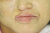
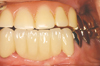
A 60-year old woman was referred from the plastic surgery department to the Department of Prosthodontics at Yonsei University Health System. She had a chief complaint of restoring her oral function and esthetics after series of plastic skin graft procedure done. During the extra oral examination it was noted that the elasticity of muscle and skin tissue around oral angular area was lost due to scars from burns and tissue grafting, which resulted in limited maximum mouth opening of 20 mm. Therefore, proper prosthodontic treatment was impossible on the right quadrants of the mouth (Fig. 1). The intraoral examination showed that all of maxillary teeth were missing except tooth number #25 and splinted gold crowns on #34 and #35, whereas fixed partial denture on #33 to #43 were present on the mandible.
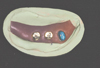
In order to restore the patient's masticatory function, prosthodontic treatment plan of using 4 implants on the left quadrants was established. Each of two implants were placed on upper and lower posterior left jaws, however no implant on anterior ridge were possible due to severe atrophy of the maxilla. Anterior cantilever type removable partial denture was planned to meet patient's esthetic demand, which consists of a telescopic crown abutment on #25 and two implant supported abutments on area of #26 and #27 in the maxillary area (Fig. 2). On the mandible, Implant-supported fixed partial denture on area of #36 and #37 was planned.
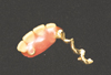
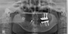
The implants (Strauman® Basel, Switzerland) (ϕ4.8×10 mm) were placed on predetermined area accompanied with sinus lift ridge augmentation procedure and proper follow up was performed during the 6 month healing period of time. Customized abutments were fabricated on implants and designed in the predetermined angle paralleling to the prepared tooth of #25 (Fig. 3). Removable partial denture was inserted to verify phonetics, esthetics and function. Key and keyway rigid attachment was used for retention and ledge type rest was used for support (Fig. 4). Panoramic view at initial placement of the prostheses was presented in Fig. 5. No complications have occurred in 5 years since the insertion of the prostheses (Fig. 6).
DISCUSSION
Limited prosthodontic treatment is due to scar contracture on the tissues and trismus. Removable type of prosthesis was selected as treatment option after considering the factors of oral hygiene maintenance and lip support of patient to improve esthetics. Because there was only one natural tooth in the maxilla, we, therefore, had to improve the unfavorable removable partial denture using implants.2 Due to the lack of anterior ridge, the implant option on anterior ridge was deemed unavailable, and natural tooth was splinted with implant to support anterior cantilevered prosthesis.
A remaining tooth was splinted to implant supported abutments. Combining implants with natural teeth is controversial because of the variations in movement during function.3 A lot of complications regarding implant and tooth-supported prosthesis may arise; including fracture of the prosthetic components, intrusion of the natural tooth, marginal bone loss, and loss of osseointegration,4 but survival rates of implant and tooth-supported fixed prostheses are comparable to implant-supported fixed prostheses.5 There was no prosthetic complication in implant-and tooth-supported fixed prosthesis in this case.
In this case, a removable partial denture with long anterior cantilever was used. Forces in the cantilevered area were transmitted to the abutments, causing tilting and rotational movements,6 but cantilevered prostheses are preferable when reduced stress is inherent.6,7 The prosthesis in this patient is expected to have a good prognosis since it is designed with hypoocclusion in anterior areas.

 XML Download