

 PDF
PDF ePub
ePub Citation
 Print
Print


INTRODUCTION
Endosseous implants have been widely used to restore missing teeth and various literatures confirmed the implants as a predictable treatment modality with high success rates.1,2 It has been advocated that after placing implant, the surgical site should be undisturbed for three to six months before fabricating prosthesis to enhance osseointegration.3 The rationale behind this approach is that preventing implant from micromovement caused by functional force during the osseointegration period is of paramount importance for the success of an implant.4 However the treatment time of implants needs to be shortened to enhance patient's comfort and satisfaction. Immediate loading with provisional or definitive prosthesis within a week after implant placement has been reported in many literatures.5-8
Lefkove and colleagues reported immediate loading of mandibular overdenture as a provisional prosthesis and acquired successful osseointegration of the implants.5 Early protocols allowed immediate loading only when initial stability of implant was guaranteed by cross-arch stabilization with splinting implants in a completely edentulous patient.5,6
Immediate function in partially edentulous patients was investigated in other studies.7,8 Schnitman et al. followed 28 immediately loaded implants in partially edentulous patients for 10 years and documented a 84.7% success rate in 10 years.7 Tarnow et al. found that 67 out of 69 immediately loaded implants in partially edentulous patients were osseointegrated and 37 out of 38 implants with conventional protocol were osseointegrated. It was concluded that immediate loading could be an appropriate treatment modality in a partially edentulous patients.8
Traditional implant protocol requires delay of implant placement until proper bone healing after tooth extraction occurs.9 This prolonged treatment time causes disadvantages such as unesthetic appearance, loss of masticatory function, and improper bone architecture due to the alveolar bone resorption.10 On the other hand, advantages of immediate placement into the extraction sockets includes shortened treatment time, reduction in alveolar bone resorption and enhancement of esthetics and function by preserving bone tissue.11
If the implant is immediately placed and loaded, treatment time is dramatically decreased and soft tissue esthetics can be improved by provisional prosthesis.12 Anneroth et al. reported of no histological and radiographic difference between immediately placed implants and implants placed after a period of healing time in monkeys.13 The same result was documented in a human study by Paolantonio et al.14
If the implant is immediately placed into the extraction socket and immediately loaded with provisional restoration, better esthetic results can be achieved by interproximal soft tissue support.15 Colomina16 and Chaushu et al.17 reported that immediate placement and immediate loading can be a predictable treatment modality in properly selected cases. A success rate of 97.4% was reported by Roberto and Bo in 2007 after immediate placement and immediate loading in maxilla.18
Misch suggested that functional force should not be applied to provisional prosthesis during bone healing in an immediate loading procedure.19 However, when canine is treated with immediate loading implant for the patients having canine protected occlusion, the occlusion scheme is not established.
This clinical report describes the immediate placement and immediate loading of implants placed for a patient having a canine protected occlusion.
CASE REPORT
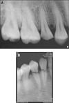
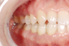
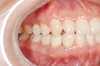
A 27-year-old woman visited for the treatment of fractured teeth. She previously suffered from right mandibular condyle neck fracture and subgingival fractures of maxillary right second premolar, mandibular right canine and premolar' crowns (Figs. 1, 2). Treatment of trismus caused by the condyle fracture was followed by extraction of fractured teeth, and the immediate placement and function of implants with provisional prosthesis were planned. After obtaining 40 mm of mouth opening, a preliminary impression was taken. Diagnostic waxing was conducted on the diagnostic cast and it was duplicated. Soft tissue model and soft shell template with 1mm thickness (Copyplast, Scheu dental GmbH, Iserlohn, Germany) were fabricated for preparing provisional prostheses according to expected soft tissue contour after the extraction. Provisional prostheses were prefabricated with quick setting acrylic resin (Tokuso Curefast, Tokuyama Dental Co., Tokyo, Japan) by using the soft shell template and the soft tissue model. Implant surgery was carried out by an oral surgeon in the presence of a prosthodontist.
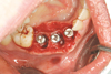
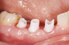
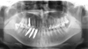
After teeth extraction, ∅4×13 mm internal connection implants (Full Osseotite Certain, The Biomet 3i, Palm Beach Gardens, FL, USA) were placed in the maxillary and mandibular right second premolars and ∅4×15 mm implants were placed in the mandibular right canine and first premolar (Fig. 3).
Autogenous bone was harvested from the maxillary right tuberosity and grafted to bony defects in the extraction sockets of the mandibular right canine and premolars after mixing with 0.25 g of xenograft bone (Bio-Oss, Geistlich Pharma AG, Wolhusen, Switzerland).
Before the flap closure a implant-level impression was taken with transfer type impression coping using light body and mono-phase polyvinylsiloxane (Aquasil Ultra, Dentsply Caulk, Milford, DE, USA) (Fig. 4). Dental stone (Silky-Rock, Whip-Mix Co. Louisville, KY, USA) was poured to save time needed for setting.
Temporary cylinders were connected and milled to modify the path and height after mounting the casts. Internal surfaces of prefabricated provisional prostheses were removed and relined to adapt to the abutments. Occlusion was adjusted on the semi-adjustable articulator. The patient had a very long and sharp upper canine and didn't want to adjust its form. Thus, it was impossible to fabricate esthetical provisional restoration that avoids canine protected occlusion during lateral excursion.
In addition, provisional restoration of maxillary right second premolar was constructed not to function. In order to distribute lateral forces by using mandibular right canine during lateral excursion, a cement-retained provisional prosthesis was splinted by a provisional fixed partial denture in mandibular edentulous sites. Temporary cylinders and constructed provisional prostheses were connected in the mouth with 15 Ncm torque. Flap was sutured with interrupted suture and occlusion was adjusted intraorally (Fig. 5).
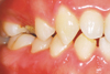
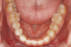
Dressing and occlusal adjustment were conducted again one day after the surgery and sutures were stitched out a week later. Occlusion was adjusted and provisional restorations were gradually reshaped to enhance soft tissue form at the follow-up appointments in two week, four week, and eight week intervals. After 4 weeks, soft tissue contour was very stable and esthetic (Fig. 6).
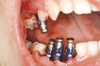
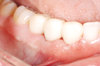
Four months after the implant placement, clinical and radiographic examination of maxilla and mandible revealed successful osseointegration. Definitive prostheses were constructed with zirconia abutment (Zirealpost, The Biomet 3i, Palm Beach Gardens, FL, USA) and pressable all-ceramic restoration (IPS-Empress II, Ivoclar Vivadent AG, Schaan, Liechtenstein) in response to the patient's high esthetic expectation. After taking implant level impression, master cast was poured and mounted using facebow transfer. Zirconia abutments were milled and definitive prostheses with canine guided occlusion were made with IPS- Empress II system. Definitive restorations were tried-in intraorally. Zirconia abutments were tightened to 20 Ncm of torque following the manufacturer's recommendation (Fig. 7). Definitive restorations were permanently cemented with resin cement (Rely-X Unicem, 3M ESPE, Seefeld, Germany) after finishing and polishing (Figs. 8, 9).
DISCUSSION
The placement of the implant immediately after extraction of the tooth has now become the reliable treatment option and is associated with preserving periodontal structure, as well as with reducing the treatment time, which ultimately benefits the patient. Usually immediate implant placement and loading is are recommended for the treatment of edentulous patient to gain maximum stabilization and anterior teeth missing patient to avoid stressful lateral force loading to implant fixture. Although functional immediate loading of provisional prosthesis in partially edentulous area is not recommended, canine tooth contacts with the opposing dentition during function is not avoidable when canine is treated with immediate loading implant for the patients with canine protected occlusion.
In this clinical case, the anatomic form of the patient's teeth was well preserved and canine guidance was clear. Moreover, the patient did not want to adjust her natural tooth. Therefore, it was impossible to avoid occlusal contact of provisional restoration during lateral excursion while maintaining esthetic form of the provisional restoration. Consequently, provisional restoration was constructed not to contact opposing dentition during maximum intercuspation and slightly guided by canines during lateral excursion. It was splinted by a provisional fixed partial denture to distribute functional force.
Clinical success in this report can be explained by the following factors. Extracted teeth sockets had been preserved without any chronic inflammation such as periodontitis. The patient concerned on esthetics of the restoration and was very cooperative with reasonable expectation, good oral hygiene maintenance and careful masticatory pattern. The results of this pilot clinical case suggest that immediate placement and immediate loading is are successful in functionally loaded implant situation if the patient's cooperation is assured.

 XML Download