

 PDF
PDF ePub
ePub Citation
 Print
Print


INTRODUCTION
The performance of definitive cements and the longevity of a restoration are affected by preparation coarseness, and the type of provisional cement, and cleaning technique used to remove provisional cement remnants.1 Due to the negative effect that residual provisional cement, and debris on prepared abutment teeth may have on the performance of definitive cement,2 all remnants must be removed prior to definitive cementation.1
Mechanical methods for removing provisional cement remnants include the use of a dental explorer, pumice or probes: however, none of these methods are fully effective; Terata3 showed that the removal of provisional cement from bovine enamel and dentin with an explorer was incomplete. Cement remnants have been observed microscopically on surfaces that appeared macroscopically clean, thus provoking a search for alternative methods for eliminating provisional cement remnants.4 For removal of debris and remnants from the dentin surface dentin surface, different cleaning agents containing ethanol, ethyl acetate, acetone, or chlorhexidine digluconate have been marketed. Kanakuri et al.5 reported that the use of a rotational brush with running water was the best method.
Button et al.6 reported that higher retentive strengths for glass ionomer and zinc polycarboxylate cements were obtained with tooth preparations cleansed with plain flour pumice than those cleansed with an explorer only.
Bachmann et al.7 investigated the bond strength of dentin bonding agents after teeth were cleaned with a scaler, a cotton pellet with pumice, and different soaps. All of the soaps tested decreased shear bond strength (SBS) values. The authors reported that the use of soap to remove remnants of provisional cements prior to adhesive cementation was not recommended for clinical use. Kanakuri et al.5 reported that the use of a rotational brush with running water was the best method. For removal of debris and remnants from the dentin surface, different cleaning agents containing ethanol, ethyl acetate, acetone, or chlorhexidine digluconate have been marketed.
Laser devices have been used in dentistry for soft-tissue surgery, root-end sealing and sterilization, and for altering enamel and dentin surfaces, in order to increase resistance to decay or to facilitate the bonding of composites.8 Because laser etching is painless and involves neither heat nor vibration, this method is attractive to patients as well as clinicians. Studies have been performed using different kinds of lasers, including Er:YAG, Nd:YAG, and CO2 lasers with attention focused particularly on Er:YAG lasers, due to their effectiveness in removing dental hard tissue.9 However, the literature contains no information about structural changes in dentin following the use of an Er:YAG laser to clean temporary cement remnants from dentin surfaces. Therefore, the purpose of this in vitro aimed to measure and compare the SBS values of ceramic discs to dentin surfaces cleaned with a dental explorer, pumice, cleaning bur and Er:YAG laser. The hypothesis tested was that the SBS would be higher after Er:YAG laser dentin cleaning with a dental explorer, pumice, or cleaning bur.
MATERIALS AND METHODS
Tooth preparation
A total of, 36 caries-free unrestored human third-molar teeth that had been stored in a 0.5% cloramin T solution at 4℃ for up to 1 month following extraction were selected as tooth specimens. Soft tissue was removed with a scaler (H6/H7, Hu-Friedy, Chicago, USA). After cleaning the teeth with pumice and then each tooth was embedded in autopolymerizing acrylic resin (Palapress, Heraeus Kulzer, Wehrheim, Germany) using a cylindrical plastic mold (20 × 20 mm). The occlusal third of each crown was sectioned with a slow-speed diamond section (Minitom, Struers GmbH Nederland) under water cooling and the exposed dentin surfaces were ground with 320-grit silicon-carbide abrasive paper under running water to ensure a smooth surface.
Cementation of provisional restorations
Provisional restorations were simulated by pouring an acrylic medium (Dentalon Plus, Wehrheim, Germany) in a Teflon mold to produce thirty-six 10 × 2 mm discs. The acrylic was prepared and polymerized according to the manufacturer's instructions. Following polymerization, discs were removed from the mold, examined for size and air bubbles, and then cemented to the dentin surfaces of tooth specimens using eugenol-free provisional cement (Cavex, Haarlem, Holland) and a special loading device (Algol Instrument Co, Hsin-Chuang, Taipei, Taiwan) under a load of 9.2 N. All specimens were stored in distilled water at 37 ± 2℃ for 2 days before use.
Experimental design
Specimens were randomly assigned to 1 of 4 groups different dentin cleaning protocols (n = 9), as follows:
Group 1 (control): Provisional cements were mechanically removed with a dental explorer using moderate pressure until the dentin surface was macroscopically clean.
Group 2: Provisional cement was removed from dentin surfaces by cleaning with a brush and pumice for 1 min using a rotary instrument at 5,000 rotations/min under water cooling.
Group 3: Provisional cement was removed from dentin surfaces by cleaning with a bur (Opticlean, Kerr, California, USA) for 1 min using a rotary instrument at 5,000 rotations/min under water cooling.
Group 4: Provisional cement was removed from dentin surfaces by cleaning with an Er:YAG laser (Fidelis Plus 3, Fotona, Ljubljana, Slovenia) under an air water spray at 200 mJ, 20 Hz a pulse duration of 100 µs, tip diameter of 800 nm and a working distance of 0.5 mm between tip and dentin surface until the dentin surface was macroscopically clean.
Ceramic disc preparation
Thirty-six 7 × 3 mm pressed lithium disilicate glass-ceramic discs (Ivoclar-Vivadent AG, Schaan, Liechtenstein) were fabricated by the lost wax technique and ingots were injected into the furnace (EP 600, Ivoclar-Vivadent, Schaan, Liechtenstein). The surfaces of the discs were ground with 220, 360, and 600 grit silicon-carbide sandpaper to standardize the bonding surface. Ground ceramic discs were air-abraded with 50 µm Al2O3 particles (Korox, Bego, Bremen, Germany) for 14 seconds from a distance of approximately 10 mm at 400 kPa with a sand-blasting device (Ar-Ge Dental, Denizli, Turkey). The discs were then cleaned in distilled water for 10 min in an ultrasonic bath (Healthsonics, Livermore, CA, USA) to ensure a contaminant-free surface.
Luting procedures
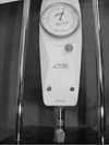
Self-adhesive luting cement (SA Cement, Kuraray, Tokyo, Japan) was used to bond the ceramic discs to the dentin surfaces. Cement was mixed and then applied to ceramic and dentin surfaces with no primer or adhesive. A special loading device was designed to apply a constant load of 9.2 N to the ceramic discs (Fig. 1). This load was used to create a uniform resin luting layer of approximately 100 µm to simulate the range of film thickness for all ceramic crowns. Initial light curing (light intensity of - 1000 mW/cm2, Elipar Freelight LED 2, 3M ESPE, MN, USA) was performed for 10 seconds. Excess cement was removed using a dental probe. The luting agent was polymerized from each direction (mesial, distal, buccal, lingual, and occlusal) with the same curing device for 40 seconds.
Shear bond strength (SBS) test
After cementation, the specimens were removed from the alignment device and stored in deionized water and thermocycled for 5,000 cycles between 5 ± 2℃ and 55 ± 2℃, with a dwell time of 20 seconds and a transfer time of 5 seconds. Finally, luted specimens were placed in a jig for SBS testing, as described under ISO/TR 11405. They were loaded to failure with a crosshead speed of 0.05 mm/min until decementation occurred, using a universal testing machine (Instron, Canton, MA, USA). SBS values were calculated and recorded using the following formula10:
SBS (MPa) = P/A
(P = maximum force (N) and A = interfacial area (mm2)).
Stereomicroscope and scanning electron microscope (SEM) evaluation
After SBS testing, the dentin surfaces of the de-bonded specimens were examined with a stereomicroscope (Wild M3B; Heerbrugg, Switzerland) at ×25 magnification to identify the mode of bond failure. The failure mode was classified according to one of three types: adhesive failure at interface (AD), cohesive fracture of resin cement (CO), or mixed fracture at the interface of resin cement (MI).
Two specimens were randomly selected from each group and prepared for SEM analysis after the SBS test. The de-bonded specimens from each group were gold sputter-coated (Bal-Tec SCD 050 Sputter Coater; Bal-Tec AG, Liechtenstein) and observed by SEM (LEO 440, Leica-Zeiss, Cambridge, UK).
Statistical analyses
All data sets were subjected to normality tests using the Kolmogorov Smirnov method; data are presented as medians and 25th and 75th percentiles (for skewed data). One-way ANOVA and Tukey HSD tests were used to perform multiple comparisons with a level of P>.05 significance level. All analyses were performed with the statistical package for scientists (Sigmastat, Aspire Software International, WA, USA) Windows version 3.10.
RESULTS
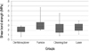
The mean SBS values and the results of one-way ANOVA are shown in Fig. 2. The dentin cleaning methods did not significantly affect the SBS of resin cement to dentin (P>.05): dental explorer (2.04 ± 0.78 MPa), pumice (3.30 ± 1.49 MPa), cleaning bur (2.60 ± 1.50 MPa), and laser (2.59 ± 0.92 MPa).
Fracture patterns
The type and frequency of failures of the specimens are presented in Table 1. Two types of failures were predominant: adhesive and mixed. Adhesive failures were the most commonly observed in all groups. However, the specimens in the groups cleaned with the dental explorer or laser showed mixed failures.
SEM observation
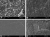
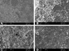
On the dentin surfaces of the specimens cleaned with the dental explorer or cleaning bur, significantly more temporary cement remnants were found versus the other groups. The dentin surfaces of the specimens cleaned with a brush and pumice showed tubule obliteration. Laser irradiation at 200 mJ with a pulse repetition rate of 20 Hz provided the cleanest dentin surface, with open dentin tubules and the absence of cracked areas (Fig. 3D). After the SBS test, mix fractures observed resin cement debris was observed on all dentin surfaces (Fig. 3) except the group cleaned with the dental explorer, which had smooth and clean surfaces (Fig. 4)
DISCUSSION
A durable and predictable bond strength between dental materials and teeth is important for clinical success.11 One problem associated with ill-fitting restorations is related to remnants of temporary cement on abutment surfaces.5 This is the first study to report on the use of the Er:YAG laser as a cleaning method. The results did not support the research hypothesis that the SBS between resin cement and dentin would be higher after cleaning with an Er:YAG laser when compared to cleaning with a dental explorer, pumice, or cleaning bur.
Carvalho et al.12 showed that application of eugenol-containing provisional cement decreased the bond strength between resin cement and dentin. Some authors have indicated that eugenol-containing materials have adverse effects, including altered wettability and reactivity of dentin.1 For this reason, in the current study eugenol-free temporary cement was used as the temporary cement.
Yap et al.13 observed elevated bond strength and resin infiltration to dentin after mechanical removal of remnants using an ultrasonic scaler followed by cleaning of dentin surfaces with a pumice-water slurry. However, in the present study, SEM observation and SBS tests results gave contradictory findings. SEM showed that a dental explorer alone was unable to remove all temporary cement from the dentin surfaces and that further cleaning was necessary. However, no significant differences were observed in SBS values.
Some cleaning protocols have been found to alter dentin surface texture.14 However, reducing roughness is not the only factor in improving bond strength. Factors such as the chemical composition of the dentin surface may have a stronger influence on adhesion, and taken together with surface roughness and physical parameters such as capillary action will be decisive for the dentin surface free energy and consequently adhesive diffusion into the deminerilized dentin surface.3 SEM observation following SBS testing in the present study showed that while laser cleaning produced effective dentin surface microstructures (Fig. 3D), resin cement particles were not present to the extent expected (Fig. 4D).
While laser application may have certain advantages, the use of an Er:YAG laser instead of conventional techniques for cleaning provisional cement was not found to significantly improve SBS. Furthermore, Er:YAG lasers are still too expensive to be cost-effective.8 According to a previous study, the use of cleaning bur also removes the dentin that is in direct contact with temporary cement, making this a reliable method for removing temporary cement, especially in inaccessible areas.3
The current study has a number of limitationsthat only one type of temporary cement and adhesive resin cement were used and that the tests were performed under in vitro conditions. It is possible that the use of different cements would have given different results.

 XML Download