

 PDF
PDF ePub
ePub Citation
 Print
Print


INTRODUCTION
Interest in dental esthetics has increased rapidly during the last few decades, among both patients and dentists.1 Because the focus of many adults has shifted toward esthetics, it becomes a primary consideration for patients seeking prosthodontic treatment. When planning treatment, dentists have to understand beauty, harmony and proportion, as perceived by society.2
Anterior occlusion relationships, malocclusions and posterior occlusion relationships are intraoral parameters that can affect facial appearance. Dental appearance is one of the most important aspects of facial attractiveness and encompasses not only tooth color, but also the position, shape, size and related aspects of teeth, such as gingival morphology and upper lip height during rest position and smile.3-5 One of the most important aspects of dental and facial esthetics is the display of anterior teeth. Esthetic is assessed by viewing the patient from the front in dynamic states, like conversation, facial expressions and smiling.
The aspect of maxillary anterior teeth plays an important role not only in dental esthetics, but also in facial esthetics.6 The maxillary central incisor is considered to be the primary reference tooth, more important than the rest of the anterior teeth in regards to the visible coronal tooth structure.7,8 For esthetic purposes, the maxillary anterior teeth must be in proportion to facial morphology.9-11
Many authors emphasized the need for achieving width-to-length proportions in the smile that harmonize with the face, and the golden proportion was suggested as guideline. The ideal maxillary central incisor should be approximately 0.8 width, compared with length, but it has been reported to vary between 0.72 and 1.24.12-15 A higher width-to-length ratio means a squarer tooth, and a lower ratio indicates a longer appearance.
It has been suggested that the width-to-length ratios of the maxillary anterior teeth were not affected by gender15. However, significant gender differences in width-to length ratios were recorded in other previous studies.13,16,17
In one study, male subjects exhibited from 0.5 to 1 mm larger tooth width than female subjects.18
Restorative dentistry frequently entails correction of tooth size discrepancies associated with various lengths and/or widths.19 Consequently, tooth dimensions may be an important aspect of esthetic reconstruction. Proper diagnosis of tooth size in each patient is critical when planning treatment for esthetic restorative dentistry.7
Currently, the perception of attractiveness is extremely influenced by media, including movies, television commercials and fashion magazines. They all entered in our life bringing facial "standards" that should convey perceptions of beauty, healthiness and fitness, mixed with feelings of social achievement, intelligence, richness and happiness. A beautiful face seems the key to success.20-22 Consequently, dentists should have a deep understanding of those quantifiable and objective facial characteristics considered "attractive" by the public.23-26
The aims of this study were to (1) determine whether celebrities considered to be beautiful differ in the width-to-length ratio of the maxillary central incisor from non-celebrities of the same age and race, and (2) compare gender differences in the width-to-length ratio of the maxillary central incisor during smiling.
MATERIALS AND METHODS
A total of 40 men and 40 women were included in this study. All of the subjects had all anterior teeth present with no caries, extreme occlusal wear, extrusion, obvious deformities or tooth mobility. They were assigned to two groups, i.e. the control and experimental group, and two different groups of adults were analyzed.
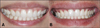
The control group consisted of 40 adults (20 male and 20 female) aged 25 to 39 years (mean age 33.2±3.8 years), selected according to criteria of dentofacial normality, with normal occlusion, no crowns, porcelain laminate veneers or composite resin restorations in the anterior maxillary segment, and no mandibular or craniocervical disorder. They were students in the School of Dentistry, Chonnam National University. These non-celebrities constituted the NON-MEDIA group (Fig. 1). Anterior tooth images of the subjects (NON-MEDIA group) were taken in a frontal view with all subjects in maximum smiling position. Each subject was seated in a dental chair with the head upright and with the occlusal plane of the maxillary teeth parallel to the floor. Digital photographic equipment (IXUS 210 IS®; Canon Inc., Tokyo, Japan) was used. The images recorded were acquired with the software driver for the digital camera of a personal computer (Xnote T290®; LG Electronics Inc., Gumi, Korea), translated to Joint Photographic Experts Group (JPEG) format and stored in database software. All photographs were taken by the same investigator to ensure standardization of the procedure.
The experimental group consisted of 40 celebrities (20 male and 20 female), selected on the basis of their soft-tissue facial appearance. The selected celebrities ranged in age from 25 to 39 years (mean age 28.9±6.2 years). These celebrities constituted the MEDIA group. Facial photographs of the MEDIA group were collected from the Internet websites (www.naver.com, www.google.co.kr). The collected photographs were with smiles showing maxillary anterior teeth from the front and with the resolution of 96 dpi or over (Fig. 2).
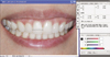
The mesiodistal and incisogingival dimensions of the maxillary central incisor were measured using a computer program for analysis, measurement and edition of photographs (Photoshop® 7.0; Adobe systems Inc., San Jose, USA). The mesiodistal dimension was measured parallel to the incisal edge. The width and length was measured at the widest mesial-distal portion and longest apical-coronal portion of the tooth (Fig. 3). The same investigator measured 80 photographs 5 times with the same method for accuracy, and the mean value was used for analysis.
Data were analyzed with the statistical software SPSS version 19.0 (SPSS Inc., Chicago, IL, USA). The limited number of subjects required assessment of normality for data distribution. Shapiro-Wilk's test was used and revealed normal distribution of the measures in width-length ratio. The independent t-test was used to compare mean measured values between the two groups (P<.05). The Student's t-test was performed to determine gender differences in the width-to-length ratio of each group. The level of significance was set at α=0.05 for all statistical evaluations.
RESULTS
For the width-to-length ratios of maxillary central incisor, mean values, standard deviations and significant differences between the two groups are listed in Table 1. Descriptive statistics (mean and standard deviation) were computed separately in the MEDIA and NON-MEDIA groups. Comparisons between mean values computed in MEDIA versus NON-MEDIA groups were performed using the Student's t-test. The significance level was set at 5% (P<.05). The MEDIA group (0.77±0.13) had significantly lower width-to-length ratio of maxillary central incisor than NON-MEDIA group (0.88±0.06).
Table 2 shows that no significant gender differences were found in the width-to-length ratio of the maxillary central incisor in MEDIA group. In NON-MEDIA group, there were marked differences between male and female subjects (Table 3). When the mean width-to-length ratio of the maxillary central incisor was compared for gender differences in all subjects, the data revealed that the mean width-to-length ratio of men was significantly higher than the corresponding value for women (Table 4).
DISCUSSION
The maxillary anterior teeth are closely related to the jaw and facial appearance, which are important for esthetics and significant in terms of dental anatomy and physical anthropology. The size and form of the maxillary anterior teeth are important for both dental and facial esthetics. The maxillary anterior teeth should be put in optimal dentolabial relations, in harmony with the overall facial appearance. The most influential factors contributing to harmonious anterior dentition are the size, shape and arrangement of the maxillary anterior teeth, particularly the maxillary central incisors, as viewed from the front.9-11,14
In a previous study on the shapes and sizes of teeth, Sterrett et al. analyzed the width, length and width-to-length ratio of the maxillary anterior teeth and investigated gender differences. They found significantly increased maxillary anterior teeth width and length measures in Caucasian males, compared to females.13 They also reported that, in Caucasians, the mean width-to-length ratio of the three anterior maxillary tooth groups was 0.81.13 Gender variations in the dimensions of the anterior teeth have been noted, with men showing wider anterior teeth than women.6,13,15,17 In Turkish population, Hasanreisoglu et al., reported that the dimensions of the central incisors varied by gender.7 They reported that, overall, men's teeth were bigger than women's. Gillen et al.15 reported that the maxillary anterior teeth of men were wider and longer than those of women. Owens et al.6 measured the width of the maxillary central incisor in several racial groups and reported variations in most of them, with men again having wider central incisors than women. In a study by Brisman,14 the width-to-length ratio of 0.75 was preferred when a variety of tooth shapes were assessed by dental students and patients.
However, most of the studies have been focused on teeth of Western population, whereas studies in Koreans have been scarce. Therefore, in order to help with diagnosis and treatment planning, the ratio of maxillary anterior teeth of Korean adults was investigated. In the present study, we evaluated the ratio by measuring the maxillary central incisor of Korean adults because, when considering the number of subjects, it was difficult to make a direct comparison between Koreans and Westerners.
Analyzing esthetics of facial appearance involves different methods, such as directly measuring with a caliper, measuring on photographs, cephalometry analysis and usage of computer images. In the present study, photographs were used. The recent advancement of using a computer program that immediately imports images taken with a digital camera is convenient and free of the burden of directly measuring on human bodies. Also, using such a program makes it possible to enlarge a photograph by its own magnification power.
The present study in Korean adults revealed that the ratio of maxillary central incisor was 0.72 to 1.24. The ratio for the MEDIA group was 0.77, whereas that of the NON-MEDIA group was 0.88, and the difference between the two was statistically significant.
The celebrities showed similar results to the esthetic criteria (golden ratio 0.8).7 It appeared that the maxillary central incisor of the NON-MEDIA group showed a more square-like form, due to the teeth having shorter length and/or greater width than those of MEDIA group. In terms of selecting the subjects, the celebrities were chosen in order to exclude subjectivity in evaluating one's physical appearance. The result could be attributed to their previous dental treatment, as they need to be seen more beautiful for their professions. In terms of gender difference, the values for men were greater than in women, as shown in other studies.
Considering the fact that both male and female celebrities are shown to the public with such ratio, in esthetic restoration of a maxillary central incisor, gender should not be important, provided that the ratio is close to 0.8 and fits one's facial appearance. Based on the result, the ratio of maxillary central incisor should be a very important clinical indicator in esthetic restoration of maxillary anterior teeth.
Because the values of the tooth width and length were measured by anterior tooth images, a little difference was found between the actual and measured values of the maxillary central incisor. This discrepancy was created by the curvature of the arch and angulation of the maxillary anterior teeth in relation to the frontal plane of the photograph. Additional research in a larger sample size, selected more systematically, is needed before extrapolating our results to the general population.


 XML Download