

 PDF
PDF ePub
ePub Citation
 Print
Print


INTRODUCTION
Aggressive periodontitis, an uncommon and destructive periodontal disease, is characterized by followings: rapid attachment loss and bone destruction in otherwise clinically healthy patient, amount of microbial deposits inconsistent with disease severity, and familial aggregation of diseased individuals.1 It usually occurs in the early decades of age. The disease has been classified into two types: localized and generalized.2 The distinction between the localized and generalized forms is based on the distribution of the periodontal destruction in the mouth. Localized aggressive periodontitis is characterized by circumpubertal onset of disease, localized first molar or incisor disease with proximal attachment loss on at least two permanent teeth, and robust serum antibody response to infecting agents. Generalized aggressive periodontitis is characterized by generalized proximal attachment loss affecting at least three other teeth than first molars and incisors, pronounced episodic nature of periodontal destruction, and poor serum antibody response to infecting agents usually affecting persons under 30 years of age.3
There was a controversy on the use of dental implants in aggressive periodontitis patients. Initially, the use of dental implants was suggested and implemented with much caution in patients with aggressive periodontitis because of an unfounded fear of bone loss. However, evidence to the contrary appears to support the use of dental implants in patients with aggressive periodontal disease.4,5 Currently, the use of dental implants must be considered in the overall treatment plan for patients with aggressive periodontitis.6 In the patient with aggressive periodontitis, the approach to restorative treatment should be made based on a single premise: extract severely compromised teeth early, and plan treatment to accommodate future tooth loss. The teeth with the best prognosis should be identified and considered when planning the restorative treatment. The lower cuspids and first premolars are generally more resistant to loss, probably because of the favorable anatomy and easier access for patient oral hygiene.6,7 The risk of further bone loss is even a greater concern to preserve bone for implant placement and treatment success.
The use of dental ceramics has been increased in young patients, because the demand for dental materials which fulfill esthetic requirements has increased. Dental ceramics with high esthetics are considered to be chemically stable with high biocompatibility. The biofilm on the prostheses stimulates the gingival inflammatory response. The growth of the biofilm result in an enhancement of the gingival crevicular fluid and subsequent clinical signs of gingivitis.8 Ceramic materials have been reported to be biocompatible and showed lower bacterial adhesion compared with metallic materials. Zirconia specimens accumulated significant less amount of biofilm than titanium specimens in vivo.9 Together with the esthetics, one of the important considerations for all-ceramic restorations is the strength of the prosthesis. Currently, CAD/CAM systems using zirconia-based ceramics for framework are available. The ceramic systems have improved mechanical properties and it is claimed that they are strong enough to produce up to four unit fixed dental prostheses to replace missing molars.10
Loss of teeth due to the aggressive periodontitis is one of the most common reasons for requiring complete denture prosthetics or full mouth rehabilitation in young patients. This clinical report describes the team approach for oral rehabilitation using dental implants and all-ceramic restorations for a young lady with a generalized aggressive periodontitis.
CASE REPORT
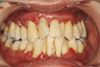
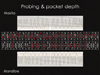
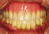
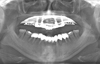
This report presents a case of aggressive periodontitis in a 23-year-old female who had previously received periodontal therapy. She was presented to the Department of Periodontics, Seoul National University Dental Hospital in 2004 with the chief complaint that her gums had been swelling (Fig. 1). She requested for dental treatment to address the issue of gum swelling and tooth mobility. Her medical history was unremarkable. Subsequent clinical and radiographic examination led to the diagnosis of generalized aggressive periodontitis (Fig. 2, 3). The patient reported of becoming aware of swelled gums and mobile teeth when she was at the age of 13. At that time, she had received scaling and root planning in conjunction with systemic antibiotics which were periodically repeated through the years with no definitive results.
In 2008, the patient was referred to the Department of Prosthodontics for evaluation and treatment planning. The objectives of treatment were patient motivation and education, improvement of oral hygiene, improvement of esthetics, and a stable and predictable outcome.
All teeth including the canine in the maxilla and left mandibular lateral incisor through right mandibular canine, and left mandibular second premolar were extracted.
Initially fixed prosthesis using implants in the maxilla were planned, but rapid and severe bone resorption after extraction was observed. Because the maxillary lip required additional support as a consequence of bone loss, the fixed prosthesis treatment plan for the maxilla was changed to an implant-supported overdenture. Facebow transfer and mounting on the casts on the articulator were performed for the diagnostic wax-up procedure.
The remaining teeth in the mandible were prepared for the fixed partial prosthesis. A provisional complete denture in the maxilla and provisional fixed partial dentures in the mandible was delivered. A computerized tomography scan with implant stent was taken to select suitable implant sites in the maxilla. In the maxilla, US II external-type implants (Osstem, Seoul, Korea) were placed at the sites of right maxillary first premolar, right maxillary lateral incisor, left maxillary first premolar, and left maxillary first premolar following a two-stage delayed-loading schedule. After the first implant surgery, the interim complete denture in the maxilla was relined with Coe-Soft™ (GC America Inc., Alsip, IL, USA).
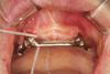
After 7 months of healing, the impression of the implants in the maxilla were made. An individual tray was fabricated. Pickup impression copings were connected, and splinted with DuraLay resin (Reliance Dental Mfg. Co., Worth, IL, USA). The functional impression technique using polyvinylsiloxane impression material was used. The occlusal plane was evaluated using the TRUBYTE™ occlusal plane plate (Dentsply, York, PA, USA). Facebow transfer and mounting the maxillary cast on the articulator was performed. A bar for the clip attachment was incorporated in the maxilla.
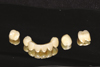
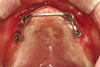
Wax denture try-in for the maxilla and zirconia framework try-in for the mandible were done. The all-ceramic restoration between the left mandibular canine and right mandibular first premolar was designed to a six unit restoration, because the lack of MD space was expected. The other all-ceramic restorations in the mandible were fabricated separately. Lower all-ceramic restorations using Lava system™ (3M ESPE, St. Paul, MN, USA) were completed (Fig. 4). Final cementation was carried out using resin cement (Multilink, Ivoclar Vivadent Inc., Lichtenstein, Germany). A Hader bar® (Attachments International Inc., San Mateo, CA, USA) for the maxillary implant-supported overdenture was fabricated (Fig. 5). Marginal fits of the Hader bar were evaluated with one-screw test, screw resistance test, Fit Checker II (GC Corporation, Tokyo, Japan) and periapical radiograph. After delivery of the final prostheses, soft tissue profiles were evaluated in the frontal and lateral view (Fig. 6, 7). A daily maintenance care by patient's effort was instructed using interdental cleaning aids (Fig. 8). A regular maintenance program was instituted with periodontal recall every 3 months following delivery of the definitive restorations.
DISCUSSION
Team approach involving prosthodontists and periodontists is required to rehabilitate patients with severe complicated periodontal situations in the planning and treatment process. In this particular aggressive periodontitis patient, an interdisciplinary approach was essential to evaluate, diagnose, and restore the function and esthetic problems using a combination of prosthodontic and periodontic treatments. A periodontitis consulted with prosthodontist before and after extraction of the teeth, made it possible to discuss and change prosthetic options in this case. The implant stent was fabricated by the prosthodontist, and the prosthodontist was participated in the surgery for the implant positioning. A periodic maintenance care by prosthodotist and periodontist has been conducted to enhance the success of prostheses and soft tissue after the prosthetic reconstruction.
The long-term success of osseointegrated implants has been recorded in numerous studies.11,12 Studies13,14 revealed that the long-term implant prognosis in patients with a history of chronic periodontitis was equivalent to that in patients without periodontal disease. It was also demonstrated that osseointegrated implants in generalized aggressive periodontitis patients can be placed successfully.4 Implants in this patient with aggressive periodontitis can accommodate the successful use of prosthesis and ensure to prevent future bone loss.
Overdenture in the maxilla was chosen to restore the masticatory function of this patient because she had deficient bone to house sufficient number of implants. Also, severe bone atrophy in the anterior area left esthetic problems such as insufficient lip support if restored with the fixed dental prosthesis.
A passive fit is an important prerequisite to ensure long-term success for implant-supported prostheses, so passive fit should be evaluated when implant framework is delivered. Marginal fit of the implant framework was evaluated by the combination of several methods: alternative finger pressure, direct vision and tactile sensation, periapical radiograph, one-screw test, screw resistance test, and disclosing media using fit checker, pressure indicating paste, and disclosing wax.15 Multiple methods including periapical radiographs were used to check the passive fit of implant framework in this case.
Development of physical properties of the dental material in the ceramic systems enables all-ceramic restorations to restore the posterior area. High esthetics and suitable strength of zirconia frameworks make it more popular.10 Also, another advantage of ceramic compared to metal is its biocompatility.9 All-ceramic restorations using zirconia frameworks ensures the strength and esthetics in this young female.
CONCLUSION
This clinical case report describes a patient restored with implant-supported overdenture for the maxilla and all-ceramic restorations in the mandible. The results showed significant improvement in esthetics and function of the masticatory system. Considering the psychological problems that these patients have faced during the early stages of their life, this alternative implant treatment and esthetic restoration may provide a better opportunity to meet this patient's needs. Team approach in the evaluation and treatment planning will be necessary to improve the esthetic and functional outcomes in aggressive periodontitis patients.

 XML Download