

 PDF
PDF ePub
ePub Citation
 Print
Print


INTRODUCTION
Allergic rhinitis (AR) is a common atopic and chronic disease in childhood. The International Study of Asthma and Allergies in Childhood (ISAAC) was conducted in Korea, and the lifetime prevalence of AR was gradually increased to 16.8% in 1995 and 22.0% in 2000.1 Environmental factors as well as genetic predisposition seem to be risk factors contributing to the development of AR.2
In 1989, the hygiene hypothesis was developed for increasing allergic diseases. It proposed that a lack of exposure to microbes in early life increases the risk of allergy in later life.3,4 This was adapted to the microbiota hypothesis, namely, microbiota changes by lifestyle factors and plays a major role in the development of the immune system.5,6,7 Delivery mode influences initial microbial colonization,6 and feeding method or use of antibiotics could affect the intestinal microbiota.8,9 Moreover, alterations in the microbiota lead to dysbiosis and disease in the neonate and affect long-term health in childhood.10
Intestinal microbiota may play an essential role in T helper 1 (Th1) cell immune responses and the balance between Th1 and Th2 cell immune responses. These immune responses involve major pathogen-recognition receptors like Toll-like receptors (TLRs) with cluster of differentiation (CD) 14 of innate immunity.11,12,13 Genetic polymorphisms that shape immune responses are associated with an increased risk of developing allergic disease. We previously reported the relationships between allergic disease and TLR4 (rs1927911)/CD14 (rs2569190).14,15,16 However, genetic predisposition alone cannot fully explain the rapid rise in the incidence of allergic disease; rather, it is likely that interactions between genes and the environment explain the increased risk of developing allergic disease.17
We investigated early-life risk factors for the development of AR by altering the intestinal microbiota, which included delivery mode, feeding method, and use of antibiotics during infancy. We also determined how genetic variations associated in CD14 (rs2569190) or TLR4 (rs1927911) could interact with early-life risk factors to promote the development of allergic disease.
MATERIALS AND METHODS
Study participants
In total, 1,828 children aged 9-12 years were recruited from 9 elementary schools in 2 areas in Korea, namely, Seoul and Jeongeup. Seoul is the largest urbanized city, and Jeongeup consists of a rural town and rural villages. Of the 1,828 subjects, 1,797 (98.3%) returned the questionnaires. AR patients were defined as children who were diagnosed with AR by the physician using the questionnaire, and atopic AR patients as children who were diagnosed with AR by the physician using the questionnaire as well as by skin prick test results.
This study was approved by the Human Ethics Committees of Hallym University and the principals of the schools attended by the children. Written informed consent was obtained from the parents or guardians of the children.
Questionnaire data
Demographic factors, potential explanatory and confounding factors, and the prevalence of diseases and symptoms were assessed by a questionnaire that was given to the parents. The written questionnaire based on the ISAAC protocol was translated into Korean.18 The presence of AR in the children was determined by asking specific questions, such as "Has your child ever been diagnosed with allergic rhinitis by a physician?". Delivery mode was determined by the question, "Was your child born by vaginal delivery or caesarian section?". Feeding method was determined by the question, "Was your child breast-fed or formula-fed?". Use of antibiotics during infancy was assessed by the question, "Did your child take antibiotics more than 3 days during infancy?".
Skin prick test
Standardized allergen extracts and control solutions from Allergopharma (Reinbek, Germany) were used. The subjects were tested for the following 16 allergens: Dermatophagoides pteronyssinus, D. farinae, dog/cat epithelium, cockroach, grass, mixed tree pollen 1 and 2, Alternaria, Aspergillus, ragweed, mugwort, milk, egg white, peanut, and soybean. A positive reaction was defined as a wheal size ≥3 mm after subtracting the negative control. The skin prick test was performed on 1,510 children (84.0%). One or more positive reactions were considered to indicate atopy.
DNA collection and single nucleotide polymorphism genotyping
Genomic DNA was extracted from peripheral blood mononuclear cells, and CD14 (rs2569190), TLR4 (rs1927911) polymorphisms were screened using the TaqMan fluorogenic 5' nuclease assay (ABI, Foster City, CA, USA). The final volume in polymerase chain reaction (PCR) was 5 µL and consisted of 10 ng of genomic DNA, 2.5 µL of TaqMan Universal PCR Master Mix, and 0.13 µL of 40× Assay Mix (Assay ID C_16043997_10 and C_11722141_10). The thermal cycle conditions were as follows: 50℃ for 2 minutes to activate uracil N-glycosylase and to prevent carry-over contamination, 95℃ for 10 minutes to activate DNA polymerase, followed by 45 cycles of 95℃ for 15 seconds and 60℃ for 1 minute. All PCRs were performed by using 384-well plates and a 384-Well Veriti thermal cycler, and the endpoint fluorescent readings were performed by using an ABI 7900 HT Sequence Detection System. Duplicate samples and negative controls were included to ensure genotyping accuracy.
Statistical analysis
Multivariate logistic regression analysis was used to calculate adjusted odds ratios (aORs) for early-life factors and the association between genetic polymorphisms and the early-life factors with regard to the development of AR. The additive effect of the early-life factors in the context of genotypes was determined. All statistical methods were adjusted for predefined covariates, including sex, age, body mass index, maternal and paternal history of allergic disease, familial income, environmental tobacco smoke, and region. All statistical analyses were performed by using SPSS version 18.0 (SPSS Inc., Cary, NC, USA). The results are presented as aORs and 95% confidence intervals (95% CIs). A P value of <0.05 was considered statistically significant.
RESULTS
Population characteristics and prevalence of AR
In total, 1,540 children answered the question about physician's diagnosis of AR. AR had been diagnosed by a physician in 24% (369/1,540) of the children. The demographic data obtained from children with and without AR are presented in Table 1. Children with AR showed a higher proportion of males and higher frequencies of parental allergic history, parental income, and residence in Seoul than those without. Caesarean delivery and breast feeding were not significantly different between the 2 groups. Children with AR more frequently took antibiotics during infancy.
The prevalence of atopic AR, which was diagnosed by physicians using positive skin prick test results, was 12.8% (197/1,540).
Associations between AR and early-life risk factors
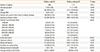
Delivery mode and feeding method were not associated with the rates of AR or atopic AR. Children who had been treated with antibiotics for more than 3 days during infancy were more frequently diagnosed with AR and atopic AR (aOR [95% CIs], 1.511 [1.222-2.037] for AR and aOR [95% CIs], 1.565 [1.078-2.272] for atopic AR) (Table 2).
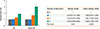
The risks of AR and atopic AR tended to increase as the number of environmental risk factors became larger (Fig. 1).
Associations between AR and genetic polymorphisms
Genotype analysis was performed on 1,299 children for CD14 and on 1,274 for TLR4. Hardy-Weinberg equilibrium tests demonstrated no significant deviation (P>0.05) for the 2 polymorphisms in control subjects. The CD14 TT, TC, and CC genotypes occurred at frequencies of 39.6% (514/1,299), 47.4% (616/1,299), and 13.0% (169/1,299), respectively, while the TLR4 CC, CT, and TT genotypes occurred at frequencies of 35.2% (449/1,274), 48.2% (614/1,274), and 16.6% (211/1,274), respectively.
There was no significant association between the genetic polymorphisms and AR/atopic AR (Supplemental Table 1).
Interactions between early-life risk factors and genetic polymorphisms in AR or atopic AR
When environmental factors were analyzed in relation to genetic polymorphisms, use of antibiotics during infancy was significantly associated with the CD14 TT genotype in AR and the TLR4 CC genotype in atopic AR. Formula feeding was associated with the CD14 CT+CC genotypes and the TLR4 CT+TT genotypes in atopic AR (Table 3).
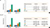
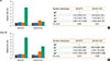
Additional analyses were performed to examine the effect of the number of risk factors on the rates of AR or atopic AR in children with different genetic polymorphisms. With regard to CD14 and TLR4 polymorphism, the risks of AR and atopic AR tended to increase as the number of environmental risk factors became larger (Figs. 2 and 3). When all the 3 early-life risk factors were present, the risks of AR and atopic AR were higher in subjects with the TLR4 CC genotype than in those with other genotypes (aOR [95% CIs], 5.127 [1.265-20.780] for AR and aOR [95% CIs], 6.078 [1.499-24.649] for atopic AR) (Fig. 2). When all the 3 early-life risk factors were present, the risks of AR and atopic AR were higher in subjects with the CD14 TT genotype than in those with other genotypes (aOR [95% CIs], 5.960 [1.421-25.002] for AR and aOR [95% CIs], 6.714 [1.440-31.312] for atopic AR) (Fig. 3). The early-life risk factors differently affected genetic polymorphisms.
DISCUSSION
In our study, use of antibiotics during infancy increased the development of AR and atopic AR. As the number of early-life risk factors became larger, the subjects were more frequently diagnosed with atopic AR in childhood. In addition, the factors interacted with polymorphisms of the TLR4 or CD14 gene when the subjects had 3 risk factors and the TLR4 CC or CD14 TT genotype. This suggests that the risk of AR and atopic AR may increase when subjects with certain genotypes were born by caesarian delivery, were formula-fed, and took antibiotics during infancy.
The 3 early-life factors are similar, which influence the intestinal microbiota and thus alter immune responses.19 In our study, the risk of AR was highest when all the 3 risk factors were present. This suggests that caesarean delivery, formula feeding, and use of antibiotics during infancy may act synergistically to promote the risk of AR, probably by influencing the intestinal microbiota in a synergistic manner.
With regard to delivery mode, neonates born by vaginal delivery are exposed to the mother's vaginal and intestinal flora as they pass through the birth canal, and consequently their bacterial communities resemble their mother's flora.7 Subjects born by caesarean section have microbial colonization later; the lack of exposure to the maternal vaginal and intestinal flora results in colonization with different species of bacteria.7,20
There have been many studies that suggest breast milk protects against allergic disease. Breast milk contains multiple immunomodulatory compounds, including IgA antibodies to microorganisms, nucleotides, oligosaccharides, antimicrobial proteins, and food proteins.21,22 Bioactive molecules in breast milk could modulate neonatal gut microbiota composition, and lactating mothers introduce their gut microbiota to infants.23
The present study showed that use of antibiotics during infancy was associated with a higher risk of developing AR. Many previous studies have also shown that early use of antibiotics promotes the development of allergic disease.24,25 Even though antibiotics are more often prescribed to children who are predisposed to develop allergic disease,26 recent epidemiologic studies have demonstrated that changes in the microbiota as a consequence of early use of antibiotics can result in the dysregulation of host immune homeostasis and an increased risk of atopy and allergic disease.27,28,29 This may reflect that early use of antibiotics shifts the composition of the microbiota, changes in metabolites, and loss of required nutrients, followed by mucosal immunity.9
In our study, the CC genotype of TLR4 rs1927911 showed a higher risk of being diagnosed with AR if subjects were born by caesarian section, were formula-fed, and took antibiotics during infancy. Several previous studies have indicated that these 3 risk factors are associated with TLRs. Vaginal delivery enhances the expressions of TLR2 and TLR4 in neonatal monocytes.30 Breast-feeding is known to influence TLR-mediated cytokine responses of neonates31 and to modulate interactions between the host immune system and the gut microbiota.23 Antibiotics targeting Gram-negative bacteria would primarily affect TLR4, and consequently depletion of the microbiota after antibiotic treatment results in reduced TLR signaling and downstream regulation of innate defenses.32
Also, some evidence indicates that the intestinal microbiota has immunomodulatory effects through activation of pattern recognition receptors, including TLRs.33 Activation of TLR signaling by intestinal commensal bacteria can induce growth factors and cytokines that are required to exert cytoprotective and reparative effects on intestinal epithelial cells.34 In addition, TLR4 recognition of lipopolysaccharide is capable of inducing CD25 and Foxp3 expression in lamina propria lymphocytes and modulates regulatory T-cell responses in the gut.35,36 Through these mechanisms, TLR signaling pathways have been implicated in protection against allergic disease.36 Therefore, alteration and depletion of the microbiota change TLR signaling, followed by downstreaming innate defense and regulatory mechanisms, consequentially increasing allergic disease.9,36,37
Even though several SNPs of TLR4 have been investigated in most studies,38,39,40 TLR4 rs1927911 has been reported in a few studies on allergy. Penders et al.38 have documented a decreased risk of sensitization in children with the rs10759931 TLR CC genotype, Kerkhof et al.40 have reported that the risk of physician-diagnosed asthma increases with increasing PM2.5 levels at the birth address in children with the TT genotype of TLR4 rs1927911. To the best of our knowledge, there is no report on TLR4 rs1927911 with AR. In our study, the subjects with CC genotype of TLR4 rs1927911 had a higher risk of AR than those with other genotypes, which is the first report on their relationship.
CD14 is involved in the recognition and signal transduction of bacterial endotoxin along with TLR4.41 Simpson et al.43 have reported the association between CD14 rs2569190 polymorphism and endotoxin exposure in relation to allergic sensitization. In children with the C allele of CD14 SNP, increasing endotoxin exposure is associated with a reduction in the risk of sensitization, whereas in those with the T allele, increasing endotoxin exposure is not so.42,43 It has been indicated that children with the TT genotype have a significantly higher mean total IgE level and a higher prevalence of atopy by the active use of mite allergen-impermeable mattress covers and that serum IgE levels are similar regardless of the use of mite allergen impermeable mattress covers in children with the TT genotype.44,45 However, it has been reported that the prevalence of atopy increases through the active multifaceted intervention program in children with CC/CT genotypes and that the risk of sensitization increases in those with CC genotypes related to E. coli colonization.38,46
The present study included a relatively large number of samples and achieved a high response rate. This is the first study to investigate whether TLR4 rs1927911 or CD14 rs2569190 polymorphisms interact with early-life environmental factors in a synergistic manner, which may have influenced the intestinal microbiota to promote the development of AR and atopic AR. The present study has some limitations. First, a question arises whether the parental recall of use of antibiotics in infancy could be reliable. Second, fecal sample analysis that depicts the effect of environmental factors on the intestinal microbiota during early childhood was not performed in the present study. Nevertheless, the present study suggests that in subjects who have different genotypes of innate immunity genes, development of AR may depend on caesarean delivery, formula feeding, and use of antibiotics during infancy. It is meaningful that some environmental factors influencing the intestinal microbiota contribute to the development of AR. Further prospective studies are needed to confirm our results.
In conclusion, the present study showed that early-life risk factors significantly interacted with polymorphisms of the TLR4 and CD14 gene in pediatric patients with AR. The results of the present study suggest that the development of AR may be influenced by interactions between early-life risk factors and that the interactions can be modified by genetic polymorphisms. Therefore, early microbial contact through vaginal delivery, breast feeding method, and use of antibiotics during infancy could be effective in modulating risk factors of AR.

 XML Download