

 PDF
PDF ePub
ePub Citation
 Print
Print


INTRODUCTION
It is well known that upper airway cough syndrome (UACS), asthma, and gastroesophageal reflux disease (GERD) commonly induce chronic cough, which persists longer than 8 weeks.1,2 Chronic cough caused by GERD is usually diagnosed by esophagogastroduodenoscopy (EGD) and 24-hour pH monitoring, and it may respond to proton pump inhibitors (PPIs), antacids, or surgery.1
Achalasia is a rare esophageal disorder caused by failure of the lower esophageal sphincter to relax.3,4 The most common symptoms of achalasia are gastrointestinal symptoms including dysphagia and regurgitation of undigested food. However, in many cases, it is accompanied by respiratory symptoms including cough and wheezing.5,6 Achalasia has been reported in unusual causes of chronic cough in pediatric patients,7,8 but it has not been reported as a cause of chronic cough in adult patients who present with chronic cough but without typical gastrointestinal symptoms. In addition, achalasia can cause symptoms similar to GERD, thus may be misdiagnosed as GERD. Here, we report a case of achalasia misdiagnosed as GERD in an adult patient with chronic cough.
CASE REPORT
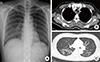
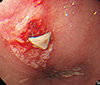
A previously healthy 40-year-old woman was admitted to the Gastrointestinal Center for cough and heartburn, which were aggravated at night. Her symptoms had been ongoing for over 4 months. She also complained of rhinorrhea and salivation during sleep, acid reflux during coughing, and intermittent dysphagia. On her first visit, her vital signs were stable and white blood cell (WBC) count was 14,440/mm3 (neutrophil 82.0%, lymphocyte 13.8%). All other laboratory data were unremarkable. Chest X-ray revealed haziness in the right middle and lower lobe, suggesting community acquired pneumonia (Fig. 1). EGD revealed multiple acute ulcers on the esophagus and chronic superficial gastritis (Fig. 2). She was treated with levofloxacin for pneumonia and with a PPI and calcium channel blocker for esophageal ulcers and GERD.
Although the chest X-ray findings for pneumonia improved, she continued to complain of nocturnal cough and rhinorrhea for 4 months after discharge. She was referred to the Allergy Clinic for further evaluation of cough and rhinorrhea.
Laboratory studies revealed that her complete blood count and differential were normal without peripheral blood eosinophilia (WBC 9,150/mm3, total eosinophil count 200/mm3). Her total serum IgE concentration was 65.1 IU/mL, and skin tests for 55 common aeroallergens were negative. A chest X-ray and paranasal sinus films did not show any abnormalities. A pulmonary function test (PFT) showed unremarkable findings (FEV1/FVC 80.7%, FEV1 2.26 L [70%], FVC 2.80 L), and a methacholine challenge test was negative.
Because she had a history of esophageal ulcers and symptoms of cough and rhinorrhea, GERD and UACS were considered to be the main causes of her symptoms. However, her symptoms did not improve and, in fact, worsened upon use of the PPI, intranasal corticosteroids, decongestants, and anti-histamines. She was readmitted for further evaluation.
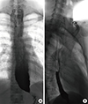
On second admission, her blood pressure was 139/93 mmHg, heart rate was 93/min, respiration rate was 20/min, and body temperature was 36.9℃. Her peripheral blood WBC count was 15,600/mm3 (neutrophils 71.6%, lymphocytes 23.1%), C-reactive protein was 4.83 mg/dL (normal 0-0.3 mg/dL), and erythrocyte sedimentation rate was 37 mm/h (normal 0-22 mm/h). EGD was performed to reexamine the esophageal ulcers and to evaluate gastroesophageal reflux severity. However, the ulcers were completely healed, and there was no evidence of GERD. Plain chest X-ray showed multiple consolidations on bilateral lung parenchyma, and chest CT exhibited multiple patchy ground glass opacities on both lungs. The dilated esophagus was distended with fluid-filled material, which suggested achalasia with aspiration pneumonia (Fig. 1). Esophagography showed symmetric esophageal narrowing and beaking appearance of the esophago-gastric junction level and mild dilatation of the proximal esophageal portion, which were consistent with characteristic findings of achalasia (Fig. 3A).
To treat achalasia, graded pneumatic dilatation was performed. A follow-up esophagography was performed 4 days after pneumatic dilatation, and the beak-shaped narrowing segment of the distal esophagus was dilated and showed good passage of the oral contrast media (Fig. 3B). Aspiration pneumonia gradually improved with third generation cephalosporin and metronidazole treatment and completely resolved soon after pneumatic dilatation. After pneumatic dilatation, she did not complain of cough or other symptoms during the 1-year follow-up period.
DISCUSSION
Cough is one of the most common symptoms for which patients seek medical care. UACS, asthma, and GERD are the most common causes of chronic cough, but other causes including laryngopharyngeal reflux disease, eosinophilic bronchitis, and the use of ACE inhibitors, should be suspected if previous treatment for such diseases do not improve the patient's symptoms.1,2
Achalasia is an uncommon cause of chronic cough. It is observed in both genders at the same frequency and occurs both in children and the elderly.3 The average age at diagnosis is during the sixth decade, and its incidence and prevalence were reported as 1.6 per 100,000 and 10.8 per 100,000 individuals, respectively, in the United States.3,9 Gastrointestinal symptoms are the primary symptoms of achalasia, and respiratory symptoms occur less frequently.6 According to previous reports,5,6,10 30%-50% of patients with achalasia have respiratory symptoms, such as cough, aspiration, and wheezing. However, most of these symptoms are accompanied by gastrointestinal symptoms, and respiratory symptoms are unlikely to precede the more typical gastrointestinal symptoms of achalasia.5,6
Cough as a primary presenting symptom of achalasia has rarely been reported and only so in the pediatric population.7,8 Furthermore, a few cases of achalasia misdiagnosed as uncontrolled asthma or as an aggravating factor for existing asthma have been reported, again predominantly in pediatric patients.11,12,13 Although cases of acute airway obstruction due to achalasia have been reported in the elderly,14 achalasia has not been reported as the primary cause of cough in adult patients who presented with chronic cough but did not have gastrointestinal symptoms.
Achalasia can cause cough and respiratory symptoms secondary to food retention in the dilated esophagus with regurgitation, and to the mass effect of the dilated esophagus that compresses trachea.5,6 Several symptoms suggest that chronic cough in our patient might be caused mainly by retained food or gastric juice regurgitation/aspiration. First, the patient experienced 2 episodes of radiologically proven aspiration pneumonia. Second, the patient's symptoms worsened at night, and reflux and aspiration occurred more readily at night. Finally, there was no evidence of intra- or extra-thoracic obstruction, suggesting a mass effect of the dilated esophagus in flow-volume curves of PFT. However, it took approximately 10 months to diagnose achalasia, since her main symptoms were cough, heartburn, and intermittent dysphagia. In addition, the initial EGD showed esophageal ulcers, which were misinterpreted as the patient's symptoms were caused by gastroesophageal regurgitation.
Although esophageal manometry is the key test for achalasia diagnosis, it may be unavailable depending on the clinical situation. In contrast, EGD has been used widely for the evaluation of chronic cough due to GERD. Endoscopic findings of achalasia may show esophageal dilatation and retention of food or secretions. However, findings might be normal in approximately 44% of patients with achalasia.15 In patients with achalasia, progressive dysphagia, regurgitation, and heartburn are the usual symptoms and the reasons for seeking medical care. Because these symptoms are very similar to GERD symptoms, achalasia is often first misdiagnosed as the more common GERD.16 Therefore, it is difficult to diagnose achalasia using EGD. When such an uncommon disease is suspected or when cough assumed to be caused by GERD does not improve upon treatment, esophagography and chest CT should be performed to exclude achalasia or other related diseases. Even if first diagnosed as GERD, another combined disease that may provoke chronic cough should be considered if the patient does not respond to GERD treatment.
Here we report a case of achalasia-induced chronic cough without typical gastrointestinal symptoms in an adult patient. This case suggests that it is important to suspect achalasia, particularly when the symptoms do not improve despite treatment for GERD. Finally, use of esophagography, chest CT, or esophageal manometry should be considered in the diagnosis of achalasia or other uncommon diseases.
 XML Download