

 PDF
PDF ePub
ePub Citation
 Print
Print


INTRODUCTION
Asthma is a chronic inflammatory airways disease characterized by airway obstruction and bronchial airway hyperresponsiveness.1 However, pulmonary function tests can be often normal even in subjects with uncontrolled asthma symptoms. Especially in the disease early stages, when performing spirometry, subjects with asthma can show normal values of forced expiratory volume in 1 second (FEV1), forced vital capacity (FVC) and FEV1/FVC ratio associated to reduced values of forced expiratory flow between 25% and 75% of vital capacity (FEF25-75). FEF25-75 is generally considered as an approximate measure of the distal airways caliber and thus its reduction represents a small airways obstruction caused by asthma inflammation.2,3 Therefore, an isolated impairment of FEF25-75 may be a marker of an early reduction of pulmonary function in asthma.4,5,6,7,8,9,10 Although there are no recommendations regarding the utility of FEF25-75 (% of the predicted) by the various guidelines,1,11,12,13 this measurement may have a clinical significance in the diagnosis and management of asthma. In this regard some studies provide evidence that impaired FEF25-75 values might predict airway AHR in subjects affected by rhinitis and/or asthma both in children and adults.14,15,16,17,18,19 Furthermore, impaired MEF50 or FEF25-75values may be considered a reliable marker of a positive bronchial reversibility after a bronchodilator both in adults and children with asthma and/or rhinitis when the baseline spirometry shows normal values of FEV1 and FEV1/FVC.7,8,9,10,20 In addition, there is evidence that FEF25-75 may be also an asthma severity marker especially in subjects with normal FEV1 and FEV1/FVC. In fact, several studies have found that FEF25-75 is significantly related with bronchial AHR6,16,18,19 and fractional exhaled nitric oxide (FENO)10,17,21,22,23,24 as well as with an increased asthma severity, a systemic use of steroids, and asthma exacerbations.20 However, the role of a low FEF25-75 in the context of normal lung function in predicting asthma diagnosis and morbidity has not been well defined yet.
On the basis of these considerations, the aim of our retrospective study was to explore in a large cohort of subjects that underwent a Mch challenge test for suggestive asthma symptoms, whether there was a relationship between baseline FEF25-75 and AHR. In particular, we aimed to evaluate if a greater impairment of FEF25-75 may be a marker of a more severe AHR. Another aim of our study was to know whether there was a FEF25-75 cut-off value to identify subjects affected by AHR.
MATERIALS AND METHODS
Subjects
For this retrospective study, we analysed the results of 4,172 consecutive Mch challenge tests performed between 2000 and 2010 in subjects with normal baseline lung function.
All subjects had performed the test because they had reported suggestive asthma symptoms (unexplained episodes of cough and/or wheezing and/or dyspnea) in order to confirm an asthma diagnosis. All patients showed normal values of FEV1, FVC and FEV1/FVC measured at baseline before the Mch test. All selected subjects reported an appearance of symptoms within 3 years before the Mch test. Subjects who showed respiratory symptoms for over 3 years were not considered for this study.
Baseline FEV1, FEV1/FVC, FVC, FEF25-75 and PD20 FEV1 obtained after each bronchoprovocation test were considered for the study. Smoking habits, age, sex and BMI were also taken into account. Subjects were arbitrarily subdivided into 3 groups on the basis of baseline FEF25-75% values with the purpose of evaluating the possible relationship between FEF25-75 on the AHR: FEF25-75≤50% or FEF25-75>50 and ≤70% or FEF25-75>70%.
No subjects were under regular asthma treatment when the test was carried out. Subjects who had taken drugs when required, were asked to avoid taking any medications before the test: β2-agonist bronchodilators and inhaled or systemic corticosteroids were suspended 24 hours and 3 weeks before the test respectively, while antihistamines were interrupted at least 10 days before the challenge. None of the subjects had suffered from airway infections or asthma exacerbations in the four weeks prior to the test. The body mass index (BMI) value of 25 was used as a cut-off to differentiate normal weight or underweight (BMI <25) subjects from those overweight or obese (BMI >25). International age and sex specific cut off points for BMI were used to subdivide subjects with age <18 years into underweight, normal or overweight-obese.25 BMI was calculated by dividing the weight in kilograms by the square of height in metres (kg/m2).The use of the data recorded in each spirometer and the study protocol were approved by the local ethical Committees.
Mch bronchoprovocation test
The Mch bronchoprovocation test was performed by using a longer dosimeter method not perfectly following guidelines26 and which has been used in our departments for over 20 years. Mch sulphate was supplied by Lofarma (Milan, Italy) and administered in aerosol form using an MEFAR MB3 dosimeter (output: 9 µL/puff; MEFAR Elettromedicali Brescia, Italy) with an MB2 ampoule model. The buffer solution was the first to be administered, followed by 40 µg of Mch, increasing the doses until PD20 FEV1 was obtained or until the maximum dose of Mch was reached. FEV1 was assessed after inhaling 40, 80, 120, 240, 400, 800, 1,600, and 2,400 or 3,200 µg of cumulative Mch doses, respectively. At the end of exhalation, during tidal breathing, patients inhaled Mch slowly and deeply in 5 seconds and then they held their breath for 5 further seconds. The interval between 2 consecutive steps was 2 minutes. FEV1 was measured at 30 and 90 seconds after nebulization. A suitable quality of FEV1 was obtained at each step. No more than 2 maneuvers after each dose were allowed, and the highest FEV1 value was taken into account. Since not all patients had used the 3,200 µg Mch dose cut-off (see another article of ours for a better explanation)27 AHR was defined by a 20% fall in FEV1 from the reference value (see below) obtained with a cumulative Mch dose <2,400 µg. Subjects who did not achieve a 20% fall in FEV1 with a Mch dose of 2,400 µg were regarded as normoreactive.
Subjects with PD20≤400 and PD20>400 µg were arbitrarily considered as affected by moderate to severe and borderline AHR respectively, with the aim of evaluating the relationship between FEF25-75 and AHR in the different levels of its severity. We arbitrarily used the 400 µg cut-off with the purpose of identifying subjects with a higher probability to be asthmatics (i.e. those with a PD20< 400 µg).
Lung function was measured with a HP 47120E Pulmonary System Desk spirometer (Hewlett Packard, Waltham, -MA, USA). FEV1 and FVC were expressed as percentages of the predicted values at baseline, whereas FEV1/FVC was reported only as a ratio (reference equation: CECA, 1971). PD20 FEV1 was assessed by linear interpolation of the dose-response curves. FEV1 measured before administering the buffer solution was taken as baseline value, while FEV1 measured after the buffer solution was used as a reference value to calculate FEV1 fall and thus PD20.
Statistical analysis
Categorical variables were expressed as number of cases and percentages. Continuous variables were expressed as mean values and standard deviations or median values and interquartile range (IQR - 25° and 75° quartiles) according to whether they were normally distributed. Nonparametric or parametric tests were performed accordingly. Comparisons of qualitative data were performed using the chi-square test, whereas comparisons of quantitative variables among different groups were conducted by the ANOVA one way test or the Kruskall-Wallis test when appropriate. Moreover, the Bonferroni test was used for multiple comparisons. Assessments of any possible differences between the different groups considered, as well as in the different classes of subjects - males, females, smoking, non-smoking, different classes of age, underweight/normal weight and overweight/obese - were searched using both Kruskal Wallis and Mann Whitney tests. Associations between FEF25-75 and PD20 in different categories and classes of subjects considered were analyzed using the Spearman correlation test.
Receiver operator characteristic (ROC) curves to examine the ability of FEF25-75% (of predicted) to predict airway AHR (defined as a PD20< 2,400 µg or a PD20<400 µg) were created by plotting sensitivity (true positive rate) versus 1-specificity. The best threshold for any test was the one which maximizes sensitivity while minimizing the false positive rate, represented by the left upper most significant value on the curve. The area under the curve (AUC) represents a measure of the test accuracy (AUC of 1.0 indicates perfect prediction while AUC of 0.50 indicates prediction no better than chance) and was calculated via numerical integration.
A logistic binary regression model, corrected for sex, age, smoking, FEV1, FVC and seasons, was applied to evaluate if FEF25-75% was an independent AHR risk factor. In order to evaluate a possible different risk of FEF25-75% on AHR in the various levels of AHR (moderate to severe and borderline AHR), 2 additional logistic regression models were performed for each group considered (those with FEF25-75<50% and FEF25-75 between 50 and 70%)In these models, the FEF25-75>70% value was considered as the referral value. P values <0.05 were considered statistically significant. The statistical package SPSS (16.0) was used for analysis.
RESULTS
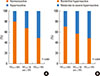
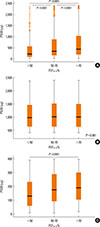
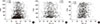
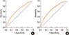
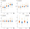
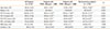
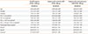
Characteristics of subjects subdivided according to their level of airways AHR are described in Table 1. Age and pulmonary function (in particular FEF25-75%) were higher in subjects with normal reactivity in comparison with those with borderline and moderate/severe AHR. This last group of subjects showed also lower percentage values of FEV1, FVC, FEV1/FVC, and FEF25-75 when compared to values measured in the group of subjects with borderline AHR. Approximately 79% of patients reported symptoms onset in the year, previous to performing the Mch test, whereas in the remaining 14% and 7% of subjects symptoms appeared 2 or three years previous to the test respectively (data not shown in tables/figures). When we subdivided all 4,172 subjects in groups on the basis of baseline FEF25-75% values (<50%, between 50 and 70% and >70%) the percentage of hyperresponsive subjects (with PD20<2,400 µg) decreased significantly (P<0.0001) when going from baseline values of FEF25-75<50% to values >70% (Fig. 1A). Also the percentages of subjects with moderate/severe AHR lowered with the increase of baseline FEF25-75% (Fig. 1B). Furthermore, also the median PD20 progressively increased in subjects with higher baseline FEF25-75% (Fig. 2A). This trend was observed only in subjects with moderate/severe AHR (Fig. 2C) but not in those with borderline AHR whose asthma diagnosis was less probable (Fig. 2B). These lower values of PD20 in subjects with FEF25-75<50% were also observed when subdividing all subjects into females, males, smokers, non-smokers, different classes of age and in underweight/normal-weight or overweight/obese patients (data not shown). In addition, the logistic regression model (Table 2) showed that baseline FEF25-75>50 and <70% of predicted (corrected for age, sex, FEV1, FVC and seasons) was a risk factor for AHR (compared to FEF25-75>70%) both in all subjects (OR: 1.39 [95%CI:1.14-1.69]; P<0.01) and also only in those affected by moderate/severe (OR: 1.35 [95%CI:1.06-1.72]; P<0.01) or borderline AHR (OR: 1.33 [95%CI:1.05-1.69]; P<0.01). This AHR risk resulted higher in subjects with baseline FEF25-75<50% (compared to those with FEF25-75>70%) especially in those affected by moderate/severe AHR (OR: 2.18 [95%CI:1.41-3.37]; P<0.0001) whose asthma diagnosis was certain. However, the application of spearman correlations found a significant but weak relationship between PD20 and FEF25-75% (r=0.189; P: 0.0001; Fig. 3A). When subjects were subdivided into borderline and moderate/severe hyperreactive, no relationships were found in the former (r=0.016; P: 0.611; Fig. 3B) whereas a significant correlation (r=0.103; P: 0.001; Fig. 3C) was observed in the latter. A small but significant relationship between PD20 and FEF25-75% was also observed in males, females, smoking, non-smoking, different classes of age, underweight/normal weight and overweight/obese (data not shown). ROC curves were used to formally compare the ability of FEF25-75% measurement to distinguish hyperresponsive subjects (defined as PD20 less than 2,400 µg or 400 µg) from subjects without AHR and to identify the optimal threshold levels for distinguishing these 2 groups.(Fig. 4A and B) The thresholds yielding the highest combined sensitivity and specificity for FEF25-75% were 75.19 and 74.95 in subjects with PD20<2,400 and <400 µg respectively. The AUC for FEF25-75% was 0.653 and 0.688 in subjects with PD20<2,400 and <400 µg respectively. We analyzed also FEV1, FVC, and FEV1/FVC % values measured in subjects with different levels of FEF25-75≤50% or FEF25-75>50 and ≤70% or FEF25-75>70% with the purpose of investigating if baseline pulmonary function was different in hyperresponsive and normoreactive subjects. On the whole, trends, both in subjects with different baseline FEF25-75% values and in those hyperresponsive or normoreactive, were similar Fig. 5. Higher values of FEV1 and FEV1/FVC were observed with the increase of FEF25-75% (Fig. 5A and B). Small differences were found in FEV1% measurements between hyperresponsive and normoreactive subjects. No differences were found in FVC% values (Fig. 5C) either among different levels of baseline FEF25-75 values or between hyperresponsive and normoreactive subjects. FEF25-75% values were similar both in non-smoking and smoking hyperreactive subjects, as well as in non-smoking and in smoking normoreactive subjects (Fig. 5D). On the contrary, different FEF25-75% values were found in normoreactive and hyperreactive subjects, both smokers and non-smokers (P<0.0001; Fig. 5D).
DISCUSSION
This study, carried out on a large number of subjects, highlights that a decrease in baseline FEF25-75% corresponds to an increase in the number of hyperresponsive subjects and levels of AHR in individuals with suggestive asthma symptoms and normal pulmonary function. This trend has been particularly observed in subjects with moderate/severe AHR where an asthma diagnosis is more probable. Furthermore, the logistic regression model confirms that a lower FEF25-75% represents an AHR risk factor. Only patients affected by a moderate/severe AHR (but not borderline), showed that a lower level of FEF25-75% was associated to a greater AHR risk. In short, this study showed that more significant small airway impairment corresponded to a more severe AHR in the early stages of asthma. However, it must be said that there is a possibility that some subjects with high values of PD20 did not result asthmatics. In fact, according to guidelines, high values of PD20 or PC20, in case of suspected asthma (as in our patients), make an asthma diagnosis less probable.26 For this reason we arbitrarily used the 400 µg cut-off to distinguish subjects with a higher from those with a lower probability to be asthmatics, reducing to minimum the possibility that at least in the group with PD20<400 µg there are not any non-asthmatics. On the other hand, it is necessary to say that airway AHR not only predicted new asthma onset but also COPD.28,29 Therefore, it is possible that part of our subjects may develop COPD and not asthma in time. However, this may regard only a small number of our patients. In fact, it seems that COPD incidence was only of 2.8 cases/1,000/year in subjects aged 20-44 years with normal lung function and respiratory symptoms (chronic cough/phlegm and dyspnea).30 Probably, COPD diagnosis would regard especially and prevalently older subjects and smokers.31 At least 25% of the latter, aged 30-60 years, will develop a clinically significant COPD over time.31 In addition, up to half of them will develop asthma-COPD overlap syndrome over time.32 Furthermore, it must be said that, as most of our patients are less than 40 years old, the symptoms they showed were definitely asthma symptoms. In fact, we know that COPD clinically manifests itself mainly after the age of 40. Therefore, only a very small number of subjects of our survey may develop COPD in time.
Results of our study are in accordance with other studies where impaired FEF25-75 values are inversely related to airway AHR both in children and adults affected by rhinitis and/or asthma.14,15,16,17,18,19 Therefore, when facing a FEF25-75 impairment associated with normal FEV1 and FEV1/FVC in patients with asthma symptoms, we are expected to think that AHR, and consequently asthma, may be present. In the early stages of asthma, higher values of FEV1 and FEV1/FVC may not reveal an airway involvement caused by an asthma inflammatory process, whereas FEF25-75 may represent an early functional airway impairment especially of peripheral airways. In such cases, a positive Mch challenge test can confirm an asthma diagnosis. Probably the normality cut-off of FEV1/VC or FEV1/FVC, indicated by guide-lines, is not representative of "normality" for all subjects.1,11,12,13 In fact, it is difficult to define a normality value which may be valid for all individuals because theoretical values, used to establish such limits, are not representative for all subjects, especially young adolescents.7,8,12,13 This is confirmed by previous studies that have shown a positive reversibility test after salbutamol (FEV1 increase >12%) in about 23-30% of subjects with FEV1>100% or FEV1/FVC >100% or FEF25-75>70% and bronchial asthma symptoms.7,8 The percentage of subjects with positive reversibility test increased to 35% when baseline FEF25-75 was <70%.7,8 Therefore, subjects with a small airway impairment measured with a FEF25-75 reduction, may already have an obstruction of proximal airways that could not be seen because of inadequate FEV1/VC or FEV1/FVC normality cut-offs. Alternatively, an isolated impairment of expiratory flows (MEF, FEF25-75) may suggest a pulmonary obstructive disease located in limited airway districts.33 This has not been confirmed by our data where we observed that the progressive reduction of FEF25-75 corresponded to a decrease also of FEV1 and FEV1/FVC. As a result, a reduction of small airways can also affect large airways. This suggests that the bronchial tree is a single structure where caliber decreases both in large and small airways at the same time. However, distal airways (<2 mm diameter) have been recognized as a predominant site of more severe inflammatory and structural changes and therefore of airflow obstruction in asthmatic patients.2,3,34 In fact, inflammatory response and remodeling in asthma is not restricted to proximal airways but can be also observed in the distal lung.34 An increased number of eosinophils and T cells (in particular CD3+) were observed in distal airways.3,34 Furthermore, a greater number of activated eosinophils was seen in the bronchioles <2 mm internal diameter and less in the ones >2 mm internal diameter, suggesting a more severe inflammatory process in the distal airways.3,34,35 In addition, as we have already said, impaired FEF25-75 values (such as less than 65 percent of predicted) were also negatively related to FeNO values10,17,21,22,23,24 and in particular to the peripheral/alveolar NO concentration parts36 thus suggesting that small airway inflammation may be responsible for peripheral airway caliber reduction. Therefore, in the earlier stages of the disease, inflammation seemed to be more severe in distal than in proximal airways thus causing a greater impairment of expiratory flows rather than volumes.34 Consequently this bronchial inflammation may be responsible for AHR and its severity. In fact, exhaled nitric oxide correlates with airway AHR in patients with mild or initial asthma and normal pulmonary function37,38,39,40,41 suggesting the presence of a link between airway inflammation (very probably located in small airway) and AHR.
On the basis of our study, and confirmed by other researches already quoted, a greater impairment of FEF25-75% seems to correspond to a more severe AHR.6,16,18,19 This is especially in line with the findings of Currie et al.42 who compared asthmatic patients with borderline Mch measured AHR to those with moderate-to-severe AHR, where the latter had significantly lower FEF25-75% values. This suggests that FEF25-75 may be also a functional marker of asthma severity. In this regard, a study conducted in children with a low FEF25-75 and normal FEV1, the first parameter was significantly associated with the use of steroids, asthma exacerbations and asthma severity.20 One more review article has shown that small airway dysfunction is associated with worse asthma control, a higher number of exacerbations, the presence of nocturnal asthma, a more severe AHR, exercise induced asthma and late-phase allergic response,43 thus confirming the possible role of FEF25-75 as a marker of asthma severity especially in subjects with normal FEV1 and FEV1/VC. However, the relationship between AHR and FEF25-75%, observed in our study, was not very significant. The relationship between PD20 and FEF25-75% was poor and ROC did not find a major cut-off of FEF25-75% to discriminate hyperreactive from normoreactive subjects. This means that FEF25-75 can be only considered an AHR risk factor and that a cut-off value of this parameter, which may allow us to distinguish hyperreactive from normoreactive subjects, does not exist. A Mch test may not be the best way to highlight the relationship between AHR and small airways impairment. In fact, airway sensitivity, detected with mannitol or adenosine monophosphate, is better related to airway inflammation (sputum eosinophils and FENO) than what found with Mch challenge.44 Furthermore, Mch reacts with muscarinic receptors prevalently located in large airways. One more explanation for the poor relationship observed between FEF25-75 and AHR may be that forced expiratory flows may have low reproducibility and high variability because they are strongly related to FVC maneuvers and because great compliance is required in performing the tests.12,13 Changes in the FVC value entail a shift in the location of the indices along the abscissa of the flow-volume curve. Probably, when performing a spirometry, the maneuver with a higher FVC value should be considered so as to have a more reliable FEF25-75 value. However, FVC variations among various maneuvers, in subjects with normal spirometry, might be low and consequently the changes of FEF25-75 could also be that low. Therefore, the evaluation of FEF25-75 may be more reliable in subjects with normal FEV1 and FVC. Whereas, in subjects with moderate to severe asthma, FVC may have a greater variability and therefore FEF25-75, may show more considerable changes and thus be poorly trustworthy.12,13
Finally, deep inhalations, performed during the dosimeter protocol for Mch challenge, have been reported to result in bronchoprotection and therefore the challenge may be falsely negative among mild asthmatics, compared to the tidal breathing protocol.45,46 This bronchoprotective effect of deep inhalation may hide a more significant relationship between AHR and small airway impairment.
In our study, subjects with normal reactivity can also have a FEF25-75% reduction. Therefore, a small airway impairment measured by a decrease of FEF25-75% should not be considered as an asthma peculiarity. This reduces also the importance of FEF25-75% to detect small airway impairment due to asthma. Only when FEF25-75 is associated to other parameters, for example typical symptoms, wheezing, AHR, reversibility to bronchodilators, atopy and specifically inflammatory markers, it may have a role in asthma management, although this has not been verified yet. Also other factors, apart from asthma, may influence a reduction of FEF25-75% in normoreactive subjects (but also in hyperreactive ones), i.e. air pollution, occupational exposure, smoking and other factors that are still unknown. However, smoking does not seem to determine any differences in FEF25-75% values both in normoreactive and hyperreactive subjects (Fig. 5D). It is already known that FEF25-75% is lower in smokers than in non-smokers.47,48 On the contrary, the lack of a higher reduction of FEF25-75% in smokers, when compared to non-smokers in our study, may be due to a shorter smoking history as our subjects were prevalently young.
In conclusion, subjects in early stages of asthma with "normal" FEV1 and FEV1/FVC, but with only a reduction in forced expiratory flows (FEF25-75), should perform a broncho-provocation test because airway AHR could be found in a considerable number of cases, thus confirming the asthma diagnosis. However, there is not a significant cut-off of FEF25-75 that may allow us to distinguish hyperreactive from normoreactive subjects because even normoreactive subjects can show lower FEF25-75 values. A greater impairment of FEF25-75 may be associated to a more severe AHR suggesting a possible role of FEF25-75 (probably together with other parameters) in the management of asthma when pulmonary function is normal.
 XML Download