

 PDF
PDF ePub
ePub Citation
 Print
Print


Abstract
Objectives
This study was performed to consider the clinical experience of an ultrasound-guided vacuum-assisted resection (Mammotome) for benign breast lesions through a core needle biopsy.
Methods
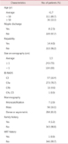
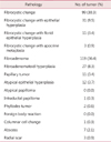
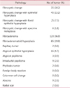
The authors carried out a core needle biopsy and Mammotome for 347 patients and investigated the pathologic results.
Figures and Tables
Fig. 1
Core needle biopy procedure, This sonogram of breast shows the core needle penestrating the central portion of the breast mass avoiding vessels via color doppler.
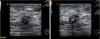
Fig. 2
Mammotome breast mass resection procedure. This each sonogram of Mammotome procedure show each step sequentially.
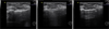
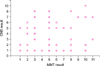
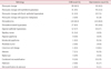
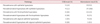
Fig. 4
The distribution between core needle biopsy results vs Mammotome results.
# Spearman rank correlation coefficient: 0.413
1. Fibrocystic change, 2. Fibrocystic change/c epithelial hyperplasia, 3. Fibrocystic change/c florid epithelial hyperplasia, 4. Fibrocystic change/c apocrine metaplasia, 5. Fibroadenoma, 6. Fibroadenomatoid hyperplasia, 7. Papillary tumor, 8. Atypical epithelial hyperplasia, 9. Atypical papilloma, 10. Intraductal papilloma, 11. Phyllodes tumor.
References
1. The Korea Central Cancer. Registry National Cancer Center. Annual report of cancer statistics in Korea in 2008. 2010. Seoul: Ministry of Health and Welfare.
2. American College of Radiology (ACR). BI-RADS-mammography. 2003. 4th ed. Reston, VA: American College of Radiology.
3. Liberman L, Drotman M, Morris EA, LaTrenta LR, Abramson AF, Zakowski MF, et al. Imaging-histologic discordance at percutaneous breast biopsy. Cancer. 2000. 89:2538–2546.
4. Lee CH, Egglin TK, Philpotts L, Mainiero MB, Tocino I. Cost-effectiveness of stereotactic core needle biopsy: analysis by means of mammographic findings. Radiology. 1997. 202:849–854.
5. Yang M, Ishida T, Takede M, Ohuchi N. Utility of an upright-type 11-gauge stereotactic vacuum-assisted biopsy device (Mammotome®) for the diagnosis of breast microcalcifications. Chin-Ger J Clin Oncol. 2009. 8:567–571.
6. Eby PR, Ochsner JE, DeMartini WB, Allison KH, Peacock S, Lehman CD. Is surgical excision necessary for focal atypical ductal hyperplasia found at stereotactic vacuum-assisted breast biopsy? Ann Surg Oncol. 2008. 15:3232–3238.
7. Darling ML, Smith DN, Lester SC, Kaelin C, Selland DL, Denison CM, et al. Atypical ductal hyperplasia and ductal carcinoma in situ as revealed by large-core needle breast biopsy: results of surgical excision. AJR Am J Roentgenol. 2000. 175:1341–1346.
8. Won B, Reynolds HE, Lazaridis CL, Jackson VP. Stereotactic biopsy of ductal carcinoma in situ of the breast using an 11-gauge vacuum-assisted device: persistent underestimation of disease. AJR Am J Roentgenol. 1999. 173:227–229.
9. Zografos GC, Zagouri F, Sergentanis TN, Nonni A, Koulocheri D, Fotou M, et al. Minimizing underestimation rate of microcalcifications excised via vacuum-assisted breast biopsy: a blind study. Breast Cancer Res Treat. 2008. 109:397–402.
10. Johnson AT, Henry-Tillman RS, Smith LF, Harshfield D, Korourian S, Brown H, et al. Percutaneous excisional breast biopsy. Am J Surg. 2002. 184:550–554.
11. March DE, Coughlin BF, Barham RB, Goulart RA, Klein SV, Bur ME, et al. Breast masses: removal of all US evidence during biopsy by using a handheld vacuum-assisted device--initial experience. Radiology. 2003. 227:549–555.
12. Fine RE, Boyd BA, Whitworth PW, Kim JA, Harness JK, Burak WE. Percutaneous removal of benign breast masses using a vacuum-assisted hand-held device with ultrasound guidance. Am J Surg. 2002. 184:332–336.
13. Cassano E, Urban LA, Pizzamiglio M, Abbate F, Maisonneuve P, Renne G, et al. Ultrasound-guided vacuum-assisted core breast biopsy: experience with 406 cases. Breast Cancer Res Treat. 2007. 102:103–110.
14. Philpotts LE, Hooley RJ, Lee CH. Comparison of automated versus vacuum-assisted biopsy methods for sonographically guided core biopsy of the breast. AJR Am J Roentgenol. 2003. 180:347–351.
15. Pfarl G, Helbich TH, Riedl CC, Wagner T, Gnant M, Rudas M, et al. Stereotactic 11-gauge vacuum-assisted breast biopsy: a validation study. AJR Am J Roentgenol. 2002. 179:1503–1507.
16. Cho N, Moon WK, Cha JH, Kim SM, Kim SJ, Lee SH, et al. Sonographically guided core biopsy of the breast: comparison of 14-gauge automated gun and 11-gauge directional vacuum-assisted biopsy methods. Korean J Radiol. 2005. 6:102–109.
17. Simon JR, Kalbhen CL, Cooper RA, Flisak ME. Accuracy and complication rates of US-guided vacuum-assisted core breast biopsy: initial results. Radiology. 2000. 215:694–697.
18. Perez-Fuentes JA, Longobardi IR, Acosta VF, Marin CE, Liberman L. Sonographically guided directional vacuum-assisted breast biopsy: preliminary experience in Venezuela. AJR Am J Roentgenol. 2001. 177:1459–1463.
19. Liberman L. Centennial dissertation. Percutaneous imaging-guided core breast biopsy: state of the art at the millennium. AJR Am J Roentgenol. 2000. 174:1191–1199.
20. Soo MS, Baker JA, Rosen EL. Sonographic detection and sonographically guided biopsy of breast microcalcifications. AJR Am J Roentgenol. 2003. 180:941–948.
21. Grady I, Gorsuch H, Wilburn-Bailey S. Ultrasound-guided, vacuum-assisted, percutaneous excision of breast lesions: an accurate technique in the diagnosis of atypical ductal hyperplasia. J Am Coll Surg. 2005. 201:14–17.
22. Duchesne N, Côté G, Dorion A, Roy M. Ultrasound guided mammotome breast biopsies. AJR Am J Roentgenol. 2001. 176:Suppl. 7.
23. Lai JT, Burrowes P, MacGregor JH. Vacuum-assisted large-core breast biopsy: complications and their incidence. Can Assoc Radiol J. 2000. 51:232–236.
24. Norton LW, Pearlman NW. Needle localization breast biopsy: accuracy versus cost. Am J Surg. 1988. 156:13B–15B.
25. Fine RE, Whitworth PW, Kim JA, Harness JK, Boyd BA, Burak WE Jr. Low-risk palpable breast masses removed using a vacuum-assisted hand-held device. Am J Surg. 2003. 186:362–367.
26. Kim MJ, Kim EK, Lee JY, Youk JH, Park BW, Kim SI, et al. Breast lesions with imaging-histologic discordance during US-guided 14G automated core biopsy: can the directional vacuum-assisted removal replace the surgical excision? Initial findings. Eur Radiol. 2007. 17:2376–2383.
27. Parker SH, Klaus AJ, McWey PJ, Schilling KJ, Cupples TE, Duchesne N, et al. Sonographically guided directional vacuum-assisted breast biopsy using a handheld device. AJR Am J Roentgenol. 2001. 177:405–408.
 XML Download