

 PDF
PDF ePub
ePub Citation
 Print
Print


Introduction
Buckwheat is a member of the Polygonaceae group of weeds and herbaceous species and not a grass, and thus not a true cereal [1]. Breeds of buckwheat vary widely, but most often used for the ingredient of buckwheat foods are Fagopyrum esculentum Moench (called 'Dan memil') and Fagopyrum tataricum Gaertn (called 'Seun memil') [2].
There is a report showing that the intake of buckwheat is helpful in lowering cholesterol level [3]. In particular, rutin in buckwheat has an excellent antioxidative activity and a capacity of reducing high blood pressure [4]. Buckwheat is also known to be good for the regulation of blood glucose, functions of the kidney, weight-control, and the prevention of intestinal cancer [2].
Meanwhile, buckwheat is known as a high allergic food in Asian countries, including Japan, China, and Korea [5]. Buckwheat allergy was studied in 1909 by Smith [6] for the first time. Smith reported one adult suffered from asthma, allergic, conjunctivitis, and rash following intake of a small amount of buckwheat. Buckwheat allergy causes a typical symptom of type I allergy, similar to soy bean and peanut allergy [2]. Buckwheat allergy may be triggered by inhalation as well, for example when using a pillow filled with buckwheat chaffs [2]. In a Korean study on food allergy, 19 of 502 children (3.8%) had benign symptoms from the skin test, and buckwheat ranked the fifth of the food antigens [7]. Studies on buckwheat allergens have been in progressing principally in Asian countries, particularly in Japan and Korea.
The susceptibility to enzymatic degradation of protein is an important step in assessing allergenicity of protein, and the correlation exists between resistance to digestion by pepsin and allergenic potential [8]. Until now, resistance to pepsin digestion has been observed in buckwheat proteins [9]. In this study, enzyme resistance of buckwheat protein in simulated gastric fluid (SGF) with pepsin, pH 1.2, and simulated intestinal fluid (SIF) with chymotrypsin, pH 7.0, was performed to obtain the relationship between allergenic potential and digestibility of buckwheat protein.
Materials and Methods
Patients' sera
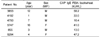
Six patients (four male and two female, ranging in age from 1 to 16 years old), sensitized to buckwheat at the Allergy Clinic of Samsung Medical Center, were included to evaluate IgE reactivity to hydrolyzed buckwheat protein. Buckwheat's IgE levels were measured using the Phadia UniCAP-system (Uppsala, Sweden). Range of specific IgE concentration is from 13 to 58.2 kUA/L (Table 1). Sera from healthy and non-atopic individuals who had never shown allergic symptoms were used as negative controls. After informed consent was received, blood samples were obtained, and the sera were frozen at -80℃ until use. This study was approved by the Samsung Medical Center Institutional Review Board.
Buckwheat protein extraction
Buckwheat powder (National Agricultural Cooperative Federation, Bong-pyung, Korea) was used in this study. Buckwheat protein was extracted as described by Tanaka et al. [9] with minor modifications. Five grams of finely ground buckwheat was extracted overnight by shaking at 4℃ in 50 ml 0.086 M NaCl (Showa, Gyoda, Japan) solution containing 0.033 M NaHCO3 (Duksan, Ansan, Korea). After centrifugation at 12,000 g for 30 min, the supernatant was filtered with 3,000 Da ultrafilter (Millipore, Cork, Ireland) to cut off proteins under 3,000 Da, and freeze-dried at -80℃. It was stored at -70℃ until use. Protein concentration was determined using Bio-Rad protein assay Kit [10].
Hydrolysis of buckwheat protein with pepsin
Hydrolysis of buckwheat proteins with pepsin was performed according to modified method of Kato et al. [11]. Simulated gastric fluid (SGF) (total vol. 40 ml) was prepared to contain approximately 1,628 unit/ml of pepsin (from porcine gastric mucosa, Sigma, St. Louis, MO, USA), with 12 M HCl (Duksan, Ansan, Korea) 33.6 µl and NaCl (Showa, Gyoda, Japan) 80 mg, pH 1.2. The protein concentration of buckwheat protein was 3 mg/ml in distilled water. To initiate hydrolysis, buckwheat protein was added to the pre-heated (37 ± 2℃) SGF (SGF: buckwheat protein = 6:1), and samples were labeled as SGF T0 (0 min), SGF P0 (0 min), SGF N0 (0 min), SGF T0.5 (0.5 min), SGF T1 (1 min), SGF T2 (2 min), SGF T5 (5 min), SGF T10 (10 min), SGF T20 (20 min), SGF T30 (30 min), SGF T60 (60 min), SGF P60 (60 min), and SGF N60 (60 min) depending on the duration of treatment. SGF P0 and P60 contained proteins only, as controls without pepsin, containing 10 mM HCl, 2 mg/ml NaCl, pH 1.2, and SGF N0 and N60 contained pepsin only, as controls without buckwheat proteins. Each specimen was quenched by adding sodium carbonate buffer (0.7 M, pH 11), then Laemmli buffer, pH 6.8 [50 mM Tris-HCl, Glycerol (Duksan, Ansan, Korea), 10% SDS, 2-mercaptoethanol (Samchun Chem., Seoul, Korea), 1% bromophenol blue (Duksan, Ansan, Korea)] was added, then, heated to 95℃ for 5 min.
Hydrolysis of buckwheat proteins with chymotrypsin
Hydrolysis of buckwheat proteins with chymotrypsin was performed according to modified method of Keum et al. [12]. The hydrolysis solution contained NaCl 2 mg/ml, and adjusted to pH 8.0. Chymotrypsin buffer contained 2.5 mg/5 ml of chymotrypsin (66 units/mg, α-chymotrypsin from bovine pancreas, Sigma, Saint Louis, MO, USA), and the concentration of buckwheat protein was 3 mg/ml in distilled water. Samples were labeled as in the pepsin experiment method above. Sample P0 and P60 contained buckwheat proteins only, as controls without chymotrypsin, containing 10 mM HCl, 2 mg/ml NaCl, pH 8.0, and N0 and N60 contained chymotrypsin only, as controls without buckwheat proteins. To initiate hydrolysis, buckwheat protein was added to the pre-heated (37 ± 2℃) chymotrypsin solution (solution:buckwheat protein = 6:1), and each specimen was drawn off at targeted duration of 1, 2, 5, 10, 20, 30, and 60 min (corresponding to specimen time points T1 through T60). Each specimen was quenched by adding sodium carbonate buffer (0.7 M, pH 11), then Laemmli buffer, pH 6.8 [50 mM Tris-HCl (Bio-Rad, Benicia, CA, USA), Glycerol, 10% SDS, 2-mercaptoethanol, 1% bromophenol blue] was added, then, heated to 9 5℃ for 5 min.
SDS-PAGE and ELISA
SDS-PAGE was performed according to the procedure described by Laemmli [13]. For densitometer, GS-800 calibrated densitometer (Bio-Rad, Benicia, CA, USA) was used. The measurement was performed in duplicate. ELISA was performed to evaluate IgE reactivity of patients' sera. Microplate, 96-well (BD Biosciences, San Jose, CA, USA), was coated with the 100 µl of buckwheat protein containing 50 µg/ml protein and incubated at 4℃ for 16 h. The protein was diluted with phosphate buffer saline (PBS) (pH 7.4). The coated plate was washed with 0.05% PBS-Tween 20 three times, and then blocked with 100 µl of 2% bovine serum albumin-phosphate buffer saline (BSA-PBS) for 1 h to prevent unspecific linking. Sera were diluted 1:80 with 2% BSA-PBS. The reaction with sera was performed for 2 h at room temperature. After washing the plate, adsorbed IgE was detected with 100 µl of Goat anti-human IgE-horseradish peroxidase (HRP) (1 µg/ml) (Kirkegaard and Perry Laboratories, Gaithersburg, MD, USA) diluted 1:2,500 with 2% BSA-PBS. The plate was further washed and developed with 100 µl of TMB peroxidase (3,3',5,5'-tetraethylbenzidine, Sigma, St. Louis, MO, USA). Absorbance was measured at 595 nm in duplicate.
The optical densities (OD) of SDS gels of each protein were measured using a reflective densitometer (Bio-Rad, Hercules, CA) and the Quantity One software (Bio-Rad), and % relative OD of hydrolyzed product with respect to that measured for the intact protein were calculated.

Results
SDS-PAGE
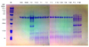
In SDS-PAGE, protein bands of 30-50, 24, 19, 16, and 10 kDa were shown (P0 and P60 lanes in Fig. 1). Among them, 24, 19, 16, and 10 kDa proteins are known as major allergens of buckwheat [9,14-18], while the other bands are not widely known as buckwheat allergens.
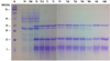
After treatment with pepsin (pH 1.2), the band of 16 kDa protein on SDS-PAGE remained till 10 min. All other protein bands disappeared in 30 sec, which indicated that the 16 kDa buckwheat protein is relatively resistant to pepsin treatment. After 60 min, all bands of proteins including the 16 kDa protein disappeared (Fig. 1). Chymotrypsin had weaker hydrolyzing power than pepsin on buckwheat proteins, and after 60 min of chymotrypsin treatment, 24 kDa protein still remained stable minutely (Fig. 2). One of the major buckwheat proteins, 16kDa, was not accurately determined for the disappearance of band, as it overlapped with the chymotrypsin band.
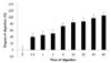
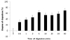
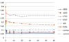
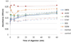
Optical density
The degree of digestion (%) graph was drawn based on the measured value with densitometer by a mathematical formula below (Fig. 3, 4). The result of densitometry on 16 kDa protein hydrolyzed with pepsin showed that it was hydrolyzed gradually (Fig. 3). It was hydrolyzed rapidly in the first 10 min, and hydrolyzed slowly after that point, remaining 30 min after the treatment. The result of densitometry on 24 kDa protein hydrolyzed with chymotrypsin showed that it was resistant to chymotrypsin (Fig. 4). Buckwheat protein, 24 kDa, was hydrolyzed to 69% in 60 min. It also showed the similar result till 10 min, as buckwheat proteins were hydrolyzed rapidly, and after that point, hydrolyzed slowly. As the 16 kDa protein band overlapped with the chymotrypsin band, it was difficult to distinguish between the two.
ELISA of buckwheat hydrolysates
Result of ELISA showed that pepsin could not hydrolyze completely the allergens of buckwheat. While 4 of 6 sera (#3853, 4192, 4750, and 855) did not show any interaction with the hydrolysates of buckwheat proteins after 30 sec hydrolysis with pepsin, the other two (sera #5747 and #5254) still interacted with the hydrolysates even after 60 min hydrolysis with pepsin. We considered the measured value, which had a gap of less than 0.05, as negative (Fig. 5). The result indicated allergenicity remained after 60 min of hydrolysis with pepsin, even though all buckwheat protein bands disappeared with pepsin treatment on SDS-PAGE.
The hydrolysates with chymotrypsin interacted with some sera (#4192, #5254, #855, #5747, #4750, #3853), indicating that it still had allergenicity as expected from the result of SDS-PAGE. It was found that chymotrypsin did not hydrolyze the allergens of buckwheat as well (Fig. 6). An increment of absorbance was observed over time.
Discussion
Traditional food allergens are heat-, enzyme-, and low pH-resistant water-soluble glycoproteins ranging in size from 10 to 70 kD and are responsible for systemic allergic reactions [19]. Several allergens have been identified in buckwheat using a combination of SDS-PAGE and immunoblotting methods with sera of buckwheat-sensitive patients. Most of the buckwheat allergens shown to bind IgE have molecular weights from about 10 to 30 kDa.
Buckwheat allergen, Fag e 1, previously known as BW24KD, is classified in 11S globulin-like legumin, and the molecular size is 22-24 kDa [16]. Until early in the 2000, buckwheat protein, 24 kDa, was considered as the major allergen according to the result that it interacted with IgE antibodies present in sera from almost all subjects (19/20) regardless of symptoms [9,17,18]. However, Tanaka et al. [9] found that 24 kDa was the cause of positive result to CAP-FEIA (CAP system fluorescein-enzyme immunoassay), not to IHR (Immediate hypersensitivity reaction). In this study, after treatment with pepsin (pH 1.2), the band of 24 kDa protein on SDS-PAGE almost disappeared, suggesting that 24 kDa protein is vulnerable to enzyme digestion and may not be a major allergen in buckwheat proteins.
Buckwheat protein, 16 kDa, is classified in 2S albumin, and has 100% homology with N-terminal amino-acid sequence of 18 kDa protein of buckwheat, 80% homology with Oriza sativa dehydrin of rice, and no homology with 24 kDa protein of buckwheat from analysis of N-terminal amino-acid sequence [9,20]. EGVRDLKE was reported as one of the 16 kDa linear IgE-binding epitopes [21]. It has been believed that the 16 kDa protein is the most allergenic among buckwheat allergens, because after pepsin treatment, the 16 kDa protein, not the 19 and 24 kDa proteins, remained undigested and preserved the capacity of IgE binding [9]. In clinical aspect, 16 kDa proteins bound with IgE antibodies present in sera from 9 of 10 patients with immediate hypersensitivity reactions, including 8 patients with anaphylaxis, but not with sera from buckwheat-specific IgE-positive subjects without immediate hypersensitivity reactions [9]. In SDS result of the present study, 16 kDa protein showed strong resistance to pepsin digestion, which was consistent with the previous study.
Buckwheat protein, 19 kDa, has specificity to sera of buckwheat allergy patient having allergic symptoms. The result of analysis of N-terminal amino-acid sequence showed 50% homology with 19 kDa α-globulin protein in rice [15]. Among buckwheat allergens, only 16 kDa (Fag e 2) and 19 kDa (Fag e 3) proteins are currently registered on IUIS Allergen Nomenclature [22]. BW10KD, 10 kDa buckwheat protein, was reported to show interaction with 57% of buckwheat allergy patients. It is also included in 2S-albumin, from the analysis result of N-terminal amino acid sequencing [14].
The results of ELISA in present study showed that after extensive digestion, allergenicity still remained, and in some case, even enhanced. The results had two patients showing Ig E reactivity to extensively hydrolysate by pepsin. Takagi et al. [23] showed similar results in hydrolysis of ovalbumin, ovomucoid, β-lactoglobulin, bovine serum albumin, and soybean trypsin inhibitor, and stated:
Some proteolytic fragments retain the same allergenic potential as the original proteins. Thus, even if proteins were rapidly digested, their proteolytic fragments could cause allergy.
It suggests that epitopes of the major buckwheat allergens were not completely hydrolyzed by pepsin, and retained allergenicity. Interestingly, the increment of absorbance was observed over time on the result of ELISA on the hydrolysates with chymotrypsin, suggesting that chymotrypsin treatment may expose the epitope to IgE of buckwheat patients' sera. Similar results were observed in the previous study of Keum et al. [12]. They treated 7S globulin of soybean with pepsin and found 50% reduction of allergenicity; however, after chymotrypsin treatment, increment of allergenicity was observed. In this study, some differences of allergenicity depending on patients' sera were observed. It is considered that each serum may interact with different epitope in buckwheat proteins, and that degree of reaction may vary.
The digestibility of a protein measured by the in vitro assay in simulated gastric fluid (SGF) or simulated intestinal fluid (SIF) is greatly influenced by the assay conditions [23]. Tanaka et al. [9] reported that 16 kDa was resistant to pepsin hydrolysis at pH 3. The 16 kDa protein did not disappear after hydrolysis for 120 min, while 19, 24 kDa proteins disappeared immediately following the hydrolysis with pepsin. Koyano et al. [24] hydrolyzed buckwheat proteins with pepsin at pH 2, and discovered that the band of 16 kDa protein disappeared gradually between 0 and 10 min. Satoh et al. [25] hydrolyzed the buckwheat proteins with pepsin at pH 1.2 and found that all of buckwheat proteins were hydrolyzed in 10 min. The result of SDS-PAGE in this study also agreed with that of Satoh et al. [25]. However, there was lack of information on allergenicity of the hydrolysates of buckwheat proteins with pepsin treatment at pH 1.2.
This study assessed the allergenic potential of buckwheat by studying the enzyme resistance of buckwheat proteins using pepsin and chymotrypsin. We found that allergenicity of buckwheat proteins did not disappear after hydrolysis of buckwheat proteins with pepsin or chymotrypsin for 60 min, suggesting that buckwheat has strong allergenic potential.
 XML Download