

 PDF
PDF ePub
ePub Citation
 Print
Print


Decompression with fusion and decompression alone are the two treatment options for patients who have a lumbar spinal stenosis with degenerative spondylolisthesis.1)
Decompression with fusion can improve back pain, radiating pain, and neurogenic intermittent claudication. In addition, it does not cause instability in patients with spondylolisthesis.2) However, this surgery can cause adjacent segmental degeneration, pseudoarthrosis and large volume blood loss during the surgery. Additionally, the hospitalization period is long.34) Recently, as the decompression surgery technique without fusion has been developed, there have been some studies that found no significant increase in instability after spinal decompression surgery without fusion through various types of microexcision, in patients who have a lumbar spinal stenosis with degenerative spondylolisthesis.567)
We used the semi-circumferential decompression (SCD) method during surgeries for patients who had a lumbar spinal stenosis with degenerative spondylolisthesis for the last 4 years. SCD is a decompressive technique to perform a total en-bloc ligamentum flavectomy under a microscope that preserves the articular facet. We have observed and analyzed relief of symptoms and occurrence of instability. In this study, we suggest the clinical effectiveness of the SCD technique for posterior decompression to treat patients who have a lumbar spinal stenosis with degenerative spondylolisthesis.
METHODS
We retrospectively analyzed the outcomes of 19 patients (mean age, 67.9 years; 2 men and 17 women) who were treated using the SCD method for a lumbar spinal stenosis with degenerative spondylolisthesis from 2010 to 2013. We excluded patients who had bilateral foraminal stenosis. The average follow-up period was 37 months (range, 25 to 56 months). All patients had radiating pain and neurologic intermittent claudication (NIC) due to a spinal stenosis. Magnetic resonance imaging showed central and lateral recess stenosis at the degenerative spondylolisthesis site in all patients. From 2010 to 2013, there was one patient with Meyerding grade II spondylolisthesis and no patient with Meyerding grade III spondylolisthesis. The follow-up period of patients with Meyerding grade II spondylolisthesis was too short (5 months). Hence, only patients with Meyerding grade I spondylolisthesis were included. Single-level degenerative spondylolisthesis was observed at L4 on 5 in 13 of 19 patients, L3 on 4 in four patients, and L5 on S1 in one patient. Double-level degenerative spondylolisthesis was observed at L3 on 4 and L4 on 5 in one patient. No instability was detected on a preoperative lumbar spine dynamic (flexion/extension) radiological study.
Technique
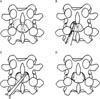
SCD is a decompressive method to remove the ligamentum flavum en-bloc. A median skin excision was used, soft tissue was detached, and the supraspinous ligament was detached from the spinous process, and moved to the side with multifidus muscle. The inferior one-quarter of the upper-level vertebral spinous process was removed, if needed, to enhance vision. The ligamentum flavum was detached from the inferior one-third of the lamina using a currette. After thinning the lamina with a high speed burr, a partial laminectomy was performed using a Kerrison rongeur. It is important that ligamentum flavum must not be removed before thinning the lamina with a high speed burr. By maintaining ligamentum flavum, we prevented damage to the dura mater while using the high speed burr. Finally, the total ligamentum flavum was removed en-bloc by detaching the ligamentum flavum from the inner side of the lamina to the inner side of the posterior facet (Figs. 1 and 2). The facet joint was preserved by leaving the superior articular facet unexcised. The detached supraspinous ligament was returned to its original position, and sutured with surrounding fascia. Ambulation was permitted beginning on postoperative day 1, and patients were encouraged to use a corset for 6 weeks.
Preoperative and postoperative symptom relief was estimated using the visual analogue scale (VAS) score and the Oswestry Disability Index (ODI) before surgery and at follow-up days. Patients were evaluated for VAS score and ODI after 3, 6 month post-surgery and every 1 year subsequently.
We compared the preoperative and last follow-up X-rays. All patients underwent dynamic (flexion/extension) X-rays. These results were used to estimate slip percentage and slip angle and assess instability and progression of lumbar degenerative spondylolisthesis. Slip percentage and slip angle was estimated using Taillard's and Boxall's methods, respectively. We estimated dynamic slip percentage (preoperative and postoperative change in the slip percentage) and dynamic slip angle (preoperative and postoperative change in the slip angle), and analyzed the occurrence of vertebral instability. The Wilcoxon signed-rank test was performed to detect postoperative changes using IBM SPSS ver. 21.0 (IBM Co., Armonk, NY, USA). A p-value less than 0.05 was considered significant.
RESULTS
Clinical Result
The mean VAS score of back pain decreased from 6.3 to 4.3, and the mean VAS score for lower leg radiating pain also decreased from 8.3 to 2.5 (p < 0.01). The average ODI score (maximum, 45 points) improved significantly from 25.3 preoperatively to 10.3 postoperatively (p < 0.01).
Radiologic Result
The change in slip percentage increased from 10% to 12.2% postoperatively, but the difference was not significant. The dynamic slip percentage did not show significant change postoperatively (5.2% vs. 5.8%). Slip angle in the SCD group also did not change (3.2° vs. 3.6°) at the last follow-up. The dynamic slip angle did not change (8.2° vs. 9.2°). These results indicated that the SCD method did not cause any vertebral instability.
DISCUSSION
Newman and Stone8) described degenerative spondylolisthesis as a disease in which a vertebral body slips anteriorly with no neural arch abnormality. Degenerative spondylolisthesis tends to be accompanied by spinal stenosis, and typically shows degeneration of the posterior facet or intervertebral disc, and ligament laxity. So far, decompression with fusion is reportedly better than decompression-only surgery.91011) In particular, a broad range of decompression techniques lead to poor outcomes, as compared to decompression with fusion.12) According to the spinal degeneration theory suggested by Kirkaldy-Willis, spinal segment stability is maintained in patients with degenerative spondylolisthesis,1314) and good surgical outcomes occur if sufficient decompression is performed preserving structures that affect posterior spinal stability. Thus, studies on decompression-only surgery with minimal excision to treat patients with degenerative spondylolisthesis were recently conducted. Weiner et al.5) reported a surgical procedure that restores the native position of the spinous process after a spinous process osteotomy and decompression to maintain stability by preserving the interspinous and supraspinous ligaments. Recently, various techniques using unilateral approach and bilateral decompression have been used in an attempt to minimize postoperative vertebral instability.67) The SCD technique has been introduced to prevent postoperative vertebral instability.15) Collectively, ours and previous results suggested that SCD technique could be applied for treatment of degenerative spondylolisthesis without instability and also for spondylolisthesis with instability preoperatively. If spinal segment stability is maintained postoperatively and sufficient decompression is achieved, surgical effect is well maintained. We carried out another study on the effect of SCD technique for spondylolisthesis with segmental instability. However, the follow-up period was short yet, studies to evaluate the effect of SCD technique for spondylolisthesis with instability after sufficient follow-up period are currently ongoing.
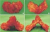
Anatomical research about ligamentum flavum by Okuda et al.16) suggested that ligamentum flavum degenerate, ossify, and calcify in patients with degenerative spondylosis, and that these changes are more severe if spondylolisthesis is present. Another research by Okuda et al.17) showed that nerve root compression is most severe in the proximal portion of the ligamentum flavum because the ligamentum flavum is thickest in this area (Fig. 2). The proximal border of the ligamentum flavum is attached to upper level vertebra at the inner surface of lamina almost horizontally and just below the pedicle; thus nerve root is compressed continuously when the ligamentum flavum is not completely removed.17) However, we can determine whether decompression is sufficient by observing the ligamentum flavum removed by en-bloc resection.
Zander et al.18) reported that a unilateral medial hemifacetectomy can induce vertebral instability, and Hamasaki et al.19) reported that a bilateral medial facetectomy for the medial one-third of the posterior facet could induce vertebral instability. Actually, in patients who have spinal stenosis with degenerative spondylolisthesis, cause of nerve root compression is ligamentum flavum hypertrophy at lateral recess, and facet hypertrophy is not cause of nerve root compression. So medial facetectomy is unnecessary for decompression of nerve root, and SCD technique can decompress nerve root sufficiently, maintaining spinal segmental stability.
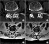
In addition, Abumi et al.20) conducted a biomechanical study demonstrating that spinal instability does not develop when the posterior facet is preserved with only the interspinous and supraspinous ligaments detached. According to these studies, the SCD technique, which decompress posteriorly by total excision of the ligamentum flavum with preservation of the supraspinous ligament and posterior facet, improves clinical outcomes and does not lead to spinal instability. Thus, the SCD technique can be applied to patients who have a spinal stenosis with degenerative spondylolisthesis (Fig. 3).
Based on analysis of clinical improvement and change in slip percentage and slip angle, we demonstrated that decompression through a total ligamentum flavectomy preserving the facet joint results in clinical improvement without causing vertebral instability.
The limitation of SCD technique is that this technique is not suitable for bilateral foraminal stenosis. There has been consensus that posterior lumbar interbody fusion is suitable for treatment of bilateral foraminal stenosis. However, the spinal stenosis with degenerative spondylolisthesis and unilateral foraminal stenosis can be treated using SCD technique and lateral fenestration technique simultaneously.
Hence, degenerative spondylolisthesis without foraminal stenosis, in which especially NIC is main symptom, is an absolute indication for SCD, and degenerative spondylolisthesis with unilateral foraminal stenosis is a relative indication for SCD. Degenerative spondylolisthesis with severe bilateral foraminal stenosis is a contraindication for SCD.
Decompression alone can reduce preoperative back pain but some preoperative back pain remains because it usually results from degeneration of facet joint degeneration or intervertebral disc. None of our patients had severe back pain that interfere with daily life activities.
Long term follow-up observations to assess instability and radiological exacerbations of spondylolisthesis are needed. Although a prospective randomized controlled clinical study is needed, our results showed that decompression surgery using the SCD technique was effective and less invasive for patients with spinal stenosis and degenerative spondylolisthesis.
In conclusion, the SCD technique, which decompress posteriorly by en-bloc total ligamentum flavectomy and preserve posterior facet, was clinically effective and does not lead to postoperative spinal instability.
 XML Download