

 PDF
PDF ePub
ePub Citation
 Print
Print


Impacted femoral neck fractures (IFNFs) are defined as fractures of the femoral neck with a close apposition of fragments based on simple radiograph and a varying degree of angulations.1) It is known that IFNFs account for 15-20% of whole femoral neck fractures (FNFs).2) Most IFNFs occur with the femoral head in valgus because there is an impaction of the fracture laterally and the femoral head trabeculae are tilted in the valgus position.1) However, Damany and Parker3) described varus IFNFs as impaction that occurs medially, such that the trabeculae are tilted into the varus. The optimal treatment of IFNFs is controversial. Some authors recommend primary operative stabilization,3-6) while others have reported satisfactory results after conservative treatment.2,7,8)
The aim of this study was to evaluate the clinical and radiologic results of IFNFs treated with multiple pinning (MP), and to determine the influence of the progression of impaction at the fracture site on clinical outcome.
METHODS
Patients
Between 2003 and 2006, 44 patients with IFNFs were diagnosed and treated with MP at an institution. Among the 44 patients, 10 who were followed for < 2 years were excluded, leaving 34 patients in the final analysis. There were 7 men and 27 women with a mean age of 65.5 years (range, 28 to 86.8 years), and all patients were followed for an average of 3.4 years (range, 2.2 to 5.9 years). This study was approved by our Institutional Review Board.
Surgical Technique and Postoperative Management
All operations were performed by one of two senior surgeons (HJK and JJY). Under anesthesia, the patient was gently placed in a supine position on the fracture table, and the fracture site was checked with fluoroscopy. After a 4-cm incision was made directly through the skin to the bone, three parallel Knowles pins were fixed from the lateral femoral cortex, just below the vastus lateralis ridge to the femoral head in an inverted triangular pattern without any attempt at fracture reduction. All patients were permitted partial weight bearing with crutches or walker assistance for at least 6 weeks postoperatively, and full weight bearing was permitted gradually after 6 weeks.
Radiographic Evaluation
Standard anteroposterior (AP) radiographs of the hip were obtained with both legs in a medial rotation of 15°. According to the institution's protocol, serial radiographs were obtained preoperatively, at 6 weeks, 3 and 6 months, and 1 year postoperatively, then at 1-year intervals until the final follow-up visit.
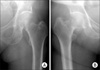
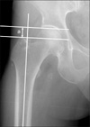
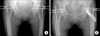
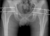
The fractures were classified as valgus or varus based on the location of the impaction and the alignment of the femoral head trabeculae on plain AP radiographs (Fig. 1). To quantify the extent of impaction of the fracture, the distance between the upper margin of the femoral head and the proximal tip of the greater trochanter (articulo-trochanteric distance, ATD)9,10) was measured on the affected side and the unaffected side using a sequential plain anteroposterior radiograph (Fig. 2). Next, the ATD of the affected side was divided by the ATD of the unaffected side for standardization, and expressed as the ATD index. To evaluate the progression of impaction at the fracture site, the percentage decrease in the ATD index between the follow-up intervals was calculated (Fig. 3). An ATD decrease in the affected side causes the ATD index to decrease, and a higher percentage decrease in the ATD index between the follow-up intervals indicates greater progression of impaction at the fracture site. A radiographic evaluation was performed before the evaluation of clinical outcome to prevent bias.
Clinical Outcome Evaluation
All patients were evaluated and were documented retrospectively using medical records. The failure of treatment was clarified as non-union and avascular necrosis (AVN), which required a major revision operation. Non-union was defined at 3 months postoperatively as follows: 1) a visible fracture line; 2) a continuing resorption of the femoral neck; or 3) re-displacement with a change in position of the fixation device.10-12) Other characteristics of the patients, including mean waiting time for surgery, preoperative Singh index score, and body mass index (BMI), were also measured to evaluate the influence on the clinical outcome of surgery.
Statistical Analysis
Data processing and statistical analyses were performed by a statistician using a SPSS ver. 17.0 (SPSS Inc., Chicago, IL, USA). An independent t-test was used to compare patient's age, BMI, Sing index, the mean percentage decrease between the follow-up intervals of the group that was not treated successfully and of the group that was treated successfully. Gender proportion, side of fracture, and the type of impacted fracture of patients in two groups were compared using the Fisher's exact test. The relative contribution of the variables to the failure of treatment was analyzed using a logistic regression analysis with a stepwise variable selection. A p < 0.05 was considered significant.
RESULTS
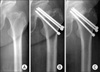
Thirty-one of 34 patients (91.2%) were classified as valgus and 3 patients (8.8%) were classified as varus. Of the 34 fractures that were followed for at least 2 years, 31 fractures (91.2%) were united. The remaining 3 patients (8.8%) complained of persistent pain because of non-union. Between 6 and 8 months after the initial surgery, 2 patients had revision surgery with a total hip arthroplasty and 1 patient had a bipolar hemi-arthroplasty. Two of the 3 non-union patients had valgus fractures and 1 patient had a varus fracture. Pain developed because of avascular necrosis in 3 patients (8.8%) 12 months after surgery, all of whom had valgus fractures and had revised to total hip arthroplasties between 15 and 22 months after the initial surgery (Fig. 4). The overall success rate was 82.4% (28 of 34 patients), and the overall failure rate was 17.6% (6 of 34 patients).
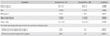
The mean percentage decrease between the preoperative ATD index and the ATD index 6 weeks after surgery was 4.5% (range, 0 to 9.7%) in the group that was treated successfully and 25.1% (range, 6.1 to 51.7%) in the group that was not treated successfully (p < 0.001). There was also a significant mean percentage decrease in the ATD index between 6 weeks and 3 months (7.0% [range, 0.7 to 16.8%] in the group that was treated successfully and 36.1% [range, 19.4 to 53.6%] in the group that was not treated successfully; p < 0.001) (Table 1). This suggests that impaction at the fracture site has progressed more within 3 months in the group that was not treated successfully than in the group that was treated successfully.
No significant association existed between the mean waiting time for surgery and the Singh index with adverse clinical outcomes after surgery. Based on an independent t-test, there was a significant difference in the BMI between the group that was treated successfully and the group that was not treated successfully (20.9 kg/m2 [range, 15.6 to 26.9 kg/m2] and 25.3 kg/m2 [range, 23.1 to 31.1 kg/m2], respectively; p = 0.037) (Table 1).
However, the odds ratio for a mean percentage decrease between the preoperative ATD index and the ATD index 6 weeks after surgery was not statistically significant (odds ratio, 1.7; 95% confidence interval, 0.8 to 3.5; p = 0.156).
There were no major complications related to the surgery. Nine of the 34 patients (26.5%) complained of mild pain and discomfort at the pin insertion site; however, only 1 patient required surgery for pin removal 2 years after the initial surgery.
DISCUSSION
During the period of the current study, 182 patients with IFNFs were treated surgically at our institution. The overall proportion of impacted fractures was 24.2% (44 of 182 patients), which is similar to previous reports.2,13-16)
Although there is no consensus in the treatment of IFNFs, our institution follows a protocol in which the IFNF is stabilized primarily because the failure rate of conservative treatment of IFNFs, including secondary displacement requiring surgery, has been reported to be as high as 46%.2,8,16-18) The overall success rate of the current study was 82.4%, which is comparable to the results of previous studies (76.1 to 92%).4-6,11,13,19-23)
The incidence of AVN of the femoral head after conservative treatment and osteosynthesis for IFNFs has been reported to be 6 to 14%1,7,8,16,18) and 1.8 to 19.5%,1,4,5,11,13,18-25) respectively. The incidence of non-union after osteosynthesis has been reported to range from 4 to 18%.4,5,11,13,19-25) In the current study, both the incidence of non-union and avascular necrosis was 8.8%.
In previous studies involving osteosynthesis for displaced femoral neck fractures, several authors have attempted to show predictors for poor outcome, such as a high degree of displacement at the fracture site,12,25,26) impairment of the blood supply,27,28) and an increase in Pauwel's angle.29) As another predictor of clinical outcome, the ATD was introduced by Nilsson et al.,30) who concluded that a decrease in the ATD > 10 mm on postoperative radiographs influenced re-displacement and non-union after surgery. At the same time, Shimizu et al.10) standardized the ATD by the diameter of the unaffected femoral head to minimize the effect of the difference in the patients' body sizes. Shimizu et al.10) measured the capital impaction index as a new indicator of an impacted fracture with excessive shortening at the fracture site and showed that the degree of capital impaction was significantly associated with unsuccessful outcomes when the capital impaction index was greater than the mean plus the standard deviation.
We also used the value of the ATD to measure the degree of impaction of fractures, but there were several differences between the aforementioned studies and the current study. First, the ATD was standardized by the ATD of the unaffected side instead of the unaffected femoral head, not only to minimize the effect of the difference in the patients' body sizes, but also to adjust for errors from the difference in hip position. With a slight flexion of the hip, the ATD of the affected side will be measured as erroneously decreased, but might be adjusted by using the ATD index (the ATD of the affected side/the ATD of the unaffected side) because both legs are usually in the same position. Second, we evaluated the progression of impaction of fractures by measuring the percentage decrease in the ATD index using sequential radiographs. There are some limitations to the use of a single value of the ATD because it cannot be applied in varus-type impacted fractures and cannot be used in case of the paradoxical increase of the ATD in valgus-type fractures (Fig. 5). In varus-type IFNFs, the ATDs are always decreased more than valgus-type fractures with the same degree of impaction and measurement of Schimizu's capital impaction index may be exaggerated, even with a small degree of impaction.10) Third, we demonstrated increased progression of impaction at the fracture site within 3 months after surgery is significantly related with a poor outcome. Calandruccio and Anderson27) reported that in non-displaced and impacted fractures, the main damage is to the vessels in the bone at the level of the fracture, whereas in displaced fractures, there may also be varying degrees of damage to the retinacular vessels. The progressive impaction or collapse of the femoral head may produce further damage to the retinacular vessels and this might be the basis for poor clinical outcomes associated with more progression of impaction in the current study. Fourth, the patients in this study were followed for a longer period of time (mean, 3.4 years; range, 2.2 to 5.9 years) than in previous studies.
There were some limitations to this study. All data were collected and analyzed retrospectively, and the number of patients was small. Further studies with a larger sample size is needed to confirm the association of the progression of impaction at the fracture site with poor clinical outcome and to ascertain the statistical difference of the clinical outcome between valgus and varus IFNFs.
In conclusion, primary stabilization with Knowles pins for impacted femoral neck fractures resulted in reasonable clinical outcomes with low morbidity. Although there was a significant difference of a mean percentage decrease in the ATD index between the group that was not treated successfully and the group that was treated successfully, we could not verify it as a risk factor for failure of treatment because the odds ratio was not statistically significant.
 XML Download