

 PDF
PDF ePub
ePub Citation
 Print
Print


INTRODUCTION
Unlike thrombolysis for myocardial ischemia, the pilot clinical studies of thrombolysis for ischemic stroke did not document dramatic, "Lazarus" or "on the table" clinical recovery during treatment.1-3 Subsequent pivotal trials of TPA have not reported any differences between the groups at 2 and 24 hours post treatment in the pre-specified end-points.4-7 However, a post-hoc analysis of the NINDS trial8 showed that by 24 hours, 27% of TPA-treated patients improved by ≥ 10 points on the National Institutes of Health Stroke Scale (NIHSS) or resolved their neurological deficit completely compared to 12% in the placebo group (p=0.002). Therefore, some patients may have experienced early clinical recovery presumably due to fast thrombus dissolution, but the overall number of these events was low.
Early clinical improvement after stroke usually occurs after arterial recanalization.9-12 A recent meta-analysis confirmed the recanalzation hypothesis by showing that the occurrence of recanalization is associated with a 4- to 5-fold increase in the odds of good final functional outcome and a 4- to 5-fold reduction in the odds of death.13 These results lend strong support to the use of restoration of vessel patency as a surrogate end point in phase II trials of pharmacological recanalization agents and in trials comparing novel to existing predicate recanalization devices in acute ischemic stroke. Since early recanalization can lead to dramatic recovery,9-12 any additional enhancement of TPA-associated thrombus dissolution can possibly produce even higher early recovery rates among patients with ischemic stroke.
The ability of an ultrasonic mechanical pressure wave to enhance thrombolysis has been documented in 1970's, 14,15 and confirmed by many in experimental models.16-20 The likely mechanism that emerged from these in vitro and in vivo experiments is the ability of ultrasound to agitate flow around and through the thrombus thus delivering more TPA to target binding sites. In stroke patients, ultrasound can promote TPA delivery to the areas with stagnant flow near occlusion.
Although low kilohertz frequencies better potentiate TPA effects,21 these systems are not available for clinical practice due to safety concerns and inability to image vasculature with this frequency/wavelength range. Meanwhile, 1-2.2 MHz frequencies can also enhance TPA-induced thrombus dissolution utilizing different mechanisms such as fluid streaming around clot surface, dis-aggregation of fibrin fibers, and creating more binding sites for TPA without heating or cavitation.22,23 This frequency range is safely used for diagnostic ultrasound examinations.
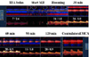
Portable diagnostic 2 MHz TCD equipment can be used the emergency room to continuously monitor TPA infusion is acute ischemic stroke patients.24 With prior training and experience in interpretation of TCD, this test, particularly in combination with urgent carotid/vertebral duplex scanning, can yield high degrees of accuracy for detection and localization of arterial occlusion as well as assessment of recanalization at bedside.24,25 In addition, TCD can be complementary to other imaging modalities such as CTA by showing real-time flow findings (real-time embolization, collateralization of flow with extracranial internal carotid artery disease, alternating flow signals indicative of steal phenomenon).26 Finally, real time flow findings during TCD-monitoring has been shown to be associated with long-term functional outcome.27,28
Once abnormal residual flow signals are identified, an ultrasound beam can be steadily focused at presumed intra-cranial thrombus location, and arterial recanalization can be monitored in real time.24 When intravenous TPA infusion was continuously monitored with 2 MHz TCD,24 we have observed early recanalization and dramatic recovery rates higher than expected.1 This non-randomized study of patients treated with TPA24 suggested potential therapeutic effect of TCD and led to a prospective randomized clinical trial.
The CLOTBUST Trial
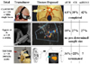
The CLOTBUST (Combined Lysis of Thrombus in Brain ischemia using transcranial Ultrasound and Systemic TPA, Fig. 1) was a phase II clinical randomized multi-center international trial with centers in Houston, Barcelona, Edmonton, Calgary.29 It had pre-specified safety and signal of efficacy end-points and a pre-determined sample size of 63 patients per group.29 All enrolled patients had an acute ischemic stroke, and were treated with a standard 0.9 mg/kg dose of intravenous TPA therapy within 3 hours of symptom onset. All patients also had MCA occlusions on pre-treatment TCD. They were randomized (1:1) to continuous TCD monitoring (Target) or placebo monitoring (Control).
Safety end-point was symptomatic brain hemorrhage (sICH) causing worsening of the neurological deficit by 4 or more NIH Stroke Scale (NIHSS) points. Primary combined activity end-point was complete recanalization on TCD or dramatic clinical recovery by the total NIHSS score ≤ 3 points, or improvement by ≥ 10 NIHSS points within 2 hours after TPA bolus. Clinical investigators were blinded to group assignment (active or sham monitoring) done by sonographers.
All projected 126 patients received TPA and were randomized 1:1 to target (median pre-treatment NIHSS 16 points) or control (NIHSS 17 points). Age, occlusion location on TCD and time to TPA bolus were similar between groups. sICH occurred in 4.8% Target and 4.8% Controls. Primary end-point was achieved by 31 (49%, Target) vs 19 (30%, Control), p=0.03. At 3 months, 42% Target and 29% Control patients achieved favorable outcomes (mRS 0-1 points), NS. This trend indicates feasibility of a pivotal phase III clinical trial that, at 274 patients per group, would be properly powered to detect this difference in outcomes at 3 months.29
Other Clinical Trials
Transcranial duplex technology was recently tested in a smaller randomized clinical trial.30 Duplex transducers are different from the ones used in CLOTBUST since they generate multiple small beams at dual emitting frequencies, one for Doppler and one for gray scale imaging (Fig. 1). One of major limitations of this technology that there are no reliable head frames for transducer fixation, and most studies are to be carried out hand-held. In addition, the mechanical index of these scanners is higher than TCD and no dose escalation study was performed to determine how little ultrasound is needs to enhance thrombolysis without safety concerns that would be outlined below.
Eggers et al. evaluated 25 patients (11 Target TPA+duplex monitoring, 14 Controls TPA alone) and reported a trend in the Target group towards higher recanalization rates, more hemorrhagic transformations (18% sICH rate), and better outcomes at 3 months compared to patients who received TPA alone.30 This study did not have a pre-determined sample size, and the results may be affected by a small number of patients enrolled. More studies are needed to evaluate the potential of transcranial duplex technology to enhance thrombolysis.
The same group and others31-33 reported provocative findings that patients who are not eligible for systemic TPA therapy may potentially benefit from continuous monitoring with ultrasound alone since, hypothetically, ultrasound may help facilitate the endogenous thrombolytic process that leads to spontaneous recanalizations in acute stroke patients. It is unclear if only partial recanalization can be induced by ultrasound alone, and if this exposure would result in a significant difference at 3 months justifying a large clinical trial. In any case, there is no clear data regarding the benefit of ultrasound monitoring without TPA and TPA treatment should not be substituted with ultrasound alone in patients otherwise eligible for thrombolytic therapy within 3 hours of symptom onset.
Furthermore, different experimental strategies are being tested in an extended time window for acute stroke treatment, and continuous exposure to ultrasound may find its application while patient may be receiving other agents such as GP IIb-IIIa antagonists or direct thrombin inhibitors or awaits intra-arterial procedures.
Ultrasound transducers were also incorporated into a catheter for intra-arterial delivery of a thrombolytic drug (EKOS Corporation). This intra-arterial device uses 1.7 - 2.1 MHz pulsed wave ultrasound with the emitting power of 400 mW, parameters similar to extracranially applied transcranial Doppler. The EKOS catheter is now being tested in phase II-III Interventional Management of Stroke (IMS) trials.34
Therapeutic, i.e. non-imaging ultrasound35 has been tested in the TRanscranial low-frequency Ultrasound-Mediated thrombolysis in Brain Ischemia (TRUMBI) trial.36 First, the investigators used a very low KHz system (<40 KHz) that produced intolerable tinnitus and was withdrawn from clinical testing (Daffertshofer M, unpublished data). It was replaced by a mid KHz system operating at 300 KHz (Fig. 1). The trial was terminated after 26 patients were enrolled with a 36% rate of symptomatic hemorrhage in the Target group and no signal of efficacy on early recanalization or clinical outcomes at 3 months.36 The trial demonstrated adverse bio-effects of mid-KHz ultrasound that promote bleeding, including brain areas not-affected by ischemia.36 Further research should determine if "standing" pressure waves and endothelial disruption may cause these adverse effects. If confirmed in vivo models, this will have implications on design of future KHz-based systems.
Microspeheres-potentiated Ultrasound-enhanced Thrombolysis
Experimental data have suggested that ultrasound-enhanced thrombolysis can be further amplified by adding gaseous microspheres,37-39 safe ultrasound contrast agents, are micron-sized lipid shells that when exposed to ultrasound, expand and produce stable cavitation with stronger reflected echoes. This is used to generate ultrasound images with better resolution. At the same time, microspheres agitate fluid where they are released by ultrasound and this is useful in drug delivery and mechanical "grinding" of a thrombus. In fact, microspheres have their own ability to lyse thrombi without a lytic drug.37
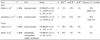
Several studies have been reported with different types of commercially available microspheres40-43 (Table). Molina et al pioneered this approach in stroke patients and reported the largest study to date that compared the CLOTBUST Target arm to the CLOTBUST Target insonation protocol combined with Levovist air microspheres (Schering AG).40 Investigators demonstrated that at 2 hours after TPA bolus the TPA+TCD+Levovist group achieved a 55% sustained recanalization rate compared to 38% in the TPA+TCD group of the CLOTBUST trial. The safety and feasibility of infusion of a new and more stable C3F8 perfultren-lipid microspheres in patients treated with ultrasound-enhanced thrombolysis has recently been reported in a small phase IIA randomized clinical trial.41 Interestingly, in 75% of patients, μS permeated to areas with no pre-treatment residual flow, and in 83%, residual flow velocity improved at median of 30 min from start of μS infusion (range 30 s-120 min) by median of 17 cm/s, or 118% above pre-treatment values (Fig. 2).
Larrue et al recently randomized patients with acute (<3 hours) middle cerebral artery main stem occlusion as demonstrated by CT or MR angiography to either transcranial duplex ultrasound continuous monitoring combined with intravenous galactose-based microspheres and rt-PA (combined treatment group), or rt-PA alone (control group).43 Their trial was prematurely discontinued on the basis of safety reasons since a high rate of asymptomatic intracerebral hemorrhage was demonstrated on gradient-echo MRI in the combined treatment group (78%). However, none of the intracerebral hemorrhages was symptomatic and the fact that asymptomatic hemorrhagic transformation in the setting of acute cerebral ischemia has not been associated with poor outcome both in the NINDS44 and the ECASS trial45 should be taken into account when interpreting the results of the former study. It is further unclear why data safety monitoring board was not appointed for this study and why a dose de-escalation decision was not made (i.e. reduce time of exposure to ultrasound or reduce the dose of microspheres). In its current design, the study does not allow to decide whether excessive hemorrhagic transformation rate was attributed to duplex ultrasound or microspheres since controls received just TPA and no ultrasound.
Finally, Perren et al studied the safety and feasibility of TCCD ultrasound monitoring combined with a second generation, phospholipid encapsulated sulphur hexafluoride microsphere (SonoVue) and intravenous systemic thrombolysis in patients with acute middle cerebral artery occlusion. Patients who received Microsphere-potentiated Ultrasound-enhanced Thrombolysis seemed to fare better in terms of improvement in NIHSS-score and sustained a more marked improvement in their residual blood flow in comparison to patients treated only with ultrasound-enhanced thrombolysis.42
Future Directions
Currently, an international multi-center controlled TUCSON trial of a new and more stable perfultren-lipid microspheres (MRX 801, www.imarx.com) is underway.46 A total of 72 patients with acute intracranial arterial occlusion as demonstrasted by CT or MR angiography will be randomized to Microspheres-potentiated Ultrasound-enhanced Thrombolysis (4 groups with increasing doses of perfultren-lipid microspheres) versus TPA treatment alone.
Microspheres offer a mechanical way to amplify stroke therapies, and can be developed as a new kind of drugs or devices to augment brain perfusion, drug and nutrient delivery within the existing and at an extended time window. One problem on the way to develop ultrasound and microsphere assisted stroke therapies is the need of an experienced sonographer to find intracranial thrombus, and expose its surface to residual flow in order to lodge more TPA and agitate stagnant flow. Personnel with these skills are lacking in most emergency centers. Future studies will focus on the development of an operator-independent ultrasound device that can be used by existing medical personnel regardless of their experience in diagnostic ultrasound.
 XML Download