

 PDF
PDF ePub
ePub Citation
 Print
Print


INTRODUCTION
Radial neuropathy is the third most common focal neuropathy of the upper limb.1 The radial nerve originates from the posterior cord of the brachial plexus, and runs through the spiral groove on the humerus.2 Radial neuropathy at the spiral groove leads to weak extension of the wrists and fingers, and paresthesia of the first web space. The radial nerve is frequently injured at the spiral groove, not only by physical trauma but also by compression. Acute compressive radial neuropathy, so-called Saturday night palsy, is particularly noticeable on awakening due to nerve compression during sleep. This clinical feature occasionally renders it difficult to distinguish between radial neuropathy and cerebral stroke. Electrophysiological studies, including nerve conduction study (NCS) and electromyography (EMG), are the diagnostic key to localizing the injury and confirming a compressive radial neuropathy. However, it usually takes up to about two weeks for radial neuropathy abnormalities to be detected on electrophysiological studies.3 In addition, compressive radial neuropathy is known to be a self-limiting disease with a favorable prognosis. This topic has been addressed in few case series.4,5,6 In the present study, radial motor conduction-including stimulation at Erb's point-was investigated in patients with acute compressive radial neuropathy within 14 days from clinical onset, with a view to determining the early diagnostic and prognostic value of proximal radial motor NCSs.
METHODS
Study population
Patients with acute compressive radial neuropathy were retrospectively enrolled at Kangbuk Samsung Hospital between January 2007 and October 2014. Patients were selected who fulfilled the following three conditions (as identified from their medical records): 1) sudden weakness of radial-nerve-innervated muscles that was confirmed by clinical or EMG examinations, 2) radial NCS including stimulation at Erb's point within 14 days from clinical onset, and 3) normal electrophysiological study findings for the median and ulnar nerves. The three following exclusion criteria were applied: 1) alternative etiologies of radial neuropathy such as trauma, infection, and hereditary neuropathy with liability to pressure palsy, 2) acute ischemic brain lesions on diffusion-weighted magnetic resonance imaging (MRI), and 3) polyneuropathy based on clinical findings. Finally, 39 patients were enrolled. In addition, radial nerve conduction data were collected prospectively from 39 control subjects to obtain reference values. The research protocol was approved by the Institutional Review Board of Kangbuk Samsung Hospital, Korea.
Clinical assessment
Clinical data included sex, age, body mass index (BMI), coexisting diseases, lesion site, compression time, motor weakness, and sensory change. Compression time was obtained to determine the sleep time or the time hanging over the armrest of a chair before wrist drop. The compression time was identified in 38 patients; one patient could not recall a definite compression event or time. Disease severity was measured according to the Medical Research Council (MRC) sum score, which evaluated the strength in two muscle groups: the wrist extensors and the finger extensors of the metacarpophalangeal joint. A score of between 0 and 5 was assigned to each muscle group, giving a maximum total score of 10. Patients were also interviewed by telephone to determine the clinical outcome. Thirty-three patients for whom clinical follow-up was available were able to provide information regarding their recovery time. The recovery time was also recorded on the medical records of five patients, and was not found to differ significantly from that informed by telephone interview. Diffusion-weighted MRI of the brain was performed in 14 patients.
Electrophysiological assessment
Electrophysiological studies were performed using the Viking Select EMG machine (Nicolet Biomedical, Madison, WI, USA). NCSs were performed with surface electrodes and EMGs were conducted using monopolar needle electrodes at room temperature. All studies were performed by a skilled EMG technician or neurologists with at least several years of experience. Motor and sensory NCSs of the median and ulnar nerves were performed by standard methods using surface electrodes. Normal values for the median and ulnar nerves were obtained based on the methods of Oh.7 For radial sensory nerve action potentials (SNAPs) and sensory nerve conduction velocities (SNCVs), the recording electrode was placed over the nerve as it courses through the anatomic snuffbox, with the stimulating electrodes located at 10-14 cm proximally along the radius. For radial compound muscle action potentials (CMAPs) and motor nerve conduction velocities (MNCVs), the surface recording electrode was placed about 4 cm proximal to the ulnar styloid process on the belly of the extensor indicis proprius. The radial nerve was stimulated at three sites based on the methods of Jebsen:8 1) in the forearm, about 4 cm proximal to the recording electrode along the lateral edge of the ulna, 2) in the arm, 5-6 cm proximal to the lateral epicondyle of the humerus, and 3) at Erb's point, the angle of the clavicle, and the posterior aspect of the sternocleidomastoid muscle. MNCVs were obtained between the forearm and arm in only 29 patients because MNCVs in the forearm-arm segment were not routinely performed at our laboratory between January 2007 and December 2009. In addition, needle EMG examinations of the muscles innervated by the radial nerve as well as other nerves were performed in 24 patients after at least 7 days from clinical onset. Three of these 24 patients underwent EMG as a follow-up electrophysiological study after more than 14 days from the symptom onset.
Reference values for the radial nerve were obtained by performing radial NCSs on the bilateral arms of 39 control subjects. The reference range of radial nerve conduction was defined as values within 2 SDs of the mean.
Partial conduction block was defined as a >50% decrease in negative-peak amplitude or 40% decrease in negative-peak area of the CMAPs between distal and proximal stimulations of the radial nerve, based on consensus criteria published by the American Association of Electrodiagnostic Medicine (AAEM) quality assurance committee.9
Statistical analysis
The chi-squared test was used to compare discrete variables, and the independent t-test was used to compare the means of two samples for continuous variables. Univariate and multivariate linear regression analyses were performed to determine the factors influencing the recovery time of radial neuropathy. In addition to age and sex, variables with p<0.1 on univariate analysis were entered into the multivariate linear regression analyses. Except where stated otherwise, the data are presented as mean±SD values, and differences were considered statistically significant at p≤0.05. Statistical analyses were performed with SPSS (version 18.0; SPSS Inc., Chicago, IL, USA) software.
RESULTS
Clinical assessment
Thirty-nine patients with radial neuropathy (age, 45.2±12.7 years; range, 22-73 years) were enrolled in this study, comprising 31 men and 8 women. Their BMI was 22.6±2.5 kg/m2. Lesions were in the right arm in 22 (56%) patients and in the left arm in 17 (44%). The time from clinical onset to hospital visit was 3.2±3.0 days, and the compression time was 3.9±3.1 hours. Wrist drop was first noted on awakening in 37 (95%) patients; one patient identified wrist drop after leaving his arm hanging over the arm rest of a chair for 30 minutes, and the other patient did not recall a definite compression event. Alcohol and sleeping pills were consumed before sleeping by 25 (64%) and 1 (3%) patients, respectively. Chronic alcoholism and diabetes mellitus were documented as coexistent diseases in eight (21%) and two (5%) patients, respectively. None of the 39 patients had any other metabolic disorders, such as thyroid disease or rheumatoid arthritis. The initial physical examination revealed moderate weakness of the wrist extensors (MRC grade, 2.7±1.0) and finger extensors (MRC grade, 2.4±1.2). The MRC sum score was 5.1±2.1. Sensory symptoms and signs in the distribution of the superficial radial sensory nerve were noted in 27 (69%). Diffusion-weighted MRI did not reveal any acute ischemic brain lesions in the 14 tested patients.
The 39 control subjects comprised 29 men and 10 women aged 43.2±11.3 years (range, 25-60 years) and with a BMI of 23.7±3.3 kg/m2. There were no significant differences in sex, age, and BMI between the control subjects and the patients with radial neuropathy.
Electrophysiological assessment
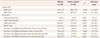
The results of the NCSs of the control subjects and radial neuropathy patients are summarized in Table 1. The reference data were obtained from 78 arms of the 39 control subjects. The decrease in negative-peak amplitude and area of the CMAP between the arm and Erb's point was greater than that between the forearm and arm in all control subjects. However, the degree of this reduction did not exceed the AAEM consensus criteria for conduction block in any of the control subjects.
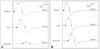
Analysis of the radial conduction studies of 39 patients with acute compressive radial neuropathy revealed that the interval from clinical onset to NCS was 5.7±4.5 days. Compared with reference data, the radial SNAP amplitude, radial SNCV, radial terminal latency, radial CMAP amplitude, and radial MNCV were abnormal in 1 (3%), 1 (3%), 3 (8%), 2 (5%), and 3 (10%) of 29 the tested patients, respectively. However, partial conduction block of the radial nerve was found in 17 (44%) patients (Fig. 1). This partial conduction block was found mainly in the arm-Erb's point segment in 15 patients, the forearm-arm segment in 1 patient, and both segments in 1 patient. In addition, ten patients exhibited partial conduction block on the test within 5 days from clinical onset, and in 2 patients on the day of onset (Supplementary Fig. 1 in the online-only Data Supplement). The clinical presentation did not differ significantly between the conduction-block group and the non-conduction-block group among the patients with acute compressive radial neuropathy (Supplementary Table 1 in the online-only Data Supplement). EMG revealed abnormal spontaneous activities in the radial-nerve-innervated muscles in 18 of the 24 tested patients.
Prognosis
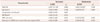
Clinical follow-up data obtained by telephone were available for 33 of the 39 patients. All 33 patients achieved complete clinical recovery without residual weakness, with a recovery time of 46.8±34.3 days. The relationships between recovery time and several adjusting factors were investigated, including compression time, BMI, MRC sum score, and the reductions in the CMAP area and the CMAP amplitude in the arm-Erb's point segment. MRC sum score, and the reductions in the CMAP area and the CMAP amplitude were significant on univariate analysis. The variance inflation factors for the reductions in the CMAP area and the CMAP amplitude were 14.148 and 13.970, respectively, indicating that there was definite multicollinearity. Therefore, subgroup analysis was performed after adjusting for the reductions in CMAP area and the CMAP amplitude; the reduction in the CMAP area in the arm-Erb's point segment was still an independent predictor for recovery time (R2=0.285) (Table 2 and Supplementary Table 2 in the online-only Data Supplement).
DISCUSSION
The present study has revealed the clinical and electrophysiological features of acute compressive radial neuropathy, a condition that is frequently caused by consuming alcohol and leaning the arm over the back of a chair. The initial muscle weakness is moderate, but the prognosis is excellent. The clinical features described herein are compatible with previous studies.4,5,10,11 This study also demonstrates the practicality and usefulness of proximal radial motor conduction in the acute phase.
In the present study, stimulation at Erb's point had early diagnostic value within 14 days from clinical onset. It is well known that routine radial NCS results, and especially SNAP, are frequently preserved.10,12 However, about half of the 39 patients in the present study exhibited partial conduction block on stimulation at Erb's point, even in the test on the day of compression. Stimulation at Erb's point also produced useful prognostic information for acute compressive radial neuropathy. In the present study, the reduction in the CMAP area between the arm and Erb's point was an independent negative predictor for recovery time; this finding is contrary to previous results. Partial conduction block is usually known to be a good prognostic factor in compressive neuropathies including radial neuropathy11,13 because the presence of partial conduction block provides evidence of segmental demyelination without axonal degeneration. NCSs were performed in this study within 14 days after compression. Partial conduction block was identified in NCSs within 5 days from clinical onset in two-thirds of the patients. In this acute phase it is difficult to distinguish between true partial conduction block and the pseudo-conduction block due to axonal conduction failure. Therefore, the significance of partial conduction block should only be related to the severity of radial nerve injury.
Medical Research Council sum score and the reduction of CMAP amplitude were also significant predictors for recovery time on univariate linear regression. In fact, the initial muscle weakness was previously reported to be an indicator of prognosis.11 The reduction in CMAP amplitude was closely associated with the reduction in the CMAP area. Therefore, these two factors may be independent predictors for recovery time, but further study is required in large numbers of patients to confirm this finding.
Several limitations of the present study need to be considered. The main limitation was the relatively small number of patients. The application of very strict inclusion criteria, including proximal radial nerve studies within 14 days from compression, made it difficult to enroll a sufficient number of patients. Furthermore, abnormal electrophysiological findings were not confirmed in radial-nerve-innervated muscles in all patients; many patients refused follow-up EMG studies due to a good clinical course and the pain associated with the tests. However, all patients in this study exhibited localized weakness of radial-nerve-innervated muscles, and 14 patients did not exhibit any brain abnormalities on diffusion-weighted MRI. The retrospective design of this study and data collection by telephone confers unavoidable bias. However, very few of the patients with radial neuropathy revisited the hospital after the diagnosis, probably due to the excellent prognosis. Finally, two technical limitations of stimulation at Erb's point have been reported previously:14 1) it activates not only the extensor indicis proprius but also all of the forearm muscles innervated by other nerves, and 2) it does not fully stimulate the brachial plexus due to technical problems. However, proper Erb's point responses were obtained from all 78 arms of the control subjects in this study. Although the reduction in the CMAP area/amplitude was more unstable in the arm-Erb's point segment than in the forearm-arm segment, the reduction in the CMAP area/amplitude did not exceed the AAEM consensus criteria of a partial conduction block.
In conclusion, the findings of the present study demonstrate that radial NCSs, which included stimulation of Erb's point, are useful for the early detection of acute compressive radial neuropathy. Furthermore, the reduction in the CMAP area between the arm and Erb's point is a useful independent predictor of recovery time.
 XML Download