

 PDF
PDF ePub
ePub Citation
 Print
Print


Dear Editor:
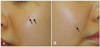
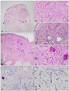
A 23-year-old woman presented with multiple, progressively growing, asymptomatic, depressed macules on face for 2 years. Physical examination revealed three yellowish to skin-colored annular atrophic macules 2~5 mm in size without elevated border on both cheeks (Fig. 1). She had no family or past history that scar might occur such as acne, chickenpox, herpes infection or prior laser treatment. Laboratory data including blood cell counts, biochemistry and urinalysis were unremarkable. Histopathology showed narrow strands of basaloid tumor cells in a fibrous stroma with adjacent embedded keratinous cysts (Fig. 2). The tumor strands were distributed from the upper to the deeper dermis. We performed additional exicisional biopsy with immunohistochemical staining in order to differentiate from malignant neoplasms including morpheic basal cell carcinoma (BCC) and microcystic adnexal carcinoma (MAC). Carcinoembryonic antigen (CEA), epithelial membrane antigen (EMA), CD23, and CK19 were negative, and CK20 showed focal positive. From these findings, she was diagnosed with desmoplastic trichoepithelioma (DTE), and remaining lesions were completely removed.
DTE is a rare benign skin appendageal neoplasm. It is a distinct variant of trichoepithelioma based upon its unique clinical and histopathological characteristics1. Clinically, it typically shows 3~8 mm diameter, annular, yellowish or skin-colored asymptomatic plaques, with an elevated border and a depressed or atrophic center on face1. It almost develops as a solitary lesion, although rare cases of patients with multiple lesions have been reported2.
Histopathologically, DTE is characterized by following features. First, epithelial strands consist of small basaloid cuboidal cells in a one to three-cell-thick layer. Second, many keratinous cysts have a peripheral border of basaloid cuboidal cells, and sometimes have several calcification foci. Finally, stroma appears as dense fibrous stroma1. Differential diagnosis includes MAC, BCC, and scar. Although MAC presents with many horn cysts and epithelial cords, it is distinguished by ductal differentiation and infiltration to the subcutis or deeper structures3. Therefore, when biopsy is superficial, MAC can be misdiagnosed as DTE3. Assessing EMA and CEA immunohistochemistry may identify ductal differentiation and CD23 expression in MAC1. CK19 demarcates follicular bulge stem cells and its positivity favors MAC over DTE4. Both DTE and morpheic BCC can exhibit basaloid cells in strands and sclerotic stroma. However, neither cellular atypia nor peripheral palisading are seen in DTE, and morpheic BCC is not usually associated with horn cyst formation1. Also, in most DTE, retained or increased CK20-positive Merkel cells are found, but not in morpheic BCC5. Scar should be considered if multiple depressed macules appear, but it can be easily differentiated based upon its histopathology.
The treatment of choice is surgical excision and Mohs micrographic surgery is recommended to achieve clear surgical margins. In our case, because multiple lesions had developed and tumor strands extended into the deep dermis, total excision was performed to exclude malignancy. Therefore, initial proper biopsy is important to make an accurate diagnosis and treatment in patients with multiple scar-like depressed macules on face.
Herein, we report a rare case of multiple DTE, which should be differentiated from malignancy histopathologically.
 XML Download