

 PDF
PDF ePub
ePub Citation
 Print
Print


INTRODUCTION
Cases of dermatitis induced by the injection of certain drugs are not rare. The most commonly reported causative drug is vitamin K1. This adverse reaction to drugs includes wheal-like erythema, pseudoscleroderma, eczematous lesion on epicutaneously exposed skin, and localized urticarial lesions1. Among them, the cutaneous adverse reaction caused by vitamin K1 is called vitamin K1 dermatitis2. In the early literature, most reactions to parenteral vitamin K were found in patients with liver disease, suggesting that the cause of the adverse reaction was related to hepatic disturbances. However, more cases have recently been described in patients without apparent liver dysfunction.
The exact mechanism of this reaction is unknown. Bruynzeel et al.2 reported about cutaneous hypersensitivity reactions to vitamin K, and they performed patch and intradermal tests with vitamin K1. Most of the tests had positive results, and the authors suggested that delayed-type hypersensitivity probably plays the key role in the mechanism. Vitamin K is mainly used in patients with hypoprothrombinemia, i.e., as an antidote to coumarin, to manage vitamin K deficiency (as an adverse effect of drugs) and the bleeding tendency caused by liver dysfunction1.
We designed a clinical study to find the cause and clinicopathologic features of injection-induced dermatitis, and to reveal whether the reaction has any relation to age, injection site, drug concentration, and time interval from the injection of the drug to the occurrence of skin lesions.
MATERIALS AND METHODS
Patients
Patients were recruited between March 2006 and March 2012 from the Department of Dermatology, Kyung Hee University Hospital at Gang-dong in Seoul, Korea. Patients who visited our clinic because of the occurrence of lesions at injection sites were recruited. The database of the recruited patients contained integrated clinical information, including their diagnosis and clinical photographs. All patients who came to our clinics because of injection-induced dermatitis were entered into the database during their first evaluation. Patients were excluded if they did not have an exact drug injection history, had other severe disease or psychological problems, or had other possible causes of dermatitis. All procedures were approved by the ethics committee of Kyung Hee University Hospital at Gang-dong.
Clinicopathologic evaluation
The medical records and clinical photographs of the patients were reviewed, as well as their clinical information including the onset age, presence of other chronic diseases, causative drug, purpose of the injection of the drug, time interval between the injection and the onset of skin lesions, sites of the lesions, extent of cutaneous involvement, and symptoms. Each patient underwent a skin biopsy on the first visit, and the lesion was evaluated by keeping photographic records with a high-resolution, 8.0-megapixel digital camera (Canon EOS 350D; Canon Inc., Tokyo, Japan).
Skin test
Patch tests were performed with the causative drugs and also with 10% vitamin K1, which is the most common drug causing dermatitis on patients' back. The patches were applied with Finn Chambers on Scanpor tape (Smart-Practice, Phoenix, AZ, USA). The result was evaluated on the second and fourth days. Intradermal tests with vitamin K1 (0.1%, 1%, and 10%) and with the causative drug in each patient were also performed. After 15 minutes, the injection site was evaluated to look for wheal growth (i.e., a small swelling of the skin). Two millimeters of growth in 15 minutes was considered a positive result. Each of the tests had at least a 2-week interval. All patients agreed to undergo skin tests and laboratory tests, if necessary.
RESULTS
Demographic data and clinical features
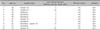
The characteristics of the study population who showed drug-induced dermatitis are shown in Table 1. There were 10 female patients with a mean age of 40.4 years.
All lesions occurred on the buttocks region. Nine patients had itching symptom, and one patient had pain sensation. None of the patients had any history of injection-induced dermatitis. All patients were in good general health, and laboratory investigations showed normal values; in particular, there were no liver function disturbances.
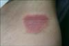
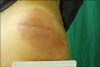
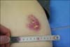
The time interval between the drug injection and the onset of skin lesions ranged from 1 day to 20 days (mean, 9.5 days). The lesions were of various clinical types: eczematous (6, 60%), sclerodermoid (2, 20%), urticarial (1, 10%), and necrosis or cellulitis like (1, 10%) (Fig. 1, 2, 3). Therefore, the eczematous type was the most common.
Histopathologic features
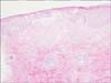
Histopathologic examination of the biopsy specimen showed spongiosis (6, 60%), intraepidermal blister (2, 20%), and necrosis (1, 10%) in the epidermis, as well as an intense perivascular infiltrate composed of predominantly lymphocytes and some eosinophils (10, 100%) in the dermis (Fig. 4, 5). Moreover, involvement of the subcutaneous fat layer (panniculitis) was observed in two patients (20%). Therefore, in this study, the most common histopathologic type of the lesions was spongiotic dermatitis.
Causative drug and skin test results
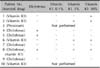
Of the 10 lesions observed, 4 (40%) were induced by diclofenac, 4 (40%) by vitamin K1, 1 (10%) by piroxicam, and 1 (10%) lesion occurred after the injection of both diclofenac and vitamin K1. The reasons for the injection of each drug in each patient were described. Four patients received an injection of diclofenac for pain control, and one patient was injected with piroxicam also for pain control. Five patients had vitamin K1 injection for anticoagulation after the occurrence of coumarin-induced prothrombin deficiency during an operation.
Patch tests and intradermal tests were performed in the patients. The patch tests with diclofenac (as is, 2.5%, 5%, and 10%) and vitamin K1 (10%) were all negative in 10 patients. Furthermore, the results of the intradermal tests are given in Table 2. Six patients had a positive reaction, consisting of erythema, induration, and vesiculation, after 1 and 2 days. Two patients had a positive reaction to diclofenac, and 3 patients reacted to vitamin K1. Moreover, one patient had positive reaction to both agents. Among four patients who had a positive reaction to vitamin K1, three patients had a reaction to a 1% concentration and one patient to a 10% concentration. None of the patients showed a positive reaction to 0.1% vitamin K1.
DISCUSSION
Cases of injection-induced dermatitis are reported occasionally. The causative agents reported previously were diclofenac, vitamin K1, docetaxel, ketoprofen, and piroxicam1234. In our present study, we focused on diclofenac and vitamin K1.
Vitamin K and its analogues are frequently used in the treatment of hypoprothrombinemia1. It is essential for the biosynthesis of prothrombin and factors VII, VIII, and X in the liver. Vitamin K exists in four different pharmacological forms. Vitamin K1 (phytomenadione) is essential for the formation of prothrombin and other coagulation factors from the liver. Therefore, the deficiency of vitamin K1 results in an easily hemorrhagic state similar to a coumarin-like anticoagulant activity; vitamin K2 (menaquinone) is synthesized in the intestine through normal bacterial action; and K3 (menadione) and K4 (menadiol) are synthetic analogues. Vitamin K4 is water soluble, whereas vitamin K1 and K3 are oil soluble. Vitamin K1 is mainly prescribed; however, vitamin K3 and K4 may also be often prescribed (orally or by injection)1235. Diclofenac is a non-steroidal anti-inflammatory drug used to treat pain, inflammatory disorders, and dysmenorrhea5.
Vitamin K1 injection is indicated in the following coagulation disorders that are due to faulty formation of factors II, VII, IX, and X resulting from vitamin K deficiency or interference with vitamin K activity: anticoagulant-induced prothrombin deficiency caused by coumarin or indanedione derivatives, prophylaxis and therapy of hemorrhagic disease of the newborn, and hypoprothrombinemia due to antibacterial therapy26. The injection route is usually used owing to its rapid effect. In our study, the patients received an injection of diclofenac for pain control and of vitamin K1 for anticoagulation after the occurrence of coumarin-induced prothrombin deficiency during an operation. Vitamin K1 has been associated with reports of severe anaphylactic/anaphylactoid reaction after intravenous administration and also with local adverse effects at the site of intramuscular or subcutaneous injection. Because of its poor solubility in water, injectable forms of vitamin K1 are prepared either with an emulsifier, polyethoxylated castor oil (Cremophor-EL; Roche Products, Dee Why, NSW, Australia) or in micelles (Mixed Micelles, Roche Products). With regard to Konakion Cremophor-EL, a total of 159 adverse events were reported to the manufacturer between 1952 and 1994 (Roche Drug Safety database)7. Information about patient ages was not available from this database. Between 1952 and 1994, an estimated 400 million adults were exposed to this preparation; between 1974 and 1993, approximately 130 million children were exposed. Of these adverse events, 48 cases were related to skin disorders that occurred at the "application site," which is the site of injection. A similar number of cases (n=52) of application-site reactions to vitamin K1 have been documented in the European and North American literature2. Two types of reaction have been described: (i) pruritic, indurated, eczematous plaques that usually develop 4~14 days after injection8 and may last several months, and (ii) sclerodermoid lesions (Texier's disease) that appear within weeks to months and persist for years. Cases of fasciitis are rarely reported9. Fasciitis may occur with or without a preceding eczematous stage. Secondary generalization of the eczematous reaction has been previously reported10.
It was previously thought that liver disease predisposed patients to adverse cutaneous reactions to vitamin K1; however, there have been several reports of a reaction occurring in the absence of liver disease261112. The association can be explained by the observation that patients with liver disease are more likely to have hypoprothrombinemia and thus are more likely to be given vitamin K1. The total dose does not seem to be a contributing factor because some patients received as little as 10 mg6811, whereas others received as much as 440 mg12. In this study, there was no patient with an evidence of preexisting hepatic disease.
The most common type in our study was the eczematous type (6, 60%), in addition to the sclerodermoid (2, 20%), urticarial (1, 10%), and necrosis or cellulitis-like (1, 10%) types. According to the literature, this erythematous eczematoid plaque is cured after a few weeks to a few months; however, some lesions occasionally persist for more than a few years and progress to sclerodermoid lesions.1314
In this study, the histopathology of most lesions showed the features of allergic contact dermatitis or subacute dermatitis corresponding to previous reports. In every 10 cases, there were perivascularly infiltrating lymphohistiocytes and eosinophils, and in 2 sclerodermoid-like cases, panniculitis was observed.
Proper diagnosis of injection-induced dermatitis could be accomplished by careful history taking and review of clinicopathologic. In the current study, the intradermal test had more positive results than the patch test. Bruynzeel et al.2 performed intradermal and patch tests with vitamin K1 in 31 patients. They obtained 12 positive results from patch tests in 17 patients, and 21 positive results from intradermal tests in 21 patients. Moreover, other previous studies also suggest that that intradermal test might be more reliable because the patch test has more false-negative results81314.
In this study, there was no association between clinical or pathological characteristics and the time interval between the drug injection and the onset of skin lesions. We could not find any difference in the time interval according to the particular clinical manifestation. Statistical analysis was impossible because there was only one patient with necrosis (or cellulitis) and only one patient with wheal. We also could not find any association between the results of the skin tests and the time interval between the drug injection and the onset of skin lesions. We randomly divided the patients according to the time interval between the drug injection and the onset of skin lesions to >7 days group and the <7 days group. No difference was found in the results of the skin tests between these two groups.
The exact pathologic mechanism of injection-induced dermatitis is not yet well known. However, many studies suggest a relation to a type IV hypersensitivity reaction. Several features of the eczematous reaction to vitamin K1 suggest that the reaction is a manifestation of delayed-type hypersensitivity, i.e., a cell-mediated immune reaction. In most cases, there is an approximate 10~14 days lag between the first dose and the appearance of the rash28101112141516, and subsequent patch and intradermal testing produces a reaction in 3~5 days and 1~2 days, respectively281314151617. A recall phenomenon has also been described in several patients2816, in which patch or intradermal testing at a distant location precipitates an eczematous flare at the original reaction sites. Lesional biopsies consistently show spongiosis of the epidermis, dermal edema, and perivascular mononuclear and eosinophilic cellular infiltrates681115, consistent with a delayed-type hypersensitivity reaction. Interestingly, although prophylaxis against hemolytic disease of the newborn with intramuscular vitamin K1 has been practiced routinely in many countries (since the 1960s in the United Kingdom)18, we could not find any reports in the English-language literature of cutaneous eruptions due to this medication in neonates. This may be a consequence of the immaturity of the neonatal immune system. However, in some cases, this injection may represent the sensitizing event (some adults had a reaction after a single injection26811). Both Finkelstein et al.8 and Sanders and Winkelmann15 took a biopsy from positive intradermal test sites in one of their patients, which showed a histologic picture compatible with a delayed-type hypersensitivity reaction. The histologic pattern, together with the mean period of 10~14 days between the administration of the drug and the occurrence of the skin lesions, all point to a type IV-mediated allergic reaction2. In our study, the time interval between injection and the onset of skin lesion ranged from 1 day to 20 days (mean interval, 9.5 days). In the study by Pang et al.19, vitamin K1 dermatitis was controlled with tacrolimus, which inhibits the proliferation of antigen-presenting T-lymphocytes and cytokines such as interleukin (IL)-2, IL-3, IL-4, IL-5, interferon-γ, and tumor necrosis factor-α. It also supports the hypothesis that a type IV hypersensitivity reaction involving T-lymphocytes contributes to its mechanism. However, in Guidetti et al.'s study20, vitamin K1 did not cause any change in the leukocyte migration inhibition test and lymphocyte blastoid transformation test; thus, the authors suggested that the mechanism is not immunologic. Therefore, the pathogenesis is still debatable.
All patients in this study were females. However, to our knowledge, thus far there is no study about the sex difference in injection-induced cutaneous adverse effects267810. We assume that this female predominance in our study was due to the frequent use of vitamin K injection in obstetrics and gynecology.
Patients with injection-induced urticarial-, eczematoid-, and scleroid-type dermatitis were treated with oral corticosteroid, antihistamine, and topical steroid. Moreover, injection-induced necrosis-type dermatitis was additionally treated with daily simple dressing and epidermal growth factor injection. The necrosis type had the longest time to recovery. In a previous study, most patients were treated with systemic and topical steroid, antihistamine, and wet dressing1278. Most patients showed good response to the treatment and had no complication. There were reports of progression of previous skin lesions into sclerodermoid lesions1314. However, in our study, there was no progression of previous skin lesions during our follow-up period. The longest follow-up duration was 3 years.
In conclusion, it seems that injection-induced dermatitis is not very rare. Vitamin K1 and diclofenac are the most common causative agents. Therefore, awareness among dermatologists about this disease and its common causative agents would be helpful in the diagnosis and treatment of injection-induced dermatitis.

 XML Download