

 PDF
PDF ePub
ePub Citation
 Print
Print


INTRODUCTION
Acne vulgaris is a common skin disorder involving the sebaceous glands that are associated with hair follicles. It has been debated whether treatment with ultraviolet (UV) radiation, visible light or infrared light worsens, improves, or has no effect on acne vulgaris. UV radiation has been found to enhance the comedogenicity of sebum1. Exposure to UV radiation, primarily UVA, can cause acneiform eruptions, which consist of multiple uniform red papular lesions. UVB, UVA, blue, blue/red, infrared light therapy, or photodynamic therapy with topical aminolevulinic acid for acne may provide some therapeutic benefits2,3,4,5,6,7,8. However, a systematic review found little evidence for or against the hypothesis that exposure to natural sunlight improves acne9. Overall, definitive evidence for the beneficial effects of sunlight exposure on acne is lacking.
Abnormal keratinization of keratinocytes, proliferation of Propionibacterium acnes, and perturbation of sebaceous gland function are all known to play important roles in the pathophysiology of acne10. We conducted this study to evaluate the effects of exposure to UVA radiation, visible light, and infrared light on cultured human sebocytes. The expression of inflammatory cytokines, matrix metalloproteinases (MMPs), and antimicrobial peptides (AMPs) was measured after exposure of cultured human sebocytes to UVA radiation and light at wavelengths of 650 nm (visible) and 830 nm (infrared). In addition, sebum production was measured in cultured human sebocytes after exposure to UVA radiation and light at wavelengths of 650 nm and 830 nm.
MATERIALS AND METHODS
Culture of human sebocytes
Primary cultures of sebocytes were performed according to the method described previously11. They were established from occipital hair follicle sebaceous glands. Sebaceous glands were dissected from hair follicles under a binocular microscope and transferred to tissue culture dishes. Sebaceous gland explants were maintained in Dulbecco's modified Eagle's medium (DMEM; HyClone Laboratories, Logan, UT, USA), supplemented with penicillin (100 U/ml), streptomycin (100 µg/ml), and 20% heat-inactivated fetal calf serum (HyClone Laboratories, Logan, UT, USA), at 37℃ in a humidified atmosphere containing 5% CO2. Explants were maintained for an initial period of 3 days in DMEM; the medium was then changed to EpiLife (MEPI500CA; Gibco BRL, Grand Island, NY, USA), supplemented with penicillin (100 U/ml), streptomycin (100 µg/ml), and fungizone (250 µg/ml). Thereafter, the medium was changed every 3 days. When the cultures became subconfluent, cells were harvested using 0.25% trypsin and 10 mM ethylenediaminetetraacetic acid in Hank's balanced salt solution and subcultured using a split ratio of 1 : 3. Only cells obtained after the second passage were used for the experiments in this study. Sebocytes in these cell populations were identified by staining with hematoxylin and eosin (Muto Pure Chemicals Co., Tokyo, Japan), Oil red O, and Nile red (Sigma, St. Louis, MO, USA), and by immunofluorescence detection of cytokeratin 1 and 7 (Chemicon, Billerica, MA, USA).
Treatment of cultured sebocytes with UVA radiation and light at wavelengths of 650 nm and 830 nm
Cultured human sebocytes were treated with increasing doses of UVA (2 J/cm2, 3 J/cm2, and 5 J/cm2) by using a UV phototherapy unit (Choongwae Machinery Co., Seoul, Korea), and with light at wavelengths of 650 nm (14 J/cm2, 29 J/cm2, and 87 J/cm2) and 830 nm (5 J/cm2, 10 J/cm2, and 30 J/cm2) by using portable light source (AinA, Daegu, Korea), before being harvested for assays evaluating the expression of genes and proteins and the production of sebum.
Reverse transcription polymerase chain reaction
Polymerase chain reaction (PCR) amplifications were performed in triplicate by using the First Strand cDNA Synthesis kit (Promega, Madison, WI, USA) and oligonucleotide primers (Genotech, Daejeon, Korea) for interleukin (IL)-1β (5'-GGG CCT CAA GGA AAA GAA TC-3', 5'-TTC TGC TTG AGA GGT GCT GA-3'), IL-6 (5'-TAC CCC CAG GAG AAG ATT CC-3', 5'-GAG GTG CCC ATG CTA CAT TT-3'), IL-8 (5'-AGA TAT TGC ACG GGA GAA-3', 5'-GAA ATA AAG GAG AAA CCA-3'), tumor necrosis factor (TNF)-α (5'-TCC TTC AGA CAC CCT CAA CC-3', 5'-GGC TAC ATG GGA ACA GCC TA-3'), MMP-1 (5'-CTG AAG GTG ATG AAG CAG CC-3', 5'-AGT CCA AGA GAA TGG CCG AG-3'), MMP-3 (5'-CTC ACA GAC CTG ACT CGG TT-3', 5'-CAC GCC TGA AGG AAG AGA TG-3'), MMP-9 (5'-CAC TGT CCA CCC CTC AGA GC-3', 5'-GCC ACT TGT CGG CGA TAA GG-3'), psoriasin (5'-TGC TGA CGA TGA TGA AGG AG-3', 5'-ATG TCT CCC AGC AAG GAC AG-3'), human β-defensin (hBD)-2 (5'-ATC AGC CAT GAG GGT CTT GT-3', 5'-GAG ACC ACA GGT GCC AAT TT-3'), hBD-3 (5'-AGC CTA GCA GCT ATG AGG ATC-3', 5'-CTT CGG CAG CAT TTT CGGCCA-3') and LL-37 (5'-GGG TAG GGC ACA CAC TAG GA-3', 5'-GGA CAG TGA CCC TCA ACC AG-3'). Image J (NIH Image, Bethesda, MD, USA) was used for quantitative analysis of band intensity in gel images. Target gene messenger RNA (mRNA) levels were normalized to that of β-actin and presented as relative ratios. The Student's t-test was used for the statistical analysis of all data. A p-value of <0.05 was considered statistically significant.
1) Extraction of total RNA
A modified acid phenol method using the TRIzol reagent (Invitrogen, Grand Island, NY, USA) was used to isolate total cellular RNA.
2) Isolation of mRNA
The Dynabeads mRNA purification kit (Dynal, Carlsbad, CA, USA) was used to isolate mRNA from total cellular RNA.
3) Reverse transcription-PCR
Synthesis of cDNA from 5 µg total RNA was performed using a cDNA synthesis kit containing ImProm-IITM reverse transcriptase and random primers (Promega). GoTaq Flexi DNA Polymerase was used for all PCR assays. The following amplification program was used, with slight modifications for specific genes as indicated: 35 cycles of denaturation at 95℃ for 1 min, followed by annealing at 56℃ for 1 min, and extension at 72℃ for 1 min. Modified conditions used for specific genes were as follows: IL-1β, 25 cycles, annealing at 56℃; psoriasin, 30 cycles, annealing at 59℃; hBD-2, 28 cycles, annealing at 57℃; hBD-3, 28 cycles, annealing at 61℃; and LL-37, 28 cycles, annealing at 61℃.
Enzyme-linked immunosorbent assay
Production of IL-1β, IL-6, IL-8, and TNF-α by cultured sebocytes was assayed using an Enzyme-linked immunosorbent assay (ELISA) kit (R&D Systems, Shanghai, China), according to the manufacturer's instructions. Samples were tested in duplicate. Briefly, 50 µl of sample was mixed with 50 µl of biotinylated antibody reagent, 100 µl of prepared streptavidin-horseradish peroxidase, and 100 µl of premixed tetramethylbenzidine substrate solution, in that order. Samples were incubated in the dark at room temperature for 30 min, after which reactions were stopped by adding 100 µl of stop solution per sample. Absorbance was measured using a VersaMax Microplate Reader (Molecular Devices, Sunnyvale, CA, USA).
Lipid assay
Cultured sebocytes were exposed to UVA radiation and light at wavelengths of 650 nm and 830 nm and were seeded in 100-mm dishes. After 5 days, when cultures reached 70% confluence, the culture medium was removed and phosphate-buffered saline (PBS) was added. Cells were resuspended in PBS and collected by centrifugation (100 g, 5 min). The supernatant was removed, and lipid extraction buffer (0.9% NaCl and 1% Triton X-100) was added to the cell pellet. After homogenization by vortexing, the mixture was centrifuged (15,700 g, 15 min), and the resulting supernatant was retained for lipid analysis. Lipid levels were measured in duplicate, using an enzymatic assay kit (ASAN Co., Seoul, Korea). Lipid measurements were normalized to sample protein levels (as determined using the Bradford method).
RESULTS
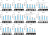
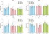
Treatment of cultured sebocytes with UVA radiation did not cause a significant increase in the expression of inflammatory cytokines, MMPs, or AMPs
The gene and protein expression of IL-1β, IL-6, and IL-8 in cultured sebocytes treated with UVA radiation (2 J/cm2, 3 J/cm2, and 5 J/cm2) did not show a significant increase compared to that observed in the control (Fig. 1, 2). However, TNF-α gene expression levels in sebocytes treated with 3 J/cm2 and 5 J/cm2 of UVA showed a significant decrease compared to that in the control (p<0.05) (Fig. 1). This was not reflected in the levels of TNF-α protein expression, which did not show a significant decrease compared to that in the control (Fig. 2). The gene expression levels of MMP-1 and MMP-3 in UVA-treated cells (2 J/cm2, 3 J/cm2, and 5 J/cm2) did not show a significant increase compared to those in the control (Fig. 1). In contrast, MMP-9 gene expression showed a significant decrease in UVA-treated cells (2 J/cm2, 3 J/cm2, and 5 J/cm2) compared to that in the control (p<0.05) (Fig. 1). The expression levels of AMPs, including psoriasin, hBD-2, hBD-3, and LL-37, in UVA-treated sebocytes (2 J/cm2, 3 J/cm2, and 5 J/cm2) did not show a significant change compared to those in the control (Fig. 1).
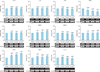
Treatment of cultured sebocytes with light at a wavelength of 650 nm did not cause a significant increase in the expression of inflammatory cytokines, MMPs, or AMPs
The gene and protein expression levels of inflammatory cytokines, including IL-1β, IL-6, IL-8, and TNF-α, in cultured sebocytes treated with light at a wavelength of 650 nm (14 J/cm2, 29 J/cm2, or 87 J/cm2) did not show a significant increase compared to those observed in the control (Fig. 2, 3). In addition, the gene expression levels of MMPs such as MMP-1, MMP-3, and MMP-9 in cultured sebocytes treated with light at a wavelength of 650 nm (14 J/cm2, 29 J/cm2, or 87 J/cm2) did not show a significant increase compared to those observed in the control (Fig. 3). Furthermore, the gene expression levels of AMPs such as psoriasin, hBD-2, hBD-3, and LL-37 in sebocytes treated with light at a wavelength of 650 nm (14 J/cm2, 29 J/cm2, or 87 J/cm2) did not show a significant change compared to those observed in the control (Fig. 3).
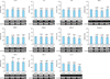
Treatment of cultured sebocytes with light at a wavelength of 830 nm did not cause a significant increase in the expression of inflammatory cytokines, MMPs, or AMPs
The gene and protein expression levels of inflammatory cytokines, including IL-1β, IL-6, IL-8, and TNF-α, in cultured sebocytes treated with light at a wavelength of 830 nm (5 J/cm2, 10 J/cm2, or 30 J/cm2) did not show a significant increase compared to those observed in the control (Fig. 2, 4). The gene expression levels of MMPs such as MMP-1, MMP-3, and MMP-9 and AMPs such as psoriasin, hBD-2, hBD-3, and LL-37 in cells treated with light at a wavelength of 830 nm (5 J/cm2, 10 J/cm2, or 30 J/cm2) did not show a significant change compared to those observed in the control (Fig. 4).
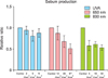
Treatment of cultured sebocytes with UVA radiation and light at wavelengths of 650 nm and 830 nm did not cause a significant decrease in sebum production
Sebum production in cultured sebocytes treated with UVA radiation (2 J/cm2, 3 J/cm2, or 5 J/cm2) did not show a significant decrease compared to that in the control (Fig. 5). Sebum production in cultured sebocytes treated with light at a wavelength of 650 nm (14 J/cm2, 29 J/cm2, or 87 J/cm2) showed a decreasing trend, as the dosage increased. An insignificant decrease in sebum production was observed in cultured sebocytes treated with light at a wavelength of 830 nm (5 J/cm2, 10 J/cm2, or 30 J/cm2) compared with that in the control (Fig. 5).
DISCUSSION
Excessive sebum production and abnormal lipid secretion from sebaceous glands contribute to the formation of primary acne lesions12. It was reported that UV (UVA and UVB) irradiation of human back skin caused increased sebum production during the first 3 days after treatment13. The effects of UVB irradiation on sebaceous gland function have been widely studied. Hyperplasia of sebaceous glands, following UVB irradiation, was observed in biopsy-based studies in a hairless murine model14. Another study reported that UVB irradiation markedly increased both cell proliferation and sebum production in cultured hamster sebocytes15. In contrast, our previous study demonstrated that UVB treatment did not increase sebum production in cultured human sebocytes16. In the present study, we examined the effects of treating cultured human sebocytes with UVA radiation, visible light, and infrared light. Similar to our previous observations with UVB irradiation, we found that these treatments did not lead to a significant decrease in sebum production.
It was also reported that UV irradiation induced the expression of inflammatory cytokines, including IL-1α, IL-6, IL-1ra, and IL-10, in human back skin, at approximately 5 days after treatment12. Interestingly, time-dependent patterns of gene induction (involving TNF-α, IL-1β, and matrilysin) have also been observed in cultured HaCaT cells irradiated with UVA/UVB17. Consistent with such reports, studies involving UVB irradiation of whole mouse skin or human skin have also reported increased expression of inflammatory cytokines such as IL-1, IL-6, and TNF-α at the mRNA level18,19. Production of inflammatory cytokines, MMPs, or AMPs by sebocytes can significantly contribute to the formation and aggravation of acne lesions20. In our previous study, UVB irradiation of sebocytes resulted in an increase in the expression of inflammatory cytokines16. In contrast, the present study found that exposure to UVA, visible light, or infrared light did not cause a significant increase in the expression of inflammatory cytokines, MMPs, or AMPs.
It has been proposed that exposure to sunlight can stimulate the production of sebum and secretion of inflammatory cytokines, thereby promoting the development or aggravation of acne lesions. In contrast, sunlight exposure has been reported to induce anti-inflammatory effects in keratinocytes, by inhibiting IL-1α and ICAM-1 (intercellular adhesion molecule 1) production21. According to Charakida et al.22, up to 70% of patients report improvement in their acne after sunlight exposure. However, since sunlight is a mixture of UV, visible, and infrared wavelengths, it is unclear which of these components might be responsible for the favorable effects on acne. Blue light appears to be the most effective, despite poor depth of skin penetration23. Red light is able to penetrate deeper, reaching the skin layer that contains the sebaceous glands24. Although the various light-based treatments appear to target P. acnes directly or indirectly, red light exposure may produce additional anti-inflammatory effects, e.g., by influencing cytokine release from macrophages25. This study examined the effects of exposure to UVA, visible light, and infrared light on cultured human sebocytes, focusing on sebum production and expression of inflammatory biomarkers.
In summary, exposure of cultured human sebocytes to UVA radiation, visible (650 nm) light, and infrared (830 nm) light did not lead to a significant increase in the expression of the inflammatory biomarkers tested or in sebum production. Light therapy for acne vulgaris is primarily aimed at targeting P. acnes. This study, therefore, provides evidence that UVA radiation, visible light, and infrared light may be used for treatment of acne, without a significant increase in the expression of inflammatory biomarkers and sebum production in sebocytes.
 XML Download