

 PDF
PDF ePub
ePub Citation
 Print
Print


Dear Editor:
Pityriasis rosea (PR) is a common acute and self-limiting inflammatory dermatosis. However, the etiology and pathogenesis of this disorder remains unknown. Studies show that a proportion of PR may be attributed to human herpes virus type 6 (HHV6) or human herpes virus type 7 (HHV7) infections or to some medications1. A recent immunohistochemical study suggests that T lymphocyte-mediated cell immunity plays an important role in the development of PR2. Considering this information, we measured the serum levels of interferon-γ (IFN-γ), interleukin (IL)-2, IL-4, and IL-10 in patients with PR, in order to determine the association of these T helper 1 (Th1) and Th2 cytokines with PR.
In total, forty-six patients (22 men and 24 women) aged 12~59 years (average age: 26.89±7.56 years) who had PR for 2~180 days were included in this study. Forty-two healthy volunteers (21 men and 21 women) aged 21~33 years (average age: 27.43±3.45 years) were recruited as controls. The healthy controls were matched with PR patients in terms of sex and mean age (p<0.05). IBM SPSS Statistics ver. 20.0 (IBM Co., Armonk, NY, USA) was used for the statistical analysis. We collected blood samples from the PR patients and healthy controls after obtaining their informed consent. Serum levels of IFN-γ, IL-2, IL-4, and IL-10 were estimated by enzyme-linked immunosorbent assay by using either a commercial immunoassay kit (R&D Systems, Minneapolis, MN, USA), or a kit from BioSource Europe, SA (Nivelles, Belgium). This study was approved by the research ethics board of Department of Dermatology, No. 1 Hospital, Anhui Medical University.
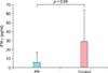
The results showed that the serum level of IFN-γ was significantly lower in PR patients (6.33±11.02 pg/ml) than in healthy controls (29.23±35.45 pg/ml) (p<0.05; Fig. 1), whereas the serum levels of IL-2, IL-4, and IL-10 were not significantly different between the two groups. Additionally, no statistical difference was observed in the serum levels of IL-2, IL-4, IL-10, and IFN-γ between the male and female patients and between patients who had PR for less than 3 weeks and those who had PR for 3 or more weeks (p>0.05).
To date, only a few studies that discuss the association of the Th1/Th2 immune response with PR have been published. A study showed that tumor necrosis factor-alpha inhibitors such as adalimumab could induce PR by downregulating the Th1 immune response3. In our study, a significant decrease in the serum level of IFN-γ was observed in patients with PR than in the healthy controls, but no statistically significant differences were observed in the levels of IL-2, IL-4, and IL-10 between the two groups. Serum IFN-γ is produced by activated CD4+ T cells and activated natural killer cells and might be the most sensitive marker of the CD4+ T cells response to HHV-6 infection4. Gangemi et al.5 found that the serum level of IL-22 was significantly higher in patients with an early stage of PR than in healthy controls and that IL-22 could limit the transmission of HHV-7 infection in PR by enhancing the production of proinflammatory and antimicrobial molecules. Moreover, diverse, fractalkine-mediated intracellular signaling pathways were involved in the pathogenesis of PR via the fractalkine receptor CX3CR1 in natural killer cells, monocytes, CD8+ T cells, and CD4+ T cells6.
The process by which CD4+ T cells produce IFN-γ and IL-22 is regulated by different signaling molecules. Recently, Qiu et al.7 first described the reciprocal relationship between IFN-γ and IL-22 production. IL-22 production is markedly enhanced when IFN-γ is absent. The detectable IL-22 mediates the up-regulation of CD27 expression in IFN-γ+ CD4+ T cells and thus affects their phenotype. Usually, acute viral infections induce an increase in the serum level of IFN-γ. Moreover, many studies have suggested the involvement of HHV infection in the pathogenesis of PR. Our findings seem to yield a contradictory result. We postulate that the decrease in the serum level of IFN-γ is most likely linked to the decreased number or impaired function of peripheral CD4+ T cells in the PR patients involved in this study.
In conclusion, we speculate that our study is the first to demonstrate a depletion in the serum level of IFN-γ and that a weakened Th1 response may contribute to the pathogenesis of PR. Further studies are needed to elucidate the alteration of CD4+ T cells in PR and whether the decreased serum level of IFN-γ and the elevated serum level of IL-22 have a synergetic influence on the development of this acute dermatosis.
 XML Download