

 PDF
PDF ePub
ePub Citation
 Print
Print


Dear Editor:
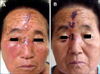
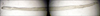
A 70-year-old Korean rural woman presented with a 7-day history of painful facial cutaneous lesions and severe ocular pain. Her general condition was good except for hypertension. Upon examination, there was erythema, vesiculobullous lesions and crusted erosions on the right side of the forehead and cheek. Also, there was right-sided periorbital edema confined to the cutaneous surface innervated by the ophthalmic division of the fifth cranial nerve (Fig. 1A). Because the Hutchinson's sign, ocular erythema, and foreign body sensation were present, she was referred to an ophthalmologist. Ophthalmologic examination showed perilimbal and conjunctival injection. No dendritic and inflammatory lesions were observed in the anterior chamber. Therefore, the diagnosis of herpes zoster ophthalmicus (HZO) was made. After admission, intravenous acyclovir (10 mg/kg) and analgesics were administered. However, despite the improvement in her cutaneous lesions and pain, the ocular pain and foreign body sensation persisted. At the second ophthalmologic examination, a chalky-white, 12.4×0.44 mm in size, thread-like worm was found in the right bulbar conjunctiva at the 8 o'clock position (Fig. 2) and it was removed. A parasitologist confirmed that the worm was an oriental eye worm. After 7 days, the initial signs and symptoms subsided. Only scattered crusted scars were observed (Fig. 1B).
HZO is defined as herpes zoster involvement of the ophthalmic division of the fifth cranial nerve. Hutchinson's sign is defined as skin lesions at the tip, side, or root of the nose, and it is a strong predictor of ocular inflammation and corneal denervation in HZO1. When the ophthalmic division is affected and Hutchinson's sign is present, unexplained painful ocular injection or visual problems can be observed. And serious visual impairment could also occur2.
Nematodes belonging to the genus Thelazia, commonly known as eye worms, infect orbital cavities and associated tissues of several mammals. The two species known to cause human thelaziasis are Thelazia callipaeda and Thelazia californiensis. Thelazia callipaeda is known as the 'oriental eye worm' because of its distribution in countries in the Far East, while Thelazia californiensis is found in the United States3,4. The final hosts of these nematodes include dogs and cats, but occasionally rabbits, monkeys, raccoons, foxes, and humans can also serve as the final host. When a fly licks the tear in the eye of a final host, the larvae enter the conjunctival sac, and become adults in 1 month after 2 molts5. The common symptoms of infestation are itching, tearing, foreign body sensation, and photophobia. Therefore, it is important to include human thelaziasis in the differential diagnosis of bacterial or allergic conjunctivitis. The optimal treatment is removal of the worm by using forceps3,4.
In this case, the cutaneous and ocular symptoms in the patient were not only due to HZO but also due to parasitic infestation. Because the Hutchinson's sign was present, we initially thought that her ocular pain was an inevitable symptom of HZO. However, these symptoms completely disappeared only after the removal of the eye worm.
In conclusion, we presented an unusual case of coinfection with HZO and oriental eye worm in a rural Korean woman. Human thelaziasis could cause ocular problems similar to HZO, too. Therefore, in cases of refractory HZO, a careful ophthalmologic examination should be considered to exclude other conditions including human thelaziasis, especially in patients living in a rural area or raising domestic animals.
 XML Download