

 PDF
PDF ePub
ePub Citation
 Print
Print


Dear Editor:
Superficial acral fibromyxoma (SAFM) is a rare, soft tissue tumor, which was initially described in 20011. SAFM is a relatively new entity, most likely representing a fibrohistiocytic neoplasm. This tumor presents as a slowly growing solitary nodule, usually confined to the dermis and subcutaneous tissue. It has a distinct predilection for finger nails and toe nails of young to middle-aged adults. Here, we report a case of SAFM occurring on the palm, which is very rare site.
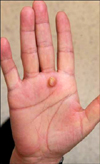
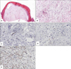
A 58-year-old Korean woman presented with a history of a nodular mass on the palm of her right hand (Fig. 1). The tumor had been present for several years and had gradually increased in size. There were no subjective symptoms, such as pain. The patient underwent shave excision of this tumor. The lesion measured 0.7×0.7 cm. It was dome-shaped, moderately circumscribed, firm, and had a uniform consistency. Histological examination showed a moderately cellular proliferation of spindled fibroblast-like and stellate cells embedded in myxoid matrix (Fig. 2A, B). The tumor cells were arranged in a loose storiform and focally fascicular pattern. Nuclear pleomorphism or mitotic activity was not present. Immunohistochemically, tumor cells were positive for CD99, negative for CD34 and epithelial membrane antigen (EMA) (Fig. 2C~E). At 6 months follow-up, there was no recurrence of the tumor.
SAFM typically is detected in adulthood as a solitary slow-growing mesenchymal mass involving the periungual and subungual regions of the fingers and toes. Hollmann et al.2 reported 124 cases of digital fibromyxoma; most tumors occurred on the digits, with the majority growing in close proximity to the nail (97% on fingers, 96% on toes). Occurrence on the palm is very rare. The histopathological characteristics of these tumors are distinctive, showing a moderately cellular dermal proliferation of spindle-shaped fibroblast-like cells in a myxoid or collagenous matrix. The differential diagnosis for SAFM includes a fibrous histiocytoma variant, dermatofibrosarcoma, cutaneous myxoma, acquired fibrokeratoma, sclerosing perineurioma, and the acral myxoinflammatory fibroblastic sarcoma. Immunohistochemical results of SAFM are controversial. Tumor cells have been reported positive for CD34 (21/23, 91.3%), EMA (18/25, 72%), and CD99 (11/13, 84.6%) in one study1, but positive for only CD34 (42/61, 68.8%) and EMA (3/40, 7.5%) in another study2. Recently, expression of CD10 was documented in a small series of 4 cases3. In our patient, tumor cells were positive only for CD99. The natural course of SAFM appears to be benign. However, transformation of a SAFM to a low-grade malignant tumor is possible. Thus, complete excision with adequate margins is mandatory. Furthermore, follow-up is necessary because of its slow growing nature and possible recurrence4.
We describe a patient with a SAFM occurred on the palm, which is the first case in Korea.
 XML Download