

 PDF
PDF ePub
ePub Citation
 Print
Print


INTRODUCTION
Basidiomycete yeasts Malassezia species (spp.) are cutaneous microflora and are considered as opportunistic pathogens. Malassezia are associated with various dermatological diseases including seborrheic dermatitis, dandruff and atopic dermatitis1,2. Among Malassezia spp., M. globosa and M. restricta were most frequently isolated from patients with dermatitis. Furthermore, M. restricta was known to be the predominant species among Korean teens and young adults3. Almost all Malassezia spp. are obligatorily lipid-dependent, which might be caused by lack of the myristic acid synthesis-the precursor of long chain fatty acids. The cell wall of Malassezia spp. contains significantly higher lipid contents than non-pathogenic fungi, such as Saccharomyces cerevisiae, implying that lipid-dependency of the fungus plays a role in virulence4. Recent genome analysis of M. globosa suggested that the absence of a gene encoding fatty acid synthase might be compensated by abundant genes encoding hydrolases that produce fatty acids. The genome of M. globosa possesses 14 lipases and 9 phospholipases, and the study showed that many of them were expressed on human scalp to use host lipids1. These results led us to investigate contributions of lipases and phospholipases in virulence of M. restricta as being the most frequently isolated Malassezia spp. within the Korean population. We searched the unannotated and incomplete M. restricta genome for a lipase and a phospholipase with M. globosa sequences and found three and one homologs respectively. The sequences of lipase and phospholipase homologs were used to design M. restricta specific primers for the current study. To investigate the expression of three lipases and a phospholipase of M. restricta, swap samples of two different body sites of at least 18 patients with seborrheic dermatitis were obtained, and the total RNA was directly extracted. Reverse transcription-polymerase chain reaction (RT-PCR) was performed and nested polymerase chain reaction (PCR) was carried out using M. restricta specific primers for lipases and phospholipases. Our results indicated that M. restricta was presented in body sites of patients and suggested that majority of the patients display expression of lipase RES_0242.
MATERIALS AND METHODS
Preparation of swap samples
Swap samples of two different body sites, forehead and cheek, from 18 patients with seborrheic dermatitis were obtained as described elsewhere5. Briefly, a rayon swap was rubbed multiple times across the body site selected. A swap was immediately placed in 1 ml Trizol (Life Technologies, Carlsbad, CA, USA) and snap-frozen in liquid nitrogen. Samples were melted after all swaps were collected and vortexed. Swaps were removed, and 0.2 g of glass beads was added. Samples were vortexed again and RNA was isolated as instructed by the manufacturer. All samples were treated with RNase-free DNase to eliminate possible genomic DNA contaminations. This study was approved by the Institutional Review Board of Konkuk University Hospital (KUH1120026).
Reverse transcription-polymerase chain reaction
Two-step RT-PCR using nested primers were used for the current study. cDNA was synthesized using RevertAid First Strand cDNA Synthesis Kit (Fermentas, Vilnis, Lithuania) and used as a template for the first round PCR. One micro-liter of the product of the first round PCR was used as template for the second round PCR using nested primers. Total 10 µl of the second round PCR was loaded on the 1.0% agarose gel, separated with electrophoresis and visualized. Gene specific primers used in the current study were listed in Table 1.
RESULTS
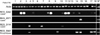
The recent genome sequencing data revealed that M. globosa possesses a total of 14 lipases and nine phospholipases. Among them, four lipases (MGL_3878, MGL_3507, MGL_0799, and MGL_0798) and two phospholipases (MGL_4252 and MGL_3326) were expressed on human scalps6. We used sequences of M. globosa lipases and phospholipases to search homologous genes in the M. restricta genome and identified three lipases and a phospholipase. Gene specific primers and nested primers were designed using sequences of these homologs (Table 1). Total of 18 patients, ten females and eight males with ages from 26 to 80 years old, with seborrheic dermatitis, were chosen to investigate whether the expression of M. restricta lipases or the phospholipases was detected on their body sites. From the 18 patients, we were able to obtain a total of 29 swap samples 12 forehead and 12 cheek swap samples from 12 patients, two forehead swap samples from two patients, and three cheek swap samples from three patients. Total RNA was extracted from the swap samples and was subsequently used as a template for cDNA synthesis. Two separate PCR, which was named as two-step nested RT-PCR in the current study, were carried out to detect expression of lipase or phospholipase homologs in M. restricta. Synthesized cDNA and gene specific primers were used for the first round PCR, and nested primers for each lipase or phospholipase homolog and products from the first round PCR were used for the second round PCR.
Using the two-step nested RT-PCR analysis, we were able to detect expression of the M. restricta ACT1 gene from the all swap samples except the cheeks of the patient 17. This result proved that M. restricta resides on the body sites of the patients tested in the current study. We also analyzed expression of lipases and phospholipases and found that 12 swap samples displayed expression of lipase RES_0242. Furthermore, our data showed that lipase RES_0571 was only expressed on the forehead of patient 13, and lipase RES_3114 was expressed on the forehead of patient 4 and on the cheeks of patient 7 and 10. A gene encoding phospholipase homolog RES_3947 was expressed on six swap samples (Fig. 1). Expression of the same lipases and phospholipases were also evaluated in the cells cultured in vitro. We found that lipase RES_0242 and lipase RES_3114 were not expressed in vitro and the results were summarized in Fig. 2. Thus, our data showed that majority of the patients displayed expression of lipase RES_0242 and suggested that the gene may play a significant role in the host environment such as contributing fatty acids production for M. restricta. Although it occurred from only three samples, lipase RES_3114 might also contribute to virulence since the gene was only expressed in vivo. Roles of phospholipase homolog RES_3947 and lipase RES_0571 were unclear because they were expressed in vitro.
DISCUSSION
Malassezia are lipophillic yeast and the recent genome sequencing analysis revealed that a fatty acid synthase gene is absent in the organisms. To compensate lipid dependency, Malassezia possess multiple lipases and phospholipases to generate lipids from the host. Among the 23 lipases and phospholipases identified from the genome of M. globosa, four lipases and two phospholipases were expressed on surface of the host6. These include lipase MGL_3057, lipase MGL_3878, lipase MGL_0799, lipase MGL_0798, phospholipase MGL_4252 and phospholipase MGL_3326. Lipase MGL_0799 was of particular interest because another independent study also identified it and indicated that the gene, designated as LIP1, is expressed on human skin. Although the whole genome sequence of M. globosa has been revealed and few studies carried out to analyze expression of lipases and phospholipases of the fungus, no information is currently available for M. restricta5,7-10. The recent study found that the most commonly identified Malassezia spp. is M. restricta rather than M. globosa in the seborrheic dermatitis patients-M. restricta was identified in 47.5% and M. globosa in 27.5%11.
In this study, we identified genes encoding lipases and phospholipases of M. restricta and investigated whether those genes are expressed on the body sites of patients with seborrheic dermatitis. Results of our study revealed that M. restricta possesses homologs of at least four lipases and a phospholipase of M. globosa, and they were expressed in some of swap samples obtained from the patients. Specifically, we found that lipase RES_0242 was detected most frequently in the swap samples while its expression was not detected in vitro. We should note that M. restricta lipase RES_0242 was the closest homolog of M. globosa Lip1, of which homolog was also identified in M. furfur7. The Lip1 protein belongs to triglyceride lipase (EC 3.1.1.3), which hydrolyzes the ester bond of triglycerides to release free fatty acids and is considered as a class 3 lipase. The protein has also been considered as a virulence factor in other pathogenic bacteria and fungi12. Lipase RES_3114 might also be interesting. Although only in three cases, our data suggested that lipase RES_3114 is regulated similarly to lipase RES_0242-it was only expressed in vivo. These data imply a possible role of lipase RES_0242, and, perhaps, lipase RES_3114 in the host environment to produce free fatty acids for the fungus. We, however, did not observe any correlation between expression of genes encoding lipases and phospholipases, and age, gender and a body site. Therefore, future study requires increased number of samples and more sensitive detection methods to evaluate expression of the genes.


 XML Download