

 PDF
PDF ePub
ePub Citation
 Print
Print


Dear Editor:
Of the several subtypes of pemphigus, pemphigus vulgaris (PV) is the most common1. PV is an autoimmune disease, is often associated with high concentrations of autoantibodies to constituents of the keratinocyte, and is characterized by loss of keratinocyte cell-cell adhesion or acantholysis. Desmoglein-3 (dsg3) has been identified as an important PV-associated autoantigen, and often an autoimmune response is directed initially against this antigen2. The acantholytic properties of dsg3 autoantibody sera of patients with PV have been described previously, but the molecular mechanisms by which these autoantibodies cause acantholysis has not been well characterized.
Heat shock proteins (HSPs) are highly conserved families of proteins acting as molecular chaperones. Hsp expression is increased in response to several environmental stresses as reviewed previously by Ghayour-Mobarhan et al.3 Hsp27 is a small HSPs with important roles in addition to being a molecular chaperone; it is a regulator of the structural integrity and stability of cell membrane, as reviewed by Kostenko and Moens4. Activation of p38 mitogen-activated protein kinase (MAPK) and subsequent phosphorylation of the Hsp27 appear to be involved in the pathogenesis of PV, leading to reorganization of the actin cytoskeleton and to keratin retraction5.
We hypothesized that because of the role of anti-Hsp27 antibodies in other autoimmune conditions and the function of Hsp27 in maintaining cell membrane integrity4, anti-Hsp27 antibodies may be involved partly in acantholysis seen in association with PV. Thus, we aimed to compare the levels of anti-Hsp27 antibody in serum of patients with PV with levels in healthy controls.
This case-control study was performed on new cases of patients with PV (who did not received immunosuppressive medication before study and their diseases were in active phase) aged 20~80 years who were recruited from the Ghaem and Imam Reza hospitals (Mashhad, Iran) between January 2007 and June 2008. Patients who had other autoimmune diseases, as well as old cases of PV (diagnosed for >1 year after the initiation of disease), were excluded; in final, 22 eligible patients were entered into the study. The diagnosis of PV was confirmed by clinical, histological and direct immunofluorescent findings. Twenty-two sex-matched healthy subjects who were not on any drug therapy, but who were referred to the Imam Reza clinic for assessment were recruited as the control group. Anthropometric parameters were assessed for all subjects. The study protocol was approved by Mashhad University of Medical Sciences Ethics Committee, and written informed consent was obtained from each participant. Serum Hsp27 antibody titers were measured using an in-house ELISA assay, as we have previously described6. The within-assay and between-assay precision was 3.5% and 5.2% respectively. Statistical analysis was performed using SPSS software (version 16, SPSS Inc., Chicago, IL, USA). Data were expressed as mean±standard deviation. Between-group comparisons were made using independent t-test (for numerical variables) or chi-square test (for categorical variables). A 2-tailed p-value of <0.05 was considered statistically significant.
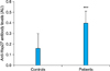
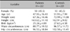
The demographic data and anthropometric parameters of both patients and controls have been summarized in Table 1. The proportion of females and age were the similar for the 2 groups (p>0.05). There was no significant difference in anthropometric parameters, including weight, height, body mass index, waist and hip circumferences (p>0.05) between the 2 groups. In the patient group, the mean Hsp27 antibody levels were 0.40±0.11 absorbency unit, while in control subjects, significantly lower levels of anti-Hsp27 antibody titers were observed (0.16±0.14 absorbency units; p<0.001) (Fig. 1).
The results support our hypothesis that anti-Hsp27 antibodies may be higher in patients with PV and could be involved in its pathogenesis. To our knowledge, this is the first study that investigates the association of anti-Hsp27 antibodies in patients with PV. Although the presence of IgG autoantibodies against dsg3 have been suggested to be the principal cause of PV2, the molecular mechanisms by which these autoantibodies cause acantholysis has not been well characterized. The immune response to some HSPs may occur due to what has been termed 'molecular mimicry' as we have previously reviewed3. Anti-Hsp27 antibodies have the potential to sequester Hsp27 antigen by forming immune complexes, and this has been previously proposed to occur following cardiovascular events7. It is possible that when keratinocytes are exposed to anti-dsg3 IgG, they release Hsp27 antigen and the antibodies that are subsequently produced form antigen-antibody immune complexes leading to clearance of Hsp27 from the circulation. It has been proposed that binding of pemphigus immunoglobulin (Ig) G to the keratinocyte activate intracellular signaling within the target keratinocyte, and this leads to acantholysis8. Recently, Berkowitz et al.9 reported that inhibitors of p38MAPK prevented PV IgG-induced phosphorylation of p38MAPK and Hsp27 leading to prevention of PV blistering disease in vivo, indicating the potential target of Hsp27 in the treatment of PV.
In conclusion, we found higher levels of anti-Hsp27 antibody titers in patients with PV, suggesting that these antibodies may have a role in the pathogenesis of PV. It would be important to study this association in a larger population sample and to investigate whether Hsp27 IgG or IgM antibodies have a pathological or protective role in the pathogenesis of PV10 and whether they are associated with disease severity, or other dermatological disorders. In vitro studies, investigating the effects of anti-Hsp27 antibodies on epithelial cells would also be of interest. It is not clear whether the Hsp27 antibody has role in the pathogenesis of PV or epiphenomenon, and this should be addressed in future studies.
 XML Download