

 PDF
PDF ePub
ePub Citation
 Print
Print


INTRODUCTION
Pityriasis versicolor is one of the most common disorders of pigmentation in the world. It is a cutaneous, superficial fungal infection characterized by skin pigmentary changes due to colonization of the stratum corneum by a lipophilic fungus in the normal flora of the skin, known as Malassezia furfur and M. globosa1-3. These Malassezia yeasts, discovered in 75~80% of healthy subjects, can cause pityriasis versicolor when the yeast converts to its mycelial form when disposed to certain predisposing factors, such as heat, moisture, occlusion, depressed cellular immunity status, and etc.4,5. Also, it has been suggested that skin pigmentary changes of pityriasis versicolor can occur as either hyperpigmented or hypopigmented lesions in accordance with interactions between Malassezia yeasts and skin characteristics, such as lipoperoxidation process, stimulus of inflammatory cell to melanocytes, and increased thickness of keratin layer6-8. However, the precise factors that enhance susceptibility to Malassezia yeasts and provoke pityriasis versicolor have yet to be completely defined.
To define these factors, we investigated skin characteristics of pityriasis versicolor patients against healthy subjects via non-invasive method, known as MPA 5®9 in this study. MPA5 is a non-invasive physiological parameter of the skin for evaluating the levels of sebum, skin hydration, transepidermal water loss (TEWL) and skin complexion. Although MPA 5® does have some advantages, such as non-invasiveness and faster detection of skin characteristics applicable to large subjects, it shows large variations depending on the specific body sites, temperature, humidity of measuring sites, and age, sex, and etc.10-14. Taking that into account, we established the normal range values of 90 healthy subjects in specific condition, and then compared the skin characteristics of pityriasis versicolor patients in the same condition before further proceeding with the study. Moreover, to investigate the correlation between specific Malassezia yeast and skin color changes in pityriasis versicolor, molecular biological evaluation of the skin lesion was carried out.
MATERIALS AND METHODS
Subjects
This study was approved by the ethnics committee of the Institutional Review Board.
The study group was composed of 63 men and 27 women of normal healthy subjects and 30 patients (21 males and 9 females) with pityriasis versicolor, who were diagnosed at the Department of Dermatology, Konkuk University Hospital from March 2008 through February 2009. The diagnosis of pityriasis versicolor was made by a positive KOH examination, showing typical hyperpigmented or hypopigmented scaly patches. History of treatment and duration of disease were not considered. Average age of normal healthy subjects was 38±11.8, and that of patients group was 32±4.4. Among the patients group, 21 patients had hyperpigmented skin lesions and 9 hypopigmented skin lesions.
The measurement of MPA 5®
1) The environment of measurement
All subjects washed off the areas to be measured with cleanser in the outpatient clinic, and was allowed to be assimilated to the conditions of constant temperature and humidity (Room humidity and room temperature 38~42%, 23~25℃) for 20 minutes before the measurement.
2) The items of measurement
(1) Skin lipid content
Skin lipid content was measured using Sebumeter® SM 815 (Courage and Khazaka, Cologne, Germany).
(2) Skin water content
Skin water content was measured by Corneometer® CM 825 (Courage and Khazaka), and the capacitance of the area of the skin with the highest coefficient of insulation was chosen for measurement, thus estimating the relative amount of water content. The average of the three measurements for each area was selected.
(3) TEWL
The amount of water evaporated from the skin surface was measured with Tewameter® TM 300 (Courage and Khazaka), and the value reflects the proportion of water vapor evaporating from the skin surface, per given unit of surface area (g/m2h) and time. Measurement was taken for one time for each area.
Molecular biological analysis of the pityriasis versicolor skin lesions
1) Sample collection and culture
Sterile cotton swabs were moistened with wash fluid containing 0.1% Triton X-100 in 0.075 M phosphate buffer (pH 7.9), and rubbed gently, with rotation. Swabbing was performed for 10 seconds on the skin lesion of pityriasis versicolor. Swabs were immediately placed in Leeming and Notman agar media and incubated at 34℃ for 14 days.
2) DNA extraction and polymerase chain reaction (PCR)
The yeasts grown in agar were harvested, and resuspended in 0.4 ml of lysis buffer (100 mM Tris-HCl pH 8.0, 1.0% SDS, 2.0% Triton X-100, 10 mM EDTA, 100 mM NaCl). Equal volumes of phenol/chloroform/isoamyl alcohol (phenol:chloroform:isoamyl alcohol=25:24:1, v/v) and glass beads (0.5 mm) were added, and the mixture was vortexed for 10 minutes. The samples were centrifuged at 12,000×g for 15 minutes. Total DNA was precipitated from the aqueous fraction with isopropranol and centrifuged at 12,000×g for 20 minutes at 4℃. The DNA pellet was washed in 70% ethanol and resuspended in sterile water.
3) 26S rDNA restriction fragment length polymorphism (26S rDNA RFLP analysis)
To amplify 26S rDNA from genomic DNA, reaction mixture contained 25 mM of each dNTP, 10X PCR buffer, 5X Q buffer, 0.5 µM primer, 0.4 µm forward primer (5'-TAACAAGGATTCCCCTAGTA-3', reverse primer (5'-ATTACGCCAGCATCCTAAG-3'), and 1.25 U Hot StarTaq polymerase in 50 µl reaction volume. 35 cycles with the following profile were programmed: denaturation for 45 seconds at 94℃; annealing for 45 seconds 52.5℃; extension for 1 minute at 72℃. After the confirmation of the amplified 26S rDNA, PCR (Veriti® 96-Well Fast Thermal Cycler. ABI) products were purified, using a LaboPass™ Gel (Cosmo, Seoul, Korea) and PCR Clean-up kit (Cosmo). Two restriction enzymes, Hha I (Koschem, Seoul, Korea) and BtsC I (NEB, London, England) were used to perform 26S rDNA-RFLP of Malassezia. In this experiment, the restriction enzyme digestion was performed with 10×PCR buffer, 10 U of the restriction enzyme, and PCR products 7.5 µl, which sum up to 20 µl. After the reaction at 37℃ for 3 hours, the electrophoresis was done with 3% (w/v) Seakem LE agarose gel (Takara Biomedicals, Otsu, Japan) by 100 volt and stained with ethidium bromide. Restriction fragments were analyzed with the size and number of DNA fragments under the UV transilluminator.
Statistical analysis
The measured values from the patient and control groups were compared and analyzed using SPSS ver 12.0 (SPSS Inc., Chicago, IL, USA). Comparative analysis of the interspecies differences and the experimentally derived values were carried out using independent samples t-test. For testing of statistical significance among the sites measurement, ANOVA test was used.
RESULTS
Skin characteristics of healthy subjects
A total of 90 healthy subjects without skin diseases (63 men [36±12.9] and 27 women [34±12.1] in their age 10~50) were included in this study, and the results obtained by non-invasive bioengineering method is as follows.
1) Skin lipid content (Sebum excretion)
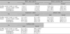
In the case of the cheek, skin lipid content was 64.2±8.6 in men, and 46.8±8.9 in women; and in the case of trunk, 62.9±22.1 in men and 43.2±19.6 in women; and in the case of the dorsum of hand, 18.7±6.3 in men and 16.0±4.7 in women. The values were significantly higher in men at the cheek and the trunk (p<0.05), and they were lower in the dorsum of hand, but not statistically significant (p>0.05). By the site, the lipid content was the highest in the cheek, followed by the trunk, and the dorsum of hand both in men and women (p<0.05) (Table 1).
2) Skin water content (Capacitance)
Skin water content was 58.3±11.9 in men and 45.2±16.5 in the case of cheek, and 52.0±19.7 in men and 40.6±20.9 in women in the case of the trunk, and in the dorsum of hand 49.3±18.2 in men and 51.1±16.4 in women, and the values were significantly higher in men compared to women in all body sites, except the dorsum of hand (p<0.05). By the body site, the values were highest in the cheek followed by the trunk and dorsum of hand in men, whereas in women, it was the highest in the dorsum of hand followed by the cheek and trunk (Table 1).
3) TEWL
TEWL was 15.3±4.2 in men and 13.2±3.8 in women in the case of the cheek, 14.6±7.9 in men and 11.4±5.1 in the case of the trunk, and 11.9±6.9 in men and 8.2±1.5 in women in the case of dorsum of hand. The values were higher in men, but not statistically significant (p>0.05). By the body site, the value was highest in the cheek, followed by the trunk and the dorsum of hand in both men and women (p<0.05) (Table 1).
4) Skin complexion (MI and EI)
MI and EI as measured by Mexameter® were 244.3±54.1/342.7±64.9 in men and 228.3±30.8/329.3±78.1 in women in the case of cheek, 224.1±58.0/320.7±83.4 in men and 211.6±82.0/304.5±106.4 in women in the case of trunk and 278.3±55.7/315.3±38.5 in men and 256.8±42.3/305.0±66.1 in women in the case of dorsum of hand. The values were higher in men in both melanin and erythema indices (p<0.05). By the body site, MI was highest in the dorsum of hand, followed by the cheek and the trunk in both men and women. In the case of EI, the values were highest in the cheek, followed by the trunk and dorsum of hand in men, and in women, they were highest in the cheek followed by the dorsum of hand and trunk (p<0.05) (Table 1). The measured values were higher in men in all body sites, a finding that was statistically significant (p<0.05). By the body site, the values were highest in the cheek, followed by the trunk and dorsum of hand in men, and in women, they were highest in the dorsum of hand followed by the cheek and trunk (Table 1).
The age ranged varied from the teens to sixties, and on comparing the average values of the patient group to the controls, the TEWL values were highest in the fifties, and in other indices, in the sixties.
Skin characteristics of pityriasis versicolor patients compared with healthy subjects
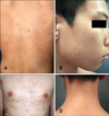
In the case of pityriasis versicolor patients group, age and sex matched 30 patients from their teens to their fifties (10~50 age [36±14.0]), who developed skin lesions in weeks to months before, hyperpigmented lesions were observed in 21 patients (38±14.2), and hypopigmented lesions in 9 patients (31±13.0) (Fig. 1). The results of comparison of the two clinical manifestations are as follows;
1) Skin lipid content (sebum excretion)
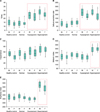
Skin lipid content in pityriasis versicolor patients with hyperpigmented lesion was 204.5±77.3 in men and 177.4±41.7 in women, and in hypopigmented lesion 211.7±81.5 in men and 177.8±40.1 in women, which was statistically significant in both types of lesions in the same sex group (p<0.05). In both types of lesions, the lipid content was higher in men (p<0.05) (Fig. 2), and the values were slightly higher in the hypopigmented lesion, which was not statistically significant (Table 2).
2) Skin water content (capacitance)
Skin water content in pityriasis versicolor patients with hyperpigmented lesion was 71.3±16.4 in men and 63.4±19.3 in women, and in the case of hypopigmented lesion, 62.8±16.9 in men and 50.5±19.9 in women, which was not a statistically significant increase in both types of lesions (p>0.05) (Table 2). The values were higher in the patient group in both types of lesions, but not statistically significant (Fig. 2).
3) TEWL
TEWL values in pityriasis versicolor patients with hyperpigmented lesion were 28.4±7.0 in men and 24.0±4.6 in women, and in the case of hypopigmented lesion, 24.1±6.2 in men and 21.7±6.7 in women. In both types of lesions, the values were higher in men but not statistically significant (p>0.05) (Table 2). The values were higher in the patient group in both types of lesions, a finding that was not statistically significant (p<0.05) (Table 2).
4) Skin complexion (MI and EI)
Melanin and erythema indices as measured by Mexameter® in pityriasis patients with hyperpigmented lesion was 318.5±41.2/474.6±70.2 in men and 307.4±32.1/451.8±71.1 in women, and in the case of hypopigmented lesion, 206.7±33.2/312.0±54.3 in men and 200.9±31.9/305.6±54.1 in women, a finding that was not statistically significant (p>0.05) (Table 2). Also, there was statistically significant increase in the case of hyperpigmented lesions only in the patient group compared to the control group (p<0.05) (Fig. 2).
The incidence of species of Malassezia yeasts in hyperpigmented and hypopigmented groups
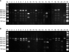
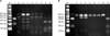
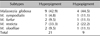
On RFLP performed to identify the difference between the incidences of species of Malassezia yeasts in pityriasis versicolor patients with hyperpigmented and hypopigmented lesions, there was no significant difference and five species, i.e., M. globosa, M. restricta, M. furfur, M. sympodialis, and M. Slooffiae were isolated (Fig. 3, 4). Among these, M. globosa was most frequently isolated in all body sites, followed by M. restricta, M. furfur, M. slooffiae, and M. sympodialis (Table 3).
DISCUSSION
Malassezia yeasts are normal flora of the skin that are discovered in 75~80% of health subjects, and are classified into 7 species, M. furfur, M. pachydermatis, M. sympodialis, M. globosa, M. obtusa, M. restricta and M. slooffiae in 1996 by Guého et al.15. Recently, on the basis of DNA-relatedness, through the molecular biology, four new species have been included: M. dermatis, M. japonica, M. nana and M. yamatoensis16-21. In Europe, M. caprae and M. equina were additionally isolated, and now Malassezia yeasts are classified into 13 species22. Among these Malassezia yeasts, M. furfur has long been identified as the fungus causing pityriasis versicolor, and recently it was known that M. globosa was also associated with a vast majority of lesions3,23,24.
The specific condition that opportunistic Malassezia yeasts induce pityriasis versicolor was reported as exogenous and endogenous factors. The exogenous factors include heat and moisture, a probable reason why the disease is more prevalent in the tropics and in the summer in more temperate climates. Another exogenous factor may be occlusion of the skin by either clothing or cosmetics2. Occlusion results in increased carbon dioxide concentration25, an altered microflora and pH range. Infection has been experimentally induced by an occlusive dressing26. Otherwise, endogenous factors account for the prevalence of the disease in temperate climates. Seborrheic dermatitis, Cushing's syndrome, immunosuppressive treatment (depressed cellular immunity), malnutrition, and hyperhidrosis (particularly flexural) have all been implicated as endogenous factors27-29.
Clinical manifestation of pityriasis versicolor is scaly hypopigmented or hyperpigmented macules observed in characteristic areas of the body, including the chest, back, abdomen, and proximal extremities4,5. The variation in pigmentation, despite being the same lesion, can be explained by the interaction between skin barrier components and Malassezia yeasts, such as lipoperoxidation process causing cytotoxic effect to make hypopigmented patches, increased keratin layer thickness or stimulation of melanocytes by inflammatory cells to make hyperpigmented patches6-8.
Noninvasive methods as the means of assessing the characteristics according to sex, age and body site are being explored by many investigators. Noninvasive methods are more objective than gross inspection and the measurement is handy and more valuable in that it leaves the skin intact9,30.
In this study, to evaluate the differences of skin characteristics between pityriasis versicolor patients and normal healthy subjects, we investigate the degree of water preservation, according to sex and age in healthy adults (Corneometer®), TEWL (Tewameter®), sebum excretion rate (Sebumeter®), MI and EI (Mexameter®) using non-invasive bioengineering methods, known as MPA 510-14.
Previous studies by Koh et al.9, Conti et al.31 measuring the physiologic factors of the skin in healthy adults differed from each other, according to sex, age and the site of measurement. In this study, the lipid content was higher in men in the body sites, such as the cheek and trunk in healthy controls, a finding that is consistent with the results obtained by Conti et al.31 and also the values of skin water content and TEWL values were higher in men, which was consistent with the results obtained by Conti et al.31. Moreover, the melanin and erythema indices as measured by Mexameter® were higher in men in all body sites, which is in agreement with results obtained by Koh et al.9.
In this study the sites of measurement in 30 pityriasis versicolor patients with hypopigmented and hyperpigmented lesions were the cheek, chest, abdomen and the back, and on measuring the values on the corresponding areas in the controls, the capacitance, reflecting the degree of water preservation, was not significantly different, but in the case of hyperpigmented lesion, TEWL, sebum excretion rate, MI & EI values differed significantly7,15,32, and in the case of hypopigmented lesion, there were statistically significant differences in TEWL and sebum excretion rate. Also, all the values were higher in the patient group, except MI and EI of pityriasis versicolor patients with hypopigmented lesions6,8.
Our results show that pityriasis versicolor skin lesion has higher humid status, higher sebum excretion rate and increased TEWL values than those of normal healthy subjects. And, there was no significant differences between hyperpigmented and hypopigmented skin lesion. These results indicate that higher humidity and sebum excretion of the skin will makes well growing status of Malassezia yeasts, and presume that interaction between Malassezia yeasts and skin barrier materials makes disruption of skin barrier causing increased TEWL. Therefore, hydration and sebum excretion rate are cause of pityriasis versicolor, and TEWL is the result of pityriasis versicolor. Moreover, given the increase in TEWL in both hyperpigmented and hypopigmented lesions, it can be thought that hyperpigmented lesions are induced by the stimulation of melanocytes by inflammatory cells, rather than increase in the thickness of the keratin layer6-8,33.
Also, on RFLP performed to identify the difference between the species of Malassezia yeasts in pityriasis versicolor patients with hyperpigmented and hypopigmented lesions, there was no significant difference and five species, i.e., M. globosa, M. restricta, M. furfur, M. sympodialis, and M. Slooffiae were isolated (Fig. 3, 4). Among these, M. globosa was most frequently isolated in all body sites, followed by M. restricta, M. furfur, M. sympodialis, and M. slooffiae. The findings did not differ significantly from previous studies on isolation of the types of Malassezia yeasts from the lesions of pityriasis versicolor24,33,34.
In turn, the species of Malassezia yeasts has not a decisive effect on the difference in skin color between hyperpigmented and hypopigmented lesions, but according to the reported literatures, the interactions between Malassezia yeasts and skin barrier materials, which makes lipoperoxidation causing hypopigmentation or stimulation of melanocytes by inflammatory cells causing hyperpigmentation. Our limitations in this study include the fact that we had a relatively low population of pityriasis versicolor patients, compared to the normal healthy subjects. Further, it is needed that the additional studies of the interactions between Malassezia yeasts and skin barrier materials, which make lipoperoxidation or stimulation of melanocytes. Future additional studies will hopefully be successful in ascertaining these differences and exact value ranges of MPA 5®.

 XML Download