

 PDF
PDF Citation
 Print
Print


INTRODUCTION
Port wine stains (PWS) are vascular ectasias that can develop thickening or nodules over time. These nodular or warty outgrowths are called blebs. Introduction of the concept of selective photothermolysis with the 577 nm and 585 nm pulsed dye laser (PDL) has revolutionized PWS treatment1. This theoretic concept of selective heating and destruction of the capillary endothelium was supported by evidence from early clinical studies with PDL. Although findings from these studies suggested that close to 100% lightening could be produced with very little risk of long-term sequelae, such as scarring and pigmentary change2,treatment of blebbed PWS using a PDL may be insufficient because penetration of the PDL exploiting wavelengths is too shallow for ablation of blebs with the usual size. We demonstrate the efficacy of a 1,064 nm long pulsed Nd:YAG laser equipped with contact cooling in treatment of blebbed PWS of four patients.
CASE REPORT
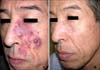
Case 1
A 53-year-old man presented with several asymptomatic violaceous nodules with surrounding erythematous flat plaques on the left cheek (V2 distribution of a trigeminal location) (Fig. 1A). The patient was treated by partial excision of blebs at other local clinics several years ago. The patient was not cosmetically satisfied with the excision scar. We first performed treatment using a 1,064 nm long pulsed Nd:YAG laser with contact cooling (COSJET SR®, Won Technology, Daejeon, Korea) at a pulse duration of 30 ms, 130 J/cm2, and a 6-mm hand piece, followed by treatment of surrounding flat lesions with a 595 nm PDL (V beam perfecta®, Candela corp, MA, USA) at a pulse duration of 1.5 ms, 10.5 J/cm2 at 8-week intervals. A 1,064 nm long pulsed Nd:YAG laser pass was used with multiple non-overlapping laser pulses and 595 nm PDL was used with an overlap of 10% to 20% of the area in adjacent spots. Topical anesthetic cream (L.M.X.4®, Ferndale Laboratories, Inc., Michigan, USA) was applied to the lesion before laser treatment. After two sessions of treatment using a 1,064 nm long pulsed Nd:YAG laser with contact cooling, blebs showed almost complete improvement. Next, after five sessions of PDL, surrounding flat lesions showed over 90% lightening (Fig. 1B). After treatment, purpura was observed as an immediate reaction; however, it was mild and transient.
Case 2
A 23-year-old man presented with four asymptomatic reddish nodules with surrounding erythematous patches on the right cheek (V2 distribution of a trigeminal location). The patient had never undergone treatment before. In the same order as above, we first administered treatment using a 1,064 nm long pulsed Nd:YAG laser with contact cooling at a pulse duration of 40 ms, 50 J/cm2, and a 10-mm hand piece and surrounding lesions were treated with four sessions using 595 nm PDL at a pulse duration of 1.5 to 3 ms, 12 J/cm2. The patient was treated with one session using a 1,064 nm long pulsed Nd:YAG laser with contact cooling for blebs and four sessions of PDL for treatment of surrounding flat lesions. All blebs had almost disappeared by the end of the session. Adverse effects were mild and well tolerated.
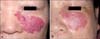
Case 3
A 60-year-old woman presented with five asymptomatic purpuric nodules with surrounding erythematous flat plaques on the left cheek (V2 distribution of a trigeminal location) (Fig. 2A). The patient had received treatment with PDL only when she was young; however, lesions remained and blebs occurred. The same as case 1, first, we administered treatment using a 1,064 nm long pulsed Nd:YAG laser with contact cooling at a pulse duration of 30 ms, 110 J/cm2, and 6-mm, and surrounding flat lesions were treated with 595 nm PDL at a pulse duration of 1.5 ms, 10.25 to 10.5 J/cm2. After three sessions of treatment using a 1,064 nm long pulsed Nd:YAG laser with contact cooling, blebs had disappeared and flat lesions showed approximately 70% lightening(Fig. 2B). An immediate purpura and edema were observed but were resolved without sequelae within 6 days.
Case 4
A 39-year-old woman presented with asymptomatic violaceous plaques with deep purple nodules over the left temple, cheek, postauricular area, and neck (V3 distribution of a trigeminal location). We administered two sessions of treatment using a 1,064 nm long pulsed Nd:YAG laser with contact cooling at a pulse duration of 30 ms, 150 J/cm2, and a 6-mm hand piece. After the end of treatment, lesions showed approximately 80% lightening. Purpura and edema formation was observed; however, it was mild and only transient.
DISCUSSION
PWS are the most common of all congenital vascular malformations, with an incidence of three per thousand live births; they occur most commonly on the face and neck, and are therefore highly visible3. These can be associated with significant cosmetic disfigurement and psychological distress. PWS persist throughout the lifetime of the patient and progress with aging, particularly showing raised, irregularly surfaced, and deeply colored skin abnormalities. Cobblestone-like, blebbed PWS occur over time.
Histologically, PWS demonstrate ecstatic dilatation of normal numbers of capillaries of diameters ranging from 10 to 150µm with normal endothelium, in the papillary and mid dermis at depths of 300 to 600µm4,5.
The most commonly used lasers employ a wavelength that selectively targets oxyhemoglobin and results in intravascular coagulation, and subsequent color fading, while leaving the surrounding skin undamaged. This process is known as selective photothermolysis1. Many clinicians consider the PDL to be the treatment of choice for port wine stains. After a series of PDL exploiting wavelengths ranging 585 to 600 nm, although almost all PWSs lighten, blebbed PWS cannot be removed completely because of the limited penetration depth. Different strategies have been employed for treatment of resistant PWS; these include repetitive PDL treatments, pulse stacking with PDL, use of intense pulsed light platforms, and use of 1,064 nm Nd:YAG6,7.
The long-pulsed Nd:YAG laser is deeply penetrating into skin and can be effective in treatment of vascular targets, such as nodular lesions in port wine stains and reticular leg veins8,9. By protecting the epidermis, cooling allows for safer use of higher radiant exposures, permits treatment of patients with darker skin types, decreases treatment pain, and enhances therapeutic outcome. More recently, sequential use of the dual-wavelength PDL (595 nm) and Nd:YAG (1,064 nm) lasers has been attempted and reported to be safe and more effective10-13. However, mass destruction of papillary dermal capillaries by short wavelength PDL may cause buildup of visible light impenetrable fibrous tissue in papillary dermis, preventing laser light at subsequent treatment sessions from penetrating to deeper capillaries14. This could potentially be remedied by initial use of a longer wavelength, more deeply penetrating laser, such as the 1,064 nm Nd:YAG, with selective cooling of the epidermis and papillary dermis, followed by laser treatment sessions with shorter wavelength, less deeply penetrating lasers, such as the PDL, for treatment of more superficial vessels.
We have treated most blebs using a 1,064 nm long pulsed Nd:YAG laser with contact cooling in four patients. Treatment of blebs with the Nd:YAG laser has been conducted at 30 ms, 120 to 150 J/cm2 and 5 to 6 mm spot and surrounding flat lesions were treated with 595 nm PDL at a pulse duration of 1.5 to 3 ms and 10 to 12 J/cm2. According to their size, most blebs require three or fewer treatment sessions at 8-week intervals. Treatments were well tolerated by four patients and showed good improvement of blebs. No complications, excepting trivial textural change, were observed. A 1,064 nm long pulsed Nd:YAG laser with contact cooling may be comparable to the dual wavelength laser in treatment of blebbed PWS.
 XML Download