

 PDF
PDF ePub
ePub Citation
 Print
Print


INTRODUCTION
In 1923, Pasini1 described a case of pigmentary atrophoderma that was both clinically and histologically unique from any other known atrophy, including localized scleroderma, under the name of progressive idiopathic atrophoderma. In 1936, in Argentina, Pierini and Vivoli2 extensively studied and defined the condition and its possible link to morphea. In 1958, the disorder was first introduced into the American dermatologic literature by Canizares et al.3, who reviewed Pierini's findings and renamed the disorder the idiopathic atrophoderma of Pasini and Pierini. They believed that the idiopathic atrophoderma of Pasini and Pierini differed sufficiently from morphea to classify it as a distinct entity. The clinical appearance of atrophoderma of Pasini and Pierini has been likened to "footprints in snow" or depressions with "cliff drop" borders4. The trunk is the most commonly involved site, especially the back and the abdomen5. The cause of atrophoderma of Pasini and Pierini is not known yet. Some authors have suggested a role for the infection with Borrelia burgdorferi6. We report a case of atrophoderma of Pasini and Pierini, associated with Borrelia burgdorferi infection and successfully treated with oral doxycycline.
CASE REPORT
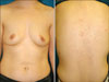
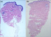
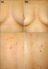
A 35-year-old woman presented with a three-month history of asymptomatic, hypopigmented, depressed patches on her chest and back. The lesions developed after a mountain climbing trip three months prior and became aggravated in the three weeks prior to her presentation at the clinic. She suffered from psoriasis vulgaris and was undergoing treatment with topical steroids and narrow-band ultraviolet B phototherapy. Physical examination revealed numerous skin lesions, composed of variably-sized round- to oval-shaped, hypopigmented patches on the intermammarial area and on the back, along with preexisting psoriatic lesions. The lesions were depressed below the level of the surrounding skin and coalesced to form large depressed areas (Fig. 1). Their distinct margins and 1~4-mm depressions gave them the typical "cliff-drop" appearance. There was no induration, sclerosis, or symptoms. Laboratory investigations, including a complete blood count, liver function tests, urinalysis, and electrolytes were all within normal limits. However, the serum IgM antibodies for Borrelia burgdorferi were positive according to three different test approaches (i.e., the indirect fluorescence assay, the emzyme-linked immunosorbent assay and Western blot) (Table 1). Skin biopsies were taken from one of the atrophic lesions and from perilesional normal skin on the back. The atrophic lesion showed no epidermal abnormalities, but had a markedly thinned dermis, compared to that of the perilesional normal skin (Fig. 2). Based on the clinical and pathologic findings, the patient was diagnosed with atrophoderma of Pasini and Pierini, associated with Borrelia burgdorferi infection. She underwent treatment with oral doxycycline 200 mg/day for three weeks, and the depressed depths of the lesions improved (Fig. 3). She took oral doxycycline 200 mg/day for an additional three weeks after that, but there was no further improvement. Her lesions were deemed stabilized, and the treatment was ended.
DISCUSSION
Atrophoderma of Pasini and Pierini is a form of dermal atrophy that manifests as either single or multiple, sharply demarcated, hyperpigmented, non-indurated patches. These patches are marked by a slight depression of the skin with an abrupt edge (i.e., the "cliff-drop" borders), usually located on the backs of adolescents or young adults. The lesions may be discrete or confluent, and the affected skin appears thinned and discolored, but the consistency and feel of the affected skin remains normal6. Distribution is often symmetric and bilateral; however, reports have described solely unilateral cases7,8. The lesions have been traditionally described as hyperpigmented; however, Saleh et al.9 described a retrospective study of 16 Lebanese patients in whom the lesions were rather hypopigmented (56%) and skin-colored (25%). The histopathologic changes, often minimal and non-diagnostic, consist of a decrease in the size of the dermal papillae, with flattening of the rete pegs. The epidermis is usually normal or slightly atrophic. Melanin is increased in the basal layer, and interstitial edema and a mild perivascular infiltrate, consisting of lymphocytes and histiocytes, may be present. The collagen bundles show varying degrees of homogenization and clumping in the mid and reticular dermis, with a normal papillary dermis. When compared with adjacent normal skin, the dermal thickness is reduced. The sweat glands and the pilosebaceous units are not affected6,10.
The cause of atrophoderma of Pasini and Pierini is not known yet11. Buechner and Rufli6 studied the sera of 26 patients with typical atrophoderma of Pasini and Pierini lesions. Ten (38%) of the 26 patients had significantly elevated titers of IgG anti-Borrelia burgdorferi antibodies (1:128 or higher). However, none of the patients had an elevated IgM titer. Six (14%) of 43 control subjects, i.e., healthy volunteers with no history or symptoms of Borrelia infection, had a reactive IgG titer.
In our case, the lesions were found after the patient had gone mountain climbing, and the IgM antibodies for Borrelia burgdorferi were positive in three different test approaches. We analyzed the serum IgM and IgG antibodies for Borrelia burgdorferi on the first and final days of oral doxycycline treatment (Table 1). The results were identical to those of the first clinic visit, i.e., there were no interval changes in either the IgM or the IgG titer. Results did not meet the positive criteria for Lyme disease, defined as an increase in IgG titer of more than four-fold, but this could be due to the antibiotic treatment. Moreover, the IgM antibodies were constantly positive in the three test approaches. Therefore, we concluded that the patient had Borrelia burgdorferi infection, which contributed to the atrophoderma of Pasini and Pierini.
No treatment is consistently effective for atrophoderma of Pasini and Pierini, but some patients respond to topical corticosteroids, antibiotics, or antimalarials. Nevertheless, the results of medical treatment with antibiotics have been inconclusive. In patients with new, early-stage, idiopathic atrophoderma of Pasini and Pierini, especially those with a positive Borrelia burgdorferi antibody titer, the standard recommended therapy for Lyme disease is suggested. A retrospective evaluation of 25 patients treated with either oral penicillin (2 million IU/d) or oral tetracycline (500 mg three times/day) for 2~3 weeks showed clinical improvement, with no new active lesions in 20 patients (80%). The same study also showed no progressive disease in four of six patients who did not receive treatment6.
In this case, the patient underwent treatment with oral doxycycline (200 mg/d) for six weeks. After the six-week treatment, there were no new lesions, and the depths of the depressions in the pre-existing lesions were improved. Herein, we report a case of atrophoderma of Pasini and Pierni associated with Borrelia burgdorferi infection, which clinically improved with oral doxycycline.

 XML Download