

 PDF
PDF ePub
ePub Citation
 Print
Print


INTRODUCTION
An increase in melanin content of cells after ultraviolet irradiation is due to increased numbers of melanocytes and the activity of tyrosinase. There are several chemical compounds that reduce melanocyte proliferation and inhibit melanogenesis, such as hydroquinine, compounds of catechol and phenol (monobenzylether of hydroquinone), trans-retinoic acid, azelaic acid, arbutin, kojic acid, pentadecenaoic acid, oleanolic acid, ursodeoxycholic acid, and medimin C. The pigment inhibitory effect has been reported with trans-retinoic acid, azelaic acid, arbutin, kojic acid, pentadecenaoic acid, oleanolic acid, ursodeoxycholic acid, and medimin C1-5; however, their use has been limited due to skin irritation and complications6-10. Recently, therapeutic demands for the treatment of hyperpigmented lesions for esthetic purposes have gradually increased in the field of dermatology. There is a greater need for drugs which are effective, simple, and with fewer adverse reactions in the prevention and treatment of hyperpigmented lesions8,10. Arbutin has been suggested to be melanogenic inhibitory compound because of its chemical and biological similarities with hydroquinone and the tyrosinase inhibitory effects. In a previous screening with mushroom tyrosinase, we showed that phytoclear-EL1, an extract from Euphorbia lathyris seeds, had melanogenic inhibitory activity11-14. There have been many reports regarding the melanogenic inhibitory effect in arbutin, but such reports in phytoclear-EL1 are rare. The aim of this study was to investigate the inhibitory effect of phytoclear-EL1 on melanogenesis in vitro and in vivo. In order to evaluate the pigment inhibitory effect of this extract, the number of melanocytes, melanin content, and tyrosinase activation were measured after this extract had been added to cultures of melanoma cells. Thirty subjects were included in this study to apply phytoclear-EL1 on areas with UVB-induced hyperpigmentation.
MATERIALS AND METHODS
In vitro experiments
Tests were subdivided as follows: 1) normal control; 2) phytoclear-EL1 (5µg/ml)-administered group; and 3) positive control (arbutin [10-3M]).
B-16 melanoma cells were sub-cultured 10 times in Dulbecco's modified Eagle medium, to which 10% fetal bovine serum (FBS) was added. Cultured B-16 melanoma cells were inoculated into each well of the 6-well tissue culture microplates (Costar, Cambridge, MA, USA) at a density of 5×10⁴ cells per well. After culturing each test group for 4 days, arbutin (Sigma Co., St. Louis, MO, USA) and phytoclear-EL1 (LG Chemical Co., Daejeon, South Korea) was added to the culture fluid. The cultures were incubated for 3 and 5 days in a 5% CO2 incubator at 37℃. Both arbutin and phytoclear-EL1 were added at concentrations of 10-3 M and 5µg/ml, respectively, to the culture fluid once every 48 hours.
Cell count and observation of morphologic changes
The cell suspensions were obtained by treating a 0.25% trypsin-EDTA solution 3 and 5 days after drug administration. Then, the cultured cells were collected and the cell count was measured using a hematocytometer. The morphologic changes were observed using a handstand phase shift microscope (Diaphot, Nikon, Japan). Five measurements were obtained for each cell count and these measured values were averaged. A total of six tests were carried out and the standard deviation and mean value for these tests were obtained.
Measurement of melanin content4,5,11,15
One ml of 1N NaOH per 1×10⁶ cells was added to the remaining cellular precipitate, and this specimen was reacted at 37℃ for 48 hours. After isolating melanin from the cells, 1 ml of distilled water was added and mixed well. Light absorption was measured at 400 nm using a UV-Vis spectrophotometer. Then, the amount of melanin was calculated by comparison with the standard straight line prepared using synthetic melanin (Sigma Chemical Co.). A total of six tests were performed and the standard deviation and mean values were obtained.
Measurement of tyrosinase activity in situ5,16
Cultured B-16 melanoma cells were plated into 60-mm culture dishes at a density of 5×10⁴ cells/ml. After 24 hours, each experimental substance was added to the culture fluid. After 48 hours, 1µCi/ml [3H]tyrosine (GE Healthcare, Cardiff, UK) and 0.1 mM L-DOPA were added. The supernatant culture fluid was obtained after 24 hours and the amount of 3H2O was obtained using a modified version of the Pomerantz charcoal adsorption. A total of six tests were performed and the standard deviation and means were obtained.
In vivo experiments
Informed consent was obtained from all the subjects after they were fully informed about the details and potential risk of the study, and the study was performed with strict adherence to the principles of the Declaration of Helsinki.
Subdivision of subjects
The tests were subdivided as follows: 1) positive control (3% arbutin), group A; 2) 0.2% phytoclear-EL1, group B; 3) placebo, group C; and 4) placebo, group D. This study included 30 healthy males who had no medical history of phototoxic or photosensitive diseases, and had no sun light exposure to the back area and the medial aspect of one proximal arm for the past 6 months. The mean age was 24.6 years. For each subject, a flat area of uniform skin tone was divided into four sections on the medial aspect of one proximal arm. The test groups were as follows: A) positive control group; B) phytoclear-EL1; and C) and D) placebo groups. A Philips TL 20W/12 UVB lamp, which emits UVB with a wavelength range of 290~320 nm, was used for the light source. UVB irradiance was measured using a UV Centra Radiometer (Osram GmbH, Berlin, Germany). A Chromameter CR-300 (Minolta Co., Osaka, Japan), with a pulsed xenon arc lamp placed in the measured area with a diameter of 8 mm as its light source, was used to gauge the light reflected perpendicularly from the surface and to analyze chromaticity of the epidermis from a diffusion light illuminating perpendicularly. The measured values were expressed as shown in the L*a*b* Coordinate System devised by Commission Internationale de l'Eclairege in 1976. In this coordinate system, L values designate lightness, while a* and b* point to chromaticity6. In this study, only L values were used to compare the extent of pigmented skin. For in vivo experiments, arbutin and phytoclear-EL1 were applied at concentrations of 3% and 0.2%, respectively.
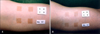
Drug application after ultraviolet irradiation (Fig. 1)
The back area of the subjects was divided into small sections (1×1 cm). Then, using a Philips UVB TL 20w/12 as the light source, the light intensity was increased from 30 mJ/cm2 to 100 mJ/cm2 with a gradual increment of 10 mJ/cm2. T minimal erythema dose (MED) of the subjects was defined as the site that had distinctive margins and complete erythema after 24 hours of irradiation. For each subject in whom a MED value was decided, the skin area on the inner surface of the arm was covered and fixed with a black cloth with 4 holes (2×2 cm). Then, 2 MED of UVB was irradiated. Two or more researchers evaluated the extent of erythema after 16~24 hours, and decided on whether re-evaluation was required. Once the maximal pigmentation value was reached using a chromameter and visual observation, each drug was applied twice daily on the areas tested. Also, the researchers and subjects had no knowledge of which experimental substance they were treated with until the evaluation was completed (double blind tests were used).
Decision on the extent of pigment suppression
The researchers and subjects rated the inhibitory effects of the experimental substance at the 3rd and 7th weeks, and classified the extent into 4 categories as follows: -, no effect; +, mild/slight effect but cannot be classified visually; ++, moderate effect with visual improvement; and +++, remarkable effect that can be identified visually. To compare the subjective results among these groups, the -, +, ++, and +++ levels were given scores of 0, 1, 2, and 3, respectively. Using a chromameter, L*, a*, and b* values were taken to measure the extent of pigmentation on the UVB-irradiated areas of these subjects before, the 3rd and 7th weeks after topical drug application. The patient's symptoms were noted on each visit.
RESULTS
In vitro experiments
1) Change in the number of B-16 melanoma cells (Fig. 2)
In contrast to the normal control, the number of B-16 melanoma cells in the 5µg/ml phytoclear-EL1 group and the positive control (10-3M arbutin) showed a significant reduction 5 days after culture (p<0.05). There was no significant difference between the positive control (10-3M arbutin) and the experimental group (5µg/ml phytoclear-EL1). No morphologic cellular differences were observed among the experimental group, normal control, and positive control.
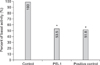
2) Change in the amount of melanin (Fig. 3)
The amount of melanin in the experimental group (5µg/ml phytoclear-EL1) and the positive control (10-3M arbutin) showed a significant decrease (p<0.05) compared to the normal control. There was no significant statistical difference between the experimental group (5µg/ml phytoclear-EL1) and the positive control (10-3M arbutin).
3) Tyrosinase activity changes (Fig. 4)
Both the 5µg/ml phytoclear-EL1 group and the positive control (10-3M arbutin) showed a significant statistical decrease (p<0.05) compared to the normal control; however, there was no significant statistical difference between 10-3M arbutin and the experimental group (5µg/ ml phytoclear-EL1).
In vivo experiments
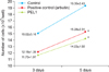
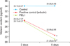
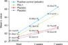
1) Objective evaluation (Fig. 5)
To determine the extent of the pigment inhibitory effect of each group after topical application of the drug, the mean L value was measured prior to topical application and at the 3rd and 7th weeks after application. ΔL denotes the difference between the L value measured before topical application and at 7 weeks. Among the mean ΔL values of the positive control (3% arbutin [group A]), the experimental group (0.2% phytoclear-EL1 [group B]), and the placebo groups (groups C and D), both the positive control (group A) and the experimental group (group B) showed a larger value as compared to the placebo groups (groups C and D; p<0.05), and the mean ΔL value of the experimental group was similar to the positive control group (3% arbutin [group A]).
2) Subjective evaluation of the researcher (Table 1, 2, 3)
In the subjective evaluation by the researchers 3 weeks after topical application, the score of the 0.2% phytoclear-EL1 group was 18, showing a significant difference from the placebo group. The group scores obtained 7 weeks after topical application were B > A ≫ C > D in decreasing order. The scores of the positive control group (3% arbutin [group A]) and the experimental group (0.2% phytoclear-EL1 [group B]) were higher than the control groups (groups C and D). The score for the experimental group (0.2% phytoclear-EL1 [group B]) was 40, which was the highest.
3) Subjective evaluations by the subjects (Table 1, 2, 3)
In the subjective evaluation by the subjects 3 weeks after topical application, the score of the subjective evaluation showed that the experimental group (0.2% phytoclear-EL1 [group B]) was 14, while the placebo (C) group was 12, showing no significant difference. However, the group scores obtained 7 weeks after topical application were A > B ≫ C > D. The score of the experimental group (0.2% phytoclear-EL1 [group B]) was 35, showing significant differences from the placebo groups (groups C and D).
4) Subjective evaluations by both researchers and subjects (Table 1, 2, 3)
Subjective evaluation scores by researchers and subjects were summed up and compared among each group. The subjective evaluation scores of the researchers and subjects 3 weeks after topical application was 32 for the experimental group (0.2% phytoclear-EL1 [group B]), showing a significant difference from that of the placebo groups (groups C and D). The group scores obtained 7 weeks after topical application were A > B ≫ C > D. The scores of the experimental group (0.2% phytoclear-EL1 [group B]) and the positive control (3% arbutin [group A]) showed higher scores than the placebo groups (groups C and D). The score of the experimental group (0.2% phytoclear-EL1 [group B]) was 75, while that of the positive control (3% arbutin [group A]) was 77, showing no significant difference. During the experimental period, there were three subjects who showed irritative symptoms and other adverse reactions. Two patients (7%) complained of pruritus and 1 patient (3%) had erythema; however, these symptoms were mild.
DISCUSSION
Epidermal hyperpigmented lesions develop by increased pigmentation due to an increase in melanin content. Such epidermal hyperpigmented lesions are divided into two categories depending on the following causes: increased melanogenesis in the pre-existing melanocyte or proliferation of the melanocyte. Epidermal hyperpigmented lesions, inflicted by increased melanogenesis, are typically due to pigmentation by means of lentigines or sunlight. The lesions caused by proliferation of the melanocyte typically include melasmas, Becker nevi, and cafe-au-lait macules, which are found in neurofibromatosis17. The treatment for such epidermal hyperpigmented lesions include surgery, chemical and laser skin peeling, as well as topical applications with hydroquinone, arbutin, and kojic acid. Nevertheless, skin irritation and safety have their limited use, thus calling for development of a treatment modality that is more effective, simple, and has less adverse reactions.
The Euphorbia lathyris L. used in this study is a 2-year-aged herbage belonging to the Class Spurge, the place of origin of which is either the Mediterranean or southwest Asia. In this study, an active ingredient of Euphorbia lathyris, which has a suppressive effect on pigments, was isolated through activity-guided fractionation after segregation by non-polar separation. This isolated substance called 5,10-diacetyl-3-benzoyllathyrol was extracted and referred to as phytoclear-EL111-14,18. This study evaluated the effect of phytoclear-EL1 on melanocyte proliferation and melanin synthesis in order to seek the mechanism of the pigment inhibitory effect of phytoclear-EL1 extracted from Euphorbia lathyris. This study also intended to determine if there would be such a pigment suppressive effect of phytoclear-EL1 in an actual clinical setting, as well as to look into its adverse reactions.
The treatment of epidermal hyperpigmented lesions with topical drugs is relatively simple and easy. Thus, various drugs that suppress pigmentation are being developed. With respect to mechanisms of drugs used to suppress or eliminate pigmentation, there are drugs that interfere with melanogenesis, reduce melanocyte proliferation, and inhibit both melanogenesis and melanocyte proliferation. The substances that have a pigment inhibitory effect in the hyperpigmented lesions include retinoid, kojic acid, arbutin, hydroquinone, pentadecenoic acid, oleanolic acid, ursodeoxycholic acid, vitamin C, catechol, phenol, medimin C, and pinellia ternata extract19. Drugs that act by both suppression of melanocytes proliferation and melanogenesis include 4-isopropylcatechol, 4-hydroxyanisole, hydroquinone, monobenzylether of hydroquinone, monoethylether of hydroquinone, and azelaic acid20-24.
Arbutin is a drug that inhibits melanogenesis and induces pigment inhibition in skin. According to Akiu et al.25, arbutin reduces tyrosinase activity in cultured B-16 melanoma cells, but does not inhibit cellular growth, while reducing melanin content. Doh et al.5 showed that the 10-3 M and 10-5 M arbutin groups decreased the melanin content and tyrosinase activity significantly in the cultured B-16 melanoma cells.
In this study, when 10-3 M arbutin was used as a positive control and compared with the 5µg/ml phytoclear-EL1 group, the melanocyte count of the phytoclear-EL1 group showed a significant reduction (p<0.05) on the 5th day, as opposed to the normal control. Phytoclear-EL1 showed an almost similar reduction in melanocyte count as the 10-3 M arbutin group. In comparing melanin content, both the 10-3 M arbutin and the 5µg/ml phytoclear-EL1 groups showed a significant reduction (p<0.05) compared to the normal control. Tyrosinase also showed the same result, confirming that phytoclear-EL1 resulted in almost the same level of suppression in terms of melanocyte proliferation, melanogenesis, and tyrosinase activity reduction as compared to arbutin (positive control).
As a subjective method of evaluation for pigment suppression in skin, the visual observation method was used, while the chromameter method was used for objective evaluation.
A chromameter is a device that analyzes color in L*, a*, and b* values. The L* value indicates lightness of black (L*=0) and white (L*=100). The a* value indicates chromaticity of red-positive values and green-negative values. The b* value indicates chromaticity of the yellow-positive and blue-negative values. In our study, the L value (lightness) was taken for evaluation of the efficacy before and after the treatment26. In this study, visual observations were carried out for each group and scores were obtained, while the L values were obtained using the chromameter. In comparing the effects of phytoclear-EL1 to that of the normal control, the mean L value differences at the starting point and 7 weeks after application showed higher values than the normal control. Arbutin, used as a positive control, applied twice topically on the skin in previous studies, were applied at concentrations of 3%, 3.5%, and 5%, and showed remarkable clinical improvement4,5. The effect of 5% and 3% arbutin was quite similar. Nevertheless, 5% arbutin showed more symptoms of skin irritation, such as erythema and pruritis. In this study, 3% arbutin was used as the positive control. The experimental group to which phytoclear-EL1 was applied showed an almost equivalent level of a skin pigment inhibitory effect as that of the positive control (arbutin). When a drug which inhibits pigmentation in vitro or in vivo is applied to the skin, concentrations which leads to a depigmentation effect without cytotoxicity and adverse skin reactions, such as skin irritation and erythema, is appropriate. In this study, 0.2% phytoclear-EL1 showed a similar pigment inhibitory effect as that of 3% arbutin, while showing mild irritation or erythema. In vivo experiment, 3 of 30 cases developed pruritis (2 cases) and erythema (1 case).
In conclusion, phytoclear-EL1 has an inhibitory effect against melanocyte proliferation and melanogenesis. Phytoclear-EL1 has a pigment inhibitory effect on the skin lesions artificially inflicted with ultraviolet B. It was revealed that topical application of 0.2% phytoclear-EL1 on human skin had a similar inhibitory effect of pigmentation as the positive control (3% arbutin).
Recently, the combined use of hydroquinone, azelaic acid, and retinoic acid has been reported to be effective in inducing a pigment inhibitory effect8,20. Thus, applying phytoclear-EL1 with arbutin, kajic acid, and retinoic acid is thought to be effective and presents an interesting area for continued research.


 XML Download