

 PDF
PDF ePub
ePub Citation
 Print
Print


Abstract
Objective
The purpose of this study was to investigate whether cortical punching could stimulate the expression of OPG, RANK, and RANKL during tooth movement by immunohistochemistry.
Methods
34 sprague-dawley rats (15 weeks old) were allocated into 3 groups: TMC group (experimental group; Tooth Movement with Corticotomy, n = 16), TM group (control group; Tooth Movement only group, n = 16), and non-treatment group (n = 2). 20 gm of orthodontic force was applied to rat incisors by inserting elastic bands. The duration of force application was 1, 4, 7 and 14 days. A microscrew (diameter 1.2 mm) was used for cortical punching of the palatal side of the upper incisors in the TMC group.
Results
Distributions of OPG, RANK, and RANKL were evaluated by immunohistochemistry. OPG, RANK and RANKL were observed on experimental and control groups. On the compression side, the degree of the expression of OPG decreased in both groups. The expression of RANK was most prominent in the experimental group of day 4. The expression of RANKL was most intensive and extensive in the experimental group of day 7. However, the expression of OPG was decreased in the experimental and control groups compared to the non treatment group. The expression of OPG, RANK and RANKL after force application were decreased at day 14.
Figures and Tables
Fig 1. A-D
OPG immunohistochemical staining photomicrographs. (Horizontal section, 4µm thick, original magnification × 400). Compression side of TMC group on day 1 (A), tension side of TMC group on day 1 (B), compression side of TM group on day 1 (C), tension side of TM group on day 1 (D). Alv means alveolar bone; PDL, periodontal ligament. Black triangle head indicates osteoclast; black diamond head, osteoblast; black arrow, OPG. OPG positive signals appear as a red color. Bar = 100µm. On the tension side, OPG expression increased from day 1 in both TMC and TM groups.
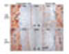
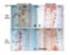
Fig 1. E-H
Compression side of TMC group on day 4 (E), tension side of TMC group on day 4 (F), compression side of TM group on day 4 (G), and tension side of TM group on day 4 (H). Alv means alveolar bone; PDL, periodontal ligament. Black triangle head indicates osteoclast; black diamond head, osteoblast; black arrow, OPG. OPG positive signals appear as a red color. Bar = 100µm.
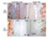
Fig 1. I-L
Compression side of TMC group on day 7 (I), tension side of TMC group on day 7 (J), compression side of TM group on day 7 (K), and tension side of TM group on day 7 (L). Alv means alveolar bone; PDL, periodontal ligament. Black triangle head indicates osteoclast; black diamond head, osteoblast; black arrow, OPG. OPG positive signals appear as a red color. L, Bar = 100µm.
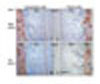
Fig 1. M-P
Compression side of TMC group on day 14 (M), tension side of TMC group on day 14 (N), compression side of TM group on day 14 (O), and tension side of TM group on day 14 (P). Alv means alveolar bone; PDL, periodontal ligament. Black triangle head indicates osteoclast; black diamond head, osteoblast; black arrow, OPG. OPG positive signals appear as a red color. Bar = 100µm.
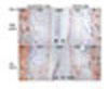
Fig 2. A-D
RANK-immunohistochemical staining photomicrographs (horizontal section, 4µm thick, original magnification × 400). Compression side of TMC group on day 1 (A), tension side of TMC group on day 1 (B), compression side of TM group on day 1 (C), and tension side of TM group on day 1 (D). Alv means alveolar bone; PDL, periodontal ligament. Black triangle head indicates osteoclast; black diamond head, osteoblast; black arrow, RANK. RANK positive signals appear as a red color. Bar = 100µm. On pressure side, RANK appeared associated with osteoclasts and peridontal ligament cells in both TMC and TM groups. On day 4, RANK expression on the compression side of TMC group was greater than that of TM group.
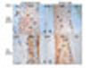
Fig 2. E-H
Compression side of TMC group on day 4 (E), tension side of TMC group on day 4 (F), compression side of TM group on day 4 (G), and tension side of TM group on day 4 (H). Alv means alveolar bone; PDL, periodontal ligament. Black triangle head indicates osteoclast; black diamond head, osteoblast; black arrow, RANK. RANK positive signals appear as a red color. Bar = 100µm.
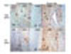
Fig 2. I-L
Compression side of TMC group on day 7 (I), tension side of TMC group on day 7 (J), compression side of TM group on day 7 (K), and tension side of TM group on day 7 (L). Alv means alveolar bone; PDL, periodontal ligament. Black triangle head indicates osteoclast; black diamond head, osteoblast; black arrow, RANK. RANK positive signals appear as a red color. Bar = 100µm.
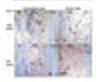
Fig 2. M-P
Compression side of TMC group on day 14 (M), tension side of TMC group on day 14 (N), compression side of TM group on day 14 (O), and tension side of TM group on day 14 (P). Alv means alveolar bone; PDL, periodontal ligament. Black triangle head indicates osteoclast; black diamond head, osteoblast; black arrow, RANK. RANK positive signals appear as a red color. Bar = 100µm.
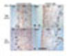
Fig 3. A-D
RANKL immunohistochemical staining photomicrographs of experimental and control groups (horizontal section, 4µm thick, original magnification × 400). Compression side of TMC group on day 1 (A), tension side of TMC group on day 1 (B), compression side of TM group on day 1 (C), and tension side of TM group on day 1 (D). Alv means alveolar bone; PDL, periodontal ligament. Black triangle head indicates osteoclast; black diamond head, osteoblast; black arrow, RANKL. RANKL positive signals appear as a red color. Bar = 100µm. On the compression side, RANKL appeared associated with osteoclasts, periodontal ligment cells and perivascular cells in both TMC and TM groups. On the compression side, RANKL expression of TMC group was more intensive and extensive than that of TM group on day 7.
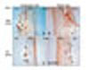
Fig 3. E-H
Compression side of TMC group on day 4 (E), tension side of TMC group on day 4 (F), compression side of TM group on day 4 (G), and tension side of TM group on day 4 (H). Alv means alveolar bone; PDL, periodontal ligament. Black triangle head indicates osteoclast; black diamond head, osteoblast; black arrow, RANKL. RANKL positive signals appear as a red color. Bar = 100µm.
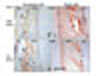
Fig 3. I-L
Compression side of TMC group on day 7 (I), tension side of TMC group on day 7 (J), compression side of TM group on day 7 (K), and tension side of TM group on day 7 (L). Alv means alveolar bone; PDL, periodontal ligament. Black triangle head indicates osteoclast; black diamond head, osteoblast; black arrow, RANKL. RANKL positive signals appear as a red color. Bar = 100µm.
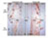
Fig 3. M-P
Compression side of TMC group on day 14 (M), tension side of TMC group on day 14 (N), compression side of TM group on day 14 (O), and tension side of TM group on day 14 (P). Alv means alveolar bone; PDL, periodontal ligament. Black triangle head indicates osteoclast; black diamond head, osteoblast; black arrow, RANKL. RANKL positive signals appear as a red color. Bar = 100µm.
References
1. Kole H. Surgical operations on the alveolar ridge to correct occlusal abnormalities. Oral Surg Oral Med Oral Pathol. 1959. 12:515–529.

2. Wilcko WM, Wilcko T, Bouquot JE, Ferguson DJ. Rapid orthodontics with alveolar reshaping: two case reports of decrowding. Int J Periodontics Restorative Dent. 2001. 21:9–19.
3. Wilcko WM, Bouquot JE, Ferguson DJ, Wilcko T. Rapid orthodontic decrowding with alveolar augmentation: case report. World J Orthod. 2003. 4:197–205.
4. Frost HM. The biology of fracture healing. An overview for clinicians. Part I. Clin Orthop Relat Res. 1989. 248:283–293.
5. Frost HM. The biology of fracture healing. An overview for clinicians. Part II. Clin Orthop Relat Res. 1989. 248:294–309.
6. Chung KL. Rapid orthodontics. 2001. Seoul: Ji-Sung;156–169.
7. Lowney JJ, Norton LA, Shafer DM, Rossomando EF. Orthodontic forces increase tumor necrosis factor alpha in the human gingival sulcus. Am J Orthod Dentofacial Orthop. 1995. 108:519–524.
8. Grieve WG 3rd, Johnson GK, Moore RN, Reinhardt RA, DuBois LM. Prostaglandin E (PGE) and interleukin-1 beta (IL-1 beta) levels in gingival crevicular fluid during human orthodontic tooth movement. Am J Orthod Dentofacial Orthop. 1994. 105:369–374.
9. Kwan Tat S, Padrines M, Theoleyre S, Heymann D, Fortun Y. IL-6, RANKL, TNF-alpha/IL-1: interrelations in bone resorption pathophysiology. Cytokine Growth Factor Rev. 2004. 15:49–60.
10. Theoleyre S, Wittrant Y, Tat SK, Fortun Y, Redini F, Heymann D. The molecular triad OPG/RANK/RANKL: involvement in the orchestration of pathophysiological bone remodeling. Cytokine Growth Factor Rev. 2004. 15:457–475.
11. Boyle WJ, Simonet WS, Lacey DL. Osteoclast differentiation and activation. Nature. 2003. 423:337–342.
12. Aubin JE, Triffit JT, Bilezikian JP. Mesenchymal stem cells and osteoblast differentiation. Principles of bone biology. 2002. Vol. 1. San Diego: Academic Press;59–81.
13. Suda T, Udagawa N, Nakamura I, Miyaura C, Takahashi N. Modulation of osteoclast differentiation by local factors. Bone. 1995. 17:suppl 2. 87S–91S.
14. Epker BN, Frost HM. Correlation of bone resorption and formation with the physical behavior of loaded bone. J Dent Res. 1965. 44:33–41.
15. Takahashi N, Akatsu T, Udaqawa N, Saaki T, Yamaguchi A, Moseley JM, et al. Osteoblastic cells are involved in osteoclast formation. Endocrinology. 1998. 123:2600–2602.
16. Heymann D, Rousselle AV. gp130 cytokine family and bone cells. Cytokine. 2000. 12:1455–1468.
17. Ren Y, Maltha JC, Kuijpers-Jagtman AM. The rat as a model for orthodontic tooth movement-a critical review and a proposed solution. Eur J Orthod. 2004. 26:483–490.
18. Ogasawara T, Yoshimine Y, Kiyoshima T, Kobayashi I, Matsuo K, Akamine A, et al. In situ expression of RANKL, RANK, osteoprotegerin and cytokines in osteoclasts of rat periodontal tissue. J Periodontal Res. 2004. 39:42–49.
19. Kanzaki H, Chiba M, Shimizu Y, Mitani H. Dual regulation of osteoclast differentiation by periodontal ligament cells through RANKL stimulation and OPG inhibition. J Dent Res. 2001. 80:887–891.
20. Nishijima Y, Yamaguchi M, Kojima T, Aihara N, Nakajima R, Kasai K. Levels of RANKL and OPG in gingival crevicular fluid during orthodotnic tooth movement and effect of compression force on releases from periodontal ligament cells in vitro. Orthod Craniofac Res. 2006. 9:63–70.
21. Kanzaki H, Chiba M, Shimizu Y, Mitani H. Periodontal ligamental cells under mechanical stress induce osteoclastogenesis by receptor activator of nuclear factor kappaB ligand up-regulation via prostaglandin E2 synthesis. J Bone Miner Res. 2002. 17:210–220.
22. Kanzaki H, Chiba M, Sato A, Miyagawa A, Arai K, Nukatsuka S, et al. Cyclical tensile force on periodontal ligament cells inhibits osteoclastogenesis through OPG induction. J Dent Res. 2006. 85:457–462.
23. Oshiro T, Shiotani A, Shibasaki Y, Sasaki T. Osteoclast induction in periodontal tissue during experimental movement of incisors in osteoprotegerin-deficient mice. Anat Rec. 2002. 266:218–225.
24. Kanzaki H, Chiba M, Arai K, Takahashi I, Haruyama N, Nishimura M, et al. Local RANKL gene transfer to the periodontal tissue accelerates orthodontic tooth movment. Gene Ther. 2006. 13:678–685.
25. Kanzaki H, Chiba M, Takahashi I, Haruyama N, Nishimura M, Mitani H. Local OPG gene transfer to periodontal tissue inhibits orthodontic tooth movement. J Dent Res. 2004. 83:920–925.
26. Hasegawa T, Yoshimura Y, KiKuiri T, Yawaka Y, Takeyama S, Matsumato A, et al. Expression of receptor activator of NF-kappa B ligand and osteoprotegerin in culture of human periodontal ligament cells. J Periodontal Res. 2002. 37:405–411.
27. Shiotani A, Shibasaki Y, Sasaki T. Localization of receptor activator of NF-kappa B ligand, RANKL, in periodontal tissues during experimental movement of rat molars. J Electron Microsc. 2001. 50:365–369.
28. Yamasaki K, Shibata Y, Fukuhara T. The effect of prostaglandins on experimental tooth movement in monkeys (Macaca fuscata). J Dent Res. 1982. 61:1444–1446.
29. Soma S, Yamashita K, Matsumoto S, Takada K. Effect of continuous infusion of PTH on orthodontic tooth movement. J Jpn Orthod Soc Abstr. 1997.
30. Takano-Yamamoto T, Kawakami M, Kobayash Y, Yamashiro T, Sakuda M. The effect of local application of 1,25-dihydroxycholecalciferol on osteoclast numbers in orthodontically treated rats. J Dent Res. 1992. 71:53–59.
31. Brudvik P, Rygh P. Root resoprtion after local injection of prostaglandin E2 during experimental tooth movement. Eur J Orthod. 1991. 13:255–263.
32. Yaffe A, Fine N, Binderman I. Regional accelerated phenomenon in the mandible following mucoperiosteal flap surgery. J Periodontol. 1994. 65:79–83.
33. Roberts WE, Huja S, Roberts JA. Bone modeling: biomechanics, molecular mechanisms, and clinical perspectives. Semin Orthod. 2004. 10:123–161.
 XML Download