

 PDF
PDF ePub
ePub Citation
 Print
Print


Abstract
Purpose
We compared the results of total ankle arthroplasty in patients with preoperative varus deformity of more than 20o with those of patients with varus deformity less than 20o.
Materials and Methods
From January 2005 to January 2013, 9 ankles with preoperative varus deformity of more than 20o (varus group) and 31 ankles with varus deformity less than 20o (control group) underwent total ankle arthroplasty. Clinical results were evaluated using the American Orthopaedic Foot and Ankle Society (AOFAS) score, and radiographic results were assessed using tibiotalar varus angle in standing anteroposterior radiographs taken preoperatively and at the last follow-up.
Results
The mean duration of clinical follow-up was 42.8 months (14∼60 months). The AOFAS score was improved by a mean 47.0 points in the varus group and 37.6 points in the control group. Statistically significant difference was observed between the two groups (p=0.041). Tibiotalar varus angle measured at the last follow-up radiograph was 2.5o in the varus group and 1.0o in the control group and the difference was not statistically significant (p=0.820).
References
1. Thomas RH, Daniels TR. Ankle arthritis. J Bone Joint Surg Am. 2003; 85:923–36.

2. Knecht SI, Estin M, Callaghan JJ, Zimmerman MB, Alliman KJ, Alvine FG, et al. The Agility total ankle arthroplasty. Seven to sixteen-year follow-up. J Bone Joint Surg Am. 2004; 86:1161–71.
3. Greisberg J, Hansen ST Jr. Ankle replacement: management of associated deformities. Foot Ankle Clin. 2002; 7:721–36.
4. Hintermann B, Valderrabano V. Total ankle replacement. Foot Ankle Clin. 2003; 8:375–405.
5. Haskell A, Mann RA. Ankle arthroplasty with preoperative coronal plane deformity: short-term results. Clin Orthop Relat Res. 2004; 424:98–103.
6. Wood PL, Deakin S. Total ankle replacement. The results in 200 ankles. J Bone Joint Surg Br. 2003; 85:334–41.
7. Valderrabano V, Pagenstert G, Hintermann B. Total ankle replacement: three component prosthesis. Tech Foot Ankle Surg. 2005; 4:42–54.
8. Hobson SA, Karantana A, Dhar S. Total ankle replacement in patients with significant preoperative deformity of the hindfoot. J Bone Joint Surg Br. 2009; 91:481–6.
9. Kim BS, Choi WJ, Kim YS, Lee JW. Total ankle replacement in moderate to severe varus deformity of the ankle. J Bone Joint Surg Br. 2009; 91:1183–90.
10. Martin DE, Kaplan PA, Kahler DM, Dussault R, Randolph BJ. Retrospective evaluation of graded stress examination of the ankle. Clin Orthop Relat Res. 1996; 328:165–70.
11. Stauffer RN, Segal NM. Total ankle arthroplasty: four years' experience. Clin Orthop Relat Res. 1981; 160:217–21.
12. Newton SE 3rd. Total ankle arthroplasty. Clinical study of fifty cases. J Bone Joint Surg Am. 1982; 64:104–11.
13. Trajkovski T, Pinsker E, Cadden A, Daniels T. Outcomes of ankle arthroplasty with preoperative coronal-plane varus deformity of 10° or greater. J Bone Joint Surg Am. 2013; 95:1382–8.
14. Valderrabano V, Pagenstert G, Horisberger M, Knupp M, Hintermann B. Sports and recreation activity of ankle arthritis patients before and after total ankle replacement. Am J Sports Med. 2006; 34:993–9.
15. Kim BS, Knupp M, Zwicky L, Lee JW, Hintermann B. Total ankle replacement in association with hindfoot fusion: outcome and complications. J Bone Joint Surg Br. 2010; 92:1540–7.
Figure 1.
Standing anteroposterior radiograph shows the measurement of ankle alignment which is defined as the angle between the anatomical axis of the tibia and a line drawn perpendicular to the talar dome (angle A). The extent of varus deformity in this case is 26o.
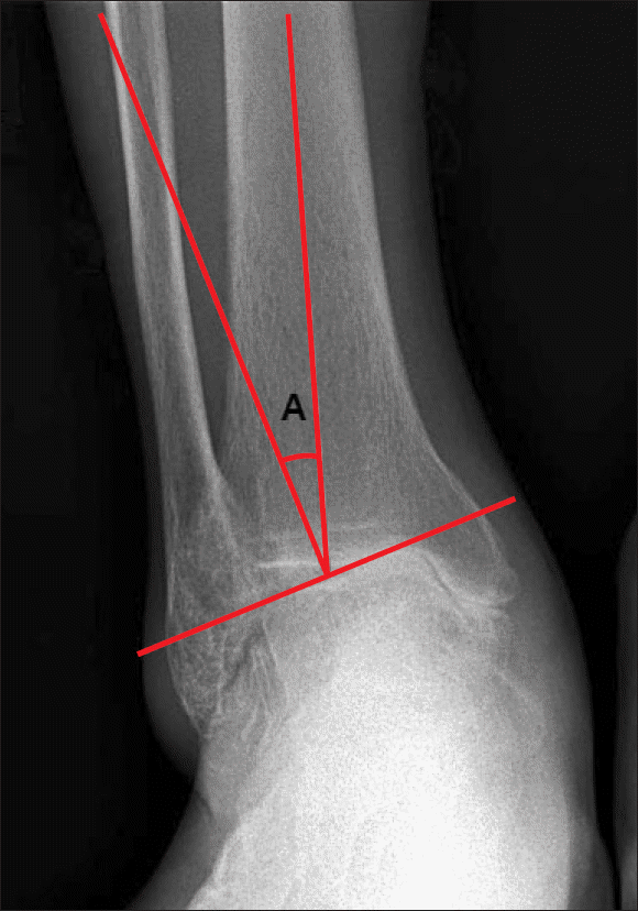
Figure 2.
Standing anteroposterior radiographs before and 44 months after total ankle arthroplasty. (A) Preoperative radiograph shows varus deformity of 28° in a 73-year-old man with post-traumatic arthritis. (B) The patient is satisfied and has a postoperative varus of 2° and an American Orthopaedic Foot and Ankle Society score of 88.
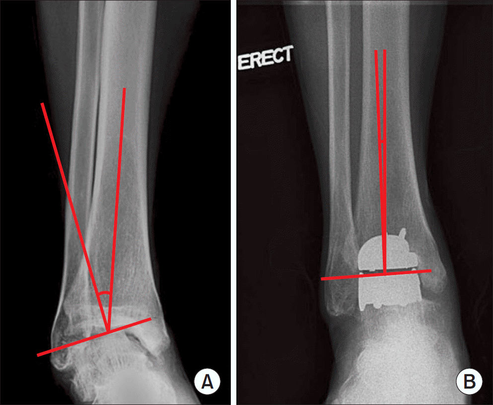
Table 1.
Patient Demographics
Table 2.
Distribution of Implants in Varus Group and Control Group
| Implant | Group I | Group II | Total |
|---|---|---|---|
| Hintegra (Newdeal SA) | 7 (77.7) | 24 (77.4) | 31 (77.5) |
| Mobility (Depuy) | 2 (22.2) | 7 (22.6) | 9 (22.2) |
| Total | 9 | 31 | 40 |
Table 3.
Clinical Results by AOFAS Score
| AOFAS score | Group I | Group II | p-value |
|---|---|---|---|
| Preoperative | 33.3±4.5 | 43.0±6.1 | 0.323 |
| Last follow-up | 80.3±6.7 | 80.6±6.5 | 0.901 |
| Improvement | 47.0±2.4 | 37.6±5.6 | 0.041 |
Table 4.
Radiographic Results
| Varus angle (o) | Group I | Group II | p-value |
|---|---|---|---|
| Preoperative | 23.9±2.8 | 7.8±3.1 | 0.038 |
| Last follow-up | 2.5±1.2 | 1.0±0.8 | 0.820 |
 XML Download