

 PDF
PDF ePub
ePub Citation
 Print
Print


Abstract
Breast ultrasound is a valuable adjunctive to mammography for the identification and characterization of palpable and nonpalpable abnormalities. With the advances of ultrasound techniques over the past 10 years, the role of ultrasound has evolved from differentiation of benign from malignant masses, evaluation of mammmographic abnormalities, and guidance modality of interventional procedure to preoperative evaluation of lesion extent, follow-up after operation, and screening method for high-risk women. It can also be used as a supplemental modality of a physical examination and mammography to increase diagnostic accuracy and to reduce unnecessary surgical biopsy. This article is to review the indication of breast ultrasound examinations as well as merits and limitations compared to mammmography.
Figures and Tables
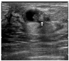
Figure 1
Invasive ductal carcinoma. Breast US shows a 1cm sized ill-defined hypoechoic mass (arrow).
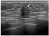
Figure 2
Automated ultrasound systems.
(A) 3-D coronal image shows architectural distortion (arrow) in 11 hour direction of the left breast, 5cm apart from nipple.
(B) Axial image shows a 1.2cm sized ill-defined hypoechoic mass (arrows).
(C) Sagittal reconstruction image shows the taller-than-wide mass (arrow). Surgery revealed invasive breast cancer.
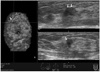
Figure 3
Elastography.
(A) A 0.7cm sized fibroadenoma shows normal strain within the mass.
(B) A 0.7cm sized invasive breast cancer shows no strain within the mass.
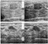
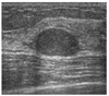
Figure 4
Breast cancer in dense breasts.
(A) Mammogram shows no definite mass. A metallic marker is attached to the palpable area (arrow) in the left breast.
(B) US images show a 1cm ill-defined hypoechoic mass (arrow) in the palpable area. This cancer is difficult to detect at mammography.
(C) US-guided core biopsy revealed invasive ductal carcinoma.
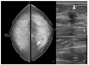
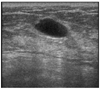
Figure 5
Intraductal papilloma. Breast US shows a complex cystic mass with echogenic solid component (arrow).
References
1. Moon WK, editor. Detection of breast cancer with mammography. 2008. 1st ed. Seoul: Ilchokak;13–42.
2. Kerlikowske K, Grady D, Barclay J, Sickles EA, Ernster V. Effect of age, breast density, and family history on the sensitivity of first screening mammography. JAMA. 1996. 276:3338.

3. Sickles EA. Breast imaging: from 1965 to the present. Radiology. 2000. 215:1–16.
4. Zonderland HM, Coerkamp EG, Hermans J, van de Vijver MJ, van Voorthuisen AE. Diagnosis of breast cancer: contribution of US as an adjunctive to mammography. Radiology. 1999. 213:412–422.
5. Feig SA, Hall FM, Ikeda DM, Mendelson EB, Rubin EC, Segel MC, Watson AB Jr, Eklund GW, Stelling CB, Jackson VP. Society of Breast Imaging residency and fellowship training curriculum. Radiol Clin North Am. 2000. 38:915–920.
6. Smith RA, Cokkinides V, Eyre HJ. Cancer screening in the United States, 2007: a review of current guidelines, practices, and prospects. CA Cancer J Clin. 2007. 57:90–104.
7. Kolb TM, Lichy J, Newhouse JH. Occult cancer in women with dense breasts: detection with screening US-diagnostic yield and tumor characteristics. Radiology. 1998. 207:191–199.
8. Cho N, Moon WK, Park JS, Cha JH, Jang M, Seong MH. Nonpalpable breast masses: evaluation by US elastography. Korean J Radiol. 2008. 9:111–118.
9. Moon WK, editor. Breast ultrasound. 2006. 1st ed. Seoul: Ilchokak;49–53.
10. Stavros AT. Breast Ultrasound. 2004. Philadelphia: Lippincott Williams & Wilkins;1–15.
11. Moy L, Slanetz PJ, Moore R, Satija S, Yeh ED, McCarthy KA, Hall D, Staffa M, Rafferty EA, Halpern E, Kopans DB. Specificity of mammography and US in the evaluation of a palpable abnormality: retrospective review. Radiology. 2002. 225:176–181.
12. Flobbe K, Bosch AM, Kessels AG, Beets GL, Nelemans PJ, von Meyenfeldt MF, van Engelshoven JM. The additional diagnostic value of ultrasonography in the diagnosis of breast cancer. Arch Intern Med. 2003. 163:1194–1199.
13. Cho N, Moon WK, Chung SY, Cha JH, Cho KS, Kim EK, Oh KK. Ductographic findings of breast cancer. Korean J Radiol. 2005. 6:31–36.
14. Park JM. Localization of breast lesions: mammographic concept. 1997. 1st ed. Seoul: Korea medical publishing company;9–36.
15. Moon WK, Im JG, Koh YH, Noh DY, Park IA. US of mammographically detected clustered microcalcifications. Radiology. 2000. 217:849–854.
16. Moon WK, Noh DY, Im JG. Multifocal, multicentric, and contralateral breast cancers: bilateral whole-breast US in the preoperative evaluation of patients. Radiology. 2002. 224:569–576.
17. Mendelson EB. Evaluation of the postoperative breast. Radiol Clin North Am. 1992. 30:107–138.
18. Shin JH, Han BK, Choe YH, Nam SJ, Park W, Im YH. Ultrasonographic detection of occult cancer in patients after surgical therapy for breast cancer. J Ultrasound Med. 2005. 24:643–649.
 XML Download