

 PDF
PDF ePub
ePub Citation
 Print
Print


Salivary gland tumors are rare in dogs and cats, with a reported overall incidence of 0.17% [1]. Most cases are reported in older patients and generally affect the mandibular and parotid glands [6,17]. Clinical signs include the presence of an asymptomatic mass in the gland that is often locally invasive [1,6,17]. An adenocarcinoma is the most common type of tumor in the salivary glands, whereas malignant mixed tumors, especially carcinosarcomas, are uncommon both in veterinary [2,16] and human medicine [1,9]. Here, we describe a case involving a cat with a carcinosarcoma in the mandibular gland.
A 9-year-old neutered female domestic short-haired cat with no past trauma or medical history besides cough and eye trauma presented to the Veterinary Medical Center of Yamaguchi University with swelling near the base of the right ear. The swelling had been discovered approximately 3 weeks earlier, at which time the area was drained of purulent matter and treated with antibiotics at the referring veterinary hospital. The purulent matter was found to primarily consist of inflammatory cells and the cat showed signs of hypersalivation, halitosis and gingivitis, which were especially severe in the right side region of the head. With the exception of the aforementioned symptoms, the cat appeared well.
Upon admission, a large fluctuated and painless subcutaneous swelling was palpated and found to contain a firm mass in the right mandibular region. In addition, the left mandibular lymph node was enlarged and the right mandibular lymph node could not be found because of the swelling. There were no neurological problems or facial nerve disorders. Hematology and biochemical analysis of the serum revealed neutrophilia (differential counting 84/100), low MCH (11.8 pg, reference range 13 to 17 pg) and MCHC (24.4 g/dl, reference range 30 to 36 g/dl) levels.
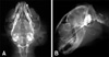
Head radiographs revealed the presence of a soft-tissue density that involved faint circular calcific opacity (Fig. 1). There was also a bony reaction in the mandible and maxilla, which was likely caused by chronic gingivitis. In addition, thoracic radiographs revealed no abnormalities.
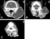
However, the contrast-enhanced computed tomography (CT) revealed the presence of a peripheral capsulated cystic area (Fig. 2A) in the right mandibular gland (Figs. 2B and C). Fine-needle aspiration revealed reddish brown exudates in the fluctuated region and the presence of some undifferentiated epithelial cells, suggesting that the mass was of a neoplastic nature. Based on these findings, this region was suspected to be a tumor that originated from the mandibular gland.
Two weeks after admission, the cystic mass was surgically removed. The mass was excised as widely as possible, and total removal of the mass was grossly achieved. In addition, the right mandibular lymph node was also removed because it showed signs of metastasis. The cat showed no major side effects following surgery. The mass contained both cystic and firm portions. Histopathological examination of the surgically removed tumor tissue revealed simultaneous proliferation of both the malignant epithelial and mesenchymal components, suggesting that the mass was a malignant mixed tumor, also known as a carcinosarcoma (Fig. 3). Histopathological analysis of the enlarged lymph node revealed that it contained metastatic tumor cells.
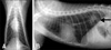
The cat received post-operative radiotherapy that was administered using an orthovoltage X-ray radiation unit (Hitachi Medico, Japan). The treatment was divided into doses of 4Gy. Four weeks after surgery, after only 2 of the radiation fractions had been administered, a firm mass (9 × 7 × 5 mm) was found in the caudal base of the right ear. The mass was surgically removed and histopathologically confirmed as a malignant mixed tumor, suggesting local recurrence of the primary tumor. The radiotherapy was continued and two additional fractions were delivered to this patient. However, thoracic radiography conducted three weeks after the second surgery strongly suggested diffuse metastasis of the tumor to the lung (Fig. 4), even though the cat remained in stable physical condition. The owner did not allow any additional treatment, and the cat died one month later. Necropsy was not performed.
In human medicine, there are three types of malignant mixed tumors of the salivary glands, carcinoma ex mixed tumors, carcinosarcomas, and metastatic mixed tumors with a benign appearance [3]. Nearly 99% of malignant mixed tumors are carcinoma ex mixed tumors, which are also known as carcinoma ex pleomorphic adenoma [11]. Based on the 60 reported cases of true malignant mixed tumors of the salivary gland in humans, the most common epithelial-origin tumor was squamous cell carcinoma or adenocarcinoma, whereas the most common non-epithelial tumor was chondrosarcoma [10]. Conversely, carcinosarcoma comprises less than 0.2% of all salivary tumors and 0.4% of malignant salivary neoplasms reported in human medicine [5]. Histologically, such tumors are characterized by the contemporary presence of epithelial (carcinomatous) and mesenchymal (sarcomatous) components [4].
In veterinary practice, tumors that originate from the salivary gland are also classified into several categories [7]. Malignant mixed tumors, carcinomas and sarcomas have been observed in veterinary cases of pleomorphic adenoma, and several case reports describing such tumors are available; however, the incidence of this type of tumor in cats is believed to be lower than in other animals. In the case reported here, the mass had adenocarcinomatous and chondrosarcoma elements (Fig. 3), which was compatible with typical histopathological findings of malignant mixed tumors in salivary glands.
The cat was referred due to swelling in the right mandibular lesion that had been rapidly enlarging without any associated facial palsy. Hammer et al. [6] reported that the most common presenting complaint for animals with salivary gland neoplasia was the discovery of a mass by the owner, followed by other signs of local invasion, including dysphagia, halitosis, and exophthalmia. However, in this case, the owner did not realize there was a problem prior to the sudden increase in the size of mandibular gland.
Salivary gland carcinosarcomas are extremely rare; therefore, there is no well-established therapeutic approach to their treatment [8]. In this case, the cat received postoperative radiotherapy until pulmonary metastasis was observed. Because it was inconvenient for the owner to visit the clinic, the dosage of the cats radiotherapy was limited to 16 Gy; therefore, it is difficult to evaluate the effectiveness of the radiotherapy in this case. However, surgical removal followed by radiotherapy seems to be the most rational approach despite the fact that the available data are not prospective or statically significant and vary widely among studies [12,13].
Salivary gland carcinosarcomas are typically aggressive. In humans, the 5-year survival rate for patients with carcinosarcoma is reportedly 0% [15]. Conversely, the survival of patients with malignant tumors is reportedly 54% [12], with an average survival that ranges from 29.3 months to 3.6 years [5,14]. Metastasis is most often to the lung and liver and the incidence of bone, brain and lymph node metastasis is low [13]. In the case described here, the mass did not invade the skull, despite the extensive tumor size, and distant metastases were found only in the lungs.
Although tumors of the salivary glands occur less frequently in animals than in humans, benign tumors in animals are rare. Therefore, it is important that the tumor be resected as early as possible to prevent it from evolving into a highly aggressive tumor [17].

 XML Download