

 PDF
PDF ePub
ePub Citation
 Print
Print


Introduction
Foot and mouth disease (FMD) is one of the most devastating diseases in cattle, swine, sheep, goats, and many wild cloven-hoofed animal species [2]. The causative agent of the disease belongs to the Aphthoviruses genus of the Picornaviridae Family.
In recent years, DNA vaccination has become one of the most promising routes for a recombinant vaccine [8,12], allowing a safe and efficient alternative to conventional vaccination. DNA vaccine technology facilitates the use of cytokines as modulators in vaccination to manipulate the immune responses. In particular, IL-1 production by mononuclear phagocytes can be triggered by macrophage-derived cytokines such as tumor necrosis factor (TNF) or Interleukin-1 (IL-1), as well as by contact with CD4+ T cells. IL-1α is a major immunoregulatory and proinflammatory cytokine that also affects the proliferation and function of fibroblast [3,6]. Recently, we observed that DNA vaccination using both IL-1α and the porcine reproductive and respiratory syndrome virus (PRRSV) ORF5 gene induced a stronger immune response compared with IL-1β administered through the intradermal route in the tail (data not shown). The DNA vaccines are generally used at high concentrations in mouse immunizations, approximately 100-200 µg per animal, as well as in a highly purified form to remove endotoxins derived from E. coli. Therefore, a low administration dose is important for clinical applications.
The aim of the study was to examine the efficiency of a DNA immunization system using plasmids at low doses in mice, and to enhance the immunogenicity against FMD by constructing plasmids containing the swine IL-1α gene in addition to the viral capsid (P1) gene including 2A or VP1 containing the major epitopes of the virus.
Materials and Methods
Construction of plasmids
The vector pSLIA, which was kindly supplied by VIDO (Vaccine and Infectious Disease Organization, Canada), is a stable mammalian expression vector that contains the CMV promoter for expression in mammalian cells. Swine IL-1α, as a molecular adjuvant, was cloned from the whole blood of pigs. The VP1 and P1-2A (P1 and 2A) cDNA from the O/SKR/2002 strain (AY312588) were amplified by a polymerase chain reaction (PCR). The sense and anti-sense primers used for VP1 were 5'-AACTGCAGATGACCACCTCCACAGGTGAGT-3' and 5'-CGGGATCCCAACAGCTGTTTCACAGGCGCC-3', respectively. The sense and anti-sense primers used for P1-2A (truncated form of 5' region) 5'-GCTCTAGAATGAACACTGGAAGCATTATCA-3' and 5'-CGGGATCCCCCAGGGTTGGGCTCGACGTCT-3', respectively. The amplified PCR products corresponding to VP1 or P12A were purified from a gel using Gene Clean Turbo kit (Q-BIO Gene, USA) and cloned into the PstI and BamHI, or XbaI and BamHI sites of pSLIA. The resulting plasmids were named pS-VP1, pSIL1A-VP1 and pSIL1A-P12A (Fig. 1).
Identification of expressed viral protein
MA104 cells, a monkey kidney cell line, were transfected using Lipofectamine plus (Gibco, USA) according to the manufacturer's instructions. The cells were incubated with bovine FMDV antiserum. After incubation, the cells were washed with PBS and incubated with the fluorescein isothiocyanate (FITC)-conjugated goat anti-bovine antibody (Cappel, USA). The cells were kept in PBS and observed by fluorescence microscopy.
For Western blotting, the MA104 cells were cultured on a tissue culture dish (100mm) and transfected with Lipofectamine plus, as described above. After 48 h of transfection, the cells were harvested using centrifugation, and disrupted with a lysis buffer and sonication. After electrophoresis in SDS-PAGE gel, the gels were transferred onto a nitrocellulose membrane, and the membrane was reacted with either the bovine FMDV antibody (NVRQS, Korea) or rabbit anti-swine IL-1α antibody (Biosource, USA). The first antibody was detected by horseradish peroxidase (HRPO)-anti-immunoglobulin conjugate and visualized by diaminobenzidine staining of the nitrocellulose membrane.
Immunizations to mice
A total of twenty specific pathogen free (SPF) C57BL/6 mice (4-8 week olds, male), which were grown according to the animal management guideline of the National Veterinary Research and Quarantine Service (NVRQS) in Korea, were divided into 4 groups (5mice/group) for the DNA immunization clinical trial. One week prior to the experiment, the mice were isolated, and kept under controlled conditions for the duration of the study. The control group mice were inoculated with pSLIA and each of the three experimental groups were administered with pS-VP1, pSIL1A-VP1 or pSIL1A-P12A, respectively. The mice were immunized three times with a low dose of the plasmids (10 µg) by a tail injection. The antibody response was examined from blood samples collected from the four groups of mice over a 7 day period before inoculation, and at 2 or 3 week intervals after inoculation.
Measurement of Anti-VP1 or anti-P12A antibody level
The immune responses after DNA vaccination were determined by indirect ELISA as described by Abu Elzein and Crowther [1]. Ninety-six well plates (Nunc, Denmark) were coated with inactive purified FMDV serotype O antigen (105 tissue culture infectious dose (TCID)50/ml) overnight and incubated with the serum samples [1 : 50 diluted in 0.1 M PBS containing 0.5% Tween 20 and 0.5% gelatin] for 1 h at 37℃. For each mouse IgG subtyping, biotinylated goat IgG specific to either mouse IgG or IgG1/IgG2a (Zymed Laboratory, USA) was reacted for 30 min at 37℃.
After the reaction with the HRP-streptavidin conjugate for 30 min at 37℃, the color reaction was developed with 100 µl/well of 2,2'-azino-bis(3-ethylbenzthiazoline-6-sulfonic acid) (ABTS) for 30 min at room temperature, and the absorbance was examined at a wavelength of 405 nm. The results are expressed as the reciprocal of the highest serum dilution resulting in a reading of two standard deviations above the value of the control sera. The serum neutralizing antibody test and liquid-phase blocking (LPB) ELISA was carried out according to the OIE Terrestrial Manual [4].
Results
VP1 and P12A proteins expressed in MA104 cells
The expression of the recombinant proteins in MA104 cells were analyzed using an immunofluorescence assay and Western blot analysis after 48 h of transfection. No bright staining was detected when the test cells were transfected with the pSLIA control vector, whereas high levels of bright fluorescent staining due to a specific reaction of the bovine antiserum and rabbit antiserum to the cytosol proteins were detected in the pS-VP1, pSIL1A-VP1 or pSIL1A-P12A transfected cells. The expression of the P12A protein appeared to be higher than that of the VP1 protein (Fig. 2).
The proteins secreted after 48 h of transfection were analyzed by SDS-PAGE. The separated proteins were transferred onto a nitrocellulose membrane. The proteins in the cell lysates of the transfected MA104 cells were immunodetected by Western blot analysis, using rabbit anti-swine IL-1α and bovine antiserum. Fig. 3 shows that the cell lysates of the pS-VP1, pSIL1A-VP1 and pSIL1A-P12A transfected MA104 cells expressed a detectable level of the recombinant proteins. The VP1, VP1-IL1α and P12A-IL1α fusion protein bands were detected with a molecular weight corresponding to 25 kDa (lane 2), 65 kDa (lane 3) and 120 kDa (lane 4), respectively.
Immunogenicity in mice
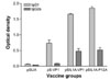
The mice (C57BL/6) were immunized through the tail with 10 µg of either pSLIA (control group), pS-VP1, pSIL1A-VP1 and pSIL1A-P12A. ELISA was used to determine the level of the humoral antibody, and the anti-VP1 antibodies one week after the third immunization. The group immunized with the recombinant plasmids encoding IL-1α showed higher titers than the group immunized without IL-1α (Fig. 4). The mice immunized with pSIL1A-P12A had a higher titer than the mice immunized with pSIL1A-VP1 (Fig. 4A). Fig. 4B shows a weak reaction in the sera of all four groups one week after the first immunization. In the pSIL1A-VP1 and pSIL1A-P12A groups, an increase in the immune response was detected in the sera one week after the second immunization. However, no increase in the immune response was detected in the pSLIA and pS-VP1 groups.
The changes in the immune response were detected by IgG subclass analysis in the sera at one week after the third immunization (Table 1). Immunizing the mice with pSIL1A-VP1 and pSIL1A-P12A produced a specific IgG1 and IgG2a response and showed an increased response by a ratio of IgG1/IgG2a (Fig. 5).
Discussion
These results show that recombinant plasmids were constructed with VP1, which contained the critical epitopes for inducing an immune response to the FMDV [13] or the capsid (P1) gene including 2A of the FMDV, which is a mixture of genes encoding structural and non-structural proteins. It was reported that the viral structural proteins induce a humoral response, whereas the non-structural proteins appear to be more effective in inducing the cellular immune system [7]. Some recombinant plasmids were also constructed to encode IL-1α in order to enhance the immune response.
The results showed that the recombinant plasmids, the expression vector encoding VP1 or capsid (P1) gene including 2A, could be transfected to MA104 cells, which then expressed VP1 or P12A. The molecular weight of the VP1 protein and the VP1 fusion protein containing IL-1α were found at the predicted molecular weight. The P12A fusion protein band was observed by 3C protease with natural processing to be a large protein without the proteolytic cleavage form. These results suggest that the pSILIA-VP1 and pSIL1A-P12A expression plasmids can transfect cells, resulting in the expression of VP1 and P12A proteins in linearized forms. In this study, these plasmids could be expressed in mammalian cells by transfection in vitro.
The recombinant proteins and synthetic peptides are often weak immunogens that require the addition of adjuvants to potentiate the immune reactivity [9]. There have been many studies on the use of cytokines as adjuvants in vaccination. Cytokines inducing IL-1 and IL-2 are frequently used in combination with viral antigens to enhance their immunogenicity [10]. IL-1 has been shown to be an effective adjuvant in several model systems, enhancing the protection induced by a viral and bacterial vaccine as well as stimulating the immunity in clinical trials, even though side effects such as fever have been reported. IL-1 molecules have diverse biological activity that are important for immune reactivity, such as inducing the cells to synthesize more of the other cytokines and activating T cell, inflammation and auto-immunity [5]. Among the various animal species, the mature IL-1α sequences are conserved in a range of approximately 70% [11].
This study demonstrated that the administration of the swine IL-1α gene significantly enhanced the immune response in mice, and the recombinant plasmids encoding IL-1α in combination could be an effective immunogen. Indirect ELISA was used to measure the antibody level in the mice immunized with pS-VP1, pSIL1A-VP1 and pSIL1A-P12A. The pSIL1A-VP1 and pSIL1A-P12A showed a similar increase in the immune response in the sera at 1 week after the 2nd immunization compared with the pSLIA group, while the pS-VP1 group showed a low immune response.
In FMD, it is important that the humoral response elicits rapid protection on account of the rapid progression of the disease. There is also the issue of having to deliver the vaccine in low doses because of the difficulty associated with the production of sufficient vaccine. For these reasons, an attempt was made to achieve enhanced immunity through intradermal route through the tail skin, which contains immune stimulating cells such as macrophages or dendritic cells.
The formation of IgG2a is typical for the Th1 response, while IgG1 is typical for Th2 response [10]. In this study, the immunization of mice with pS-VP1, pSIL1A-VP1 and pSIL1A-P12A increased the IgG1/IgG2a ratio in low dilution sera. It is possible that even though no attempt was made to detect the cytokines associated with the cellular immunity, these patterns are typical of the Th2 response. Th2 type cells have been reported to be efficient in helping induce an antibody response to protein antigens [7].
The neutralizing responses in the vaccinated group were either very low or could be not detected by the neutralizing test or LPB ELISA. This might be due to the poor conformational epitope to elicit protection against a viral challenge. In order to overcome this problem, a better strategy might be to add 3C protease to the P1 region or express multiple cistronic VP0/VP3/VP1 to mimic the immunogenicity of the conformational epitopes in the authentic structure.
DNA immunization provides a safe and efficient alternative to conventional vaccination. The plasmid DNA vaccines encoding the foot and mouth disease virus VP1 epitopes have the ability to elicit both FMDV-specific T cell proliferation and neutralizing antibody against FMD in swine [13]. Moreover, despite the difficulties associated with needle vaccination, the recombinant plasmid has other advantages, such as increased safety, reproducibility, and ease of production.
In conclusion, the VP1, P12A and IL-1α fusion genes might be useful as strong immunogens. However, research concerning protection conferred by the capsid and IL-1 co-expressed plasmids on other target animals such as swine and cattle is needed. Studies aimed at replacing a DNA vaccination strategy with an adenovirus and retrovirus gene delivery system to achieve more efficient vaccination are currently underway.

 XML Download