

 PDF
PDF ePub
ePub Citation
 Print
Print


Abstract
Distal radius fractures are a common upper extremity fracture and a considerable number of patients have a stable fracture. In the treatment of distal radius fractures, there is considerable disagreement regarding the need for a strict anatomical restoration with operation in elderly patients. Therefore, nonsurgical treatment is a still important treatment option in distal radius fractures. The radiological parameters of before or after manual reduction are important for deciding whether to perform operation or not. The radiological parameters include dorsal angulation of the articular surface, radial shortening, extent of dorsal comminution, intra-articular displacement, concomitant ulnar metaphyseal fracture, shear fracture, and fracture-dislocation of the distal radioulnar joint. In addition, clinical situations of patients, including age, activity level, underline disease, and recovery level, which the patients wish should be considered, comprehensively. For the duration of a splint or cast, three to four weeks are recommended in impacted or minimally displaced fractures and five to six weeks in displaced fractures. After reduction of the displaced fractures, patients should undergo a radiologicical examination every week to check the redisplacement or deformity of the fracture site until two or three weeks post trauma. Arm elevation is important for controlling fracture site swelling and finger exercises, including metacarpophalangeal joint motion, are needed to prevent hand stiffness. Active range of motion exercise of the wrist should be initiated immediately after removing the splint or cast.
References
1. Baron JA, Karagas M, Barrett J, et al. Basic epidemiology of fractures of the upper and lower limb among Americans over 65 years of age. Epidemiology. 7:612–618. 1996.

2. Orbay JL, Fernandez DL. Volar fixation for dorsally displaced fractures of the distal radius: a preliminary report. J Hand Surg Am. 27:205–215. 2002.
3. Bentohami A, de Burlet K, de Korte N, van den Bekerom MP, Goslings JC, Schep NW. Complications following volar locking plate fixation for distal radial fractures: a systematic review. J Hand Surg Eur Vol. 39:745–754. 2014.
4. Kodama N, Takemura Y, Ueba H, Imai S, Matsusue Y. Acceptable parameters for alignment of distal radius fracture with conservative treatment in elderly patients. J Orthop Sci. 19:292–297. 2014.
5. Palmer AK, Werner FW. Biomechanics of the distal radioulnar joint. Clin Orthop Relat Res. 187:26–35. 1984.
6. Bronstein AJ, Trumble TE, Tencer AF. The effects of distal radius fracture malalignment on forearm rotation: a cadaveric study. J Hand Surg Am. 22:258–262. 1997.
7. Knirk JL, Jupiter JB. Intra-articular fractures of the distal end of the radius in young adults. J Bone Joint Surg Am. 68:647–659. 1986.
8. Miyake T, Hashizume H, Inoue H, Shi Q, Nagayama N. Malunited Colles' fracture. Analysis of stress distribution. J Hand Surg Br. 19:737–742. 1994.
9. Kazuki K, Kusunoki M, Yamada J, Yasuda M, Shimazu A. Cineradiographic study of wrist motion after fracture of the distal radius. J Hand Surg Am. 18:41–46. 1993.
10. Fernández DL. Malunion of the distal radius: current approach to management. Instr Course Lect. 42:99–113. 1993.
11. Altissimi M, Antenucci R, Fiacca C, Mancini GB. Long-term results of conservative treatment of fractures of the distal radius. Clin Orthop Relat Res. 206:202–210. 1986.
12. Weber ER. A rational approach for the recognition and treatment of Colles' fracture. Hand Clin. 3:13–21. 1987.
13. Cooney WP. Fractures of the distal radius. A modern treatment-based classification. Orthop Clin North Am. 24:211–216. 1993.
14. Lafontaine M, Hardy D, Delince P. Stability assessment of distal radius fractures. Injury. 20:208–210. 1989.
15. Simic PM, Weiland AJ. Fractures of the distal aspect of the radius: changes in treatment over the past two decades. Instr Course Lect. 52:185–195. 2003.
16. Park MJ, Kim JP, Lee HI, Lim TK, Jung HS, Lee JS. Is a short arm cast appropriate for stable distal radius fractures in patients older than 55 years? A randomized prospective multicentre study. J Hand Surg Eur Vol. 42:487–492. 2017.
17. Martinez-Mendez D, Lizaur-Utrilla A, de-Juan-Herrero J. Intra-articular distal radius fractures in elderly patients: a randomized prospective study of casting versus volar plating. J Hand Surg Eur Vol. 43:142–147. 2018.
18. Song SW. Osteoporotic distal radius fracture-conservative treatment. J Korean Fract Soc. 21:81–86. 2008.
19. Lee JH. Surgical indications for distal radius fractures. J Korean Soc Surg Hand. 20:72–76. 2015.
20. Azzopardi T, Ehrendorfer S, Coulton T, Abela M. Unstable extraarticular fractures of the distal radius: a prospective, randomised study of immobilisation in a cast versus supplementary percutaneous pinning. J Bone Joint Surg Br. 87:837–840. 2005.
21. Wong TC, Chiu Y, Tsang WL, Leung WY, Yam SK, Yeung SH. Casting versus percutaneous pinning for extraarticular fractures of the distal radius in an elderly Chinese population: a prospective randomised controlled trial. J Hand Surg Eur Vol. 35:202–208. 2010.
22. Arora R, Lutz M, Deml C, Krappinger D, Haug L, Gabl M. A prospective randomized trial comparing nonoperative treatment with volar locking plate fixation for displaced and unstable distal radial fractures in patients sixty-five years of age and older. J Bone Joint Surg Am. 93:2146–2153. 2011.
23. Sharma H, Khare GN, Singh S, Ramaswamy AG, Kumaras-wamy V, Singh AK. Outcomes and complications of fractures of distal radius (AO type B and C): volar plating versus nonoperative treatment. J Orthop Sci. 19:537–544. 2014.
24. Young BT, Rayan GM. Outcome following nonoperative treatment of displaced distal radius fractures in low-demand patients older than 60 years. J Hand Surg Am. 25:19–28. 2000.
25. Jaremko JL, Lambert RG, Rowe BH, Johnson JA, Majumdar SR. Do radiographic indices of distal radius fracture reduction predict outcomes in older adults receiving conservative treatment? Clin Radiol. 62:65–72. 2007.
26. Kyung MG, Chung HW, Kim JS, et al. Closed reduction and cast immobilization for the treatment of distal radius fracture: does dorsal metaphyseal comminution predict radiographic and functional outcomes? J Korean Soc Surg Hand. 18:29–36. 2013.
27. Kim JM, Seo HJ, Jeon YD, Lee HM, Son JH. Comparative study of outcomes between operative and nonoperative treatment of unstable distal radius fracture in the elderly patients. J Korean Soc Surg Hand. 20:43–50. 2015.
28. The Korean Society for Surgery of the Hand: Surgery of the hand. Seoul: Panmuneducation;p. 14–15. 192-193, 283–285,. 2014.
29. Lee JH, Hong SH, Kim YJ, Back JH, Lee JS. Effect of different splints on displacement after closed reduction of the distal radius fractures: a comparison of short arm double splint and sugar-tong splint. J Korean Soc Surg Hand. 20:104–109. 2015.
30. Frykman G. Fracture of the distal radius including sequelae: shoulder-hand-finger syndrome, disturbance in the distal radioulnar joint and impairment of nerve function. A clinical and experimental study. Acta Orthop Scand. (Suppl 108):3. 1967.
31. Cho JH, Park DY, Kim JY, Han KJ. A comparison of sugar tong splint and radial gutter short arm splint after closed reduction of distal radius fracture. J Korean Soc Surg Hand. 14:194–198. 2009.
32. Bong MR, Egol KA, Leibman M, Koval KJ. A comparison of immediate postreduction splinting constructs for controlling initial displacement of fractures of the distal radius: a prospective randomized study of long-arm versus short-arm splinting. J Hand Surg Am. 31:766–770. 2006.
33. Grafstein E, Stenstrom R, Christenson J, et al. A prospective randomized controlled trial comparing circumferential casting and splinting in displaced Colles fractures. CJEM. 12:192–200. 2010.
34. Lichtman DM, Bindra RR, Boyer MI, et al. American Academy of Orthopaedic Surgeons clinical practice guideline on: the treatment of distal radius fractures. J Bone Joint Surg Am. 93:775–778. 2011.
35. McAuliffe TB, Hilliar KM, Coates CJ, Grange WJ. Early mobilisation of Colles' fractures. A prospective trial. J Bone Joint Surg Br. 69:727–729. 1987.
36. Jung MS, Sung SC, Choi IH. Textbook of fractures. 3rd ed.Seoul, Gunja: 309–336;2008.
37. Bucholz RW, Rockwood CA, Green DP. Rockwood and Green's fractures in adults. 7th ed.Philadelphia, PA: Wolters Kluwer Health/Lippincott Williams & Wilkins;2010.
Fig. 1.
Successful conservative treatment of a displaced distal radius fracture in a twenty year old woman. (A) Immediate post-trauma wrist anteroposterior and lateral X-ray. Dorsally displaced intra-articular distal radius fracture in the right wrist. (B) Wrist X-ray after manual reduction. Volar tilt of the distal radius and radial length were restored. (C) Wrist X-ray at post traumatic three months. The fracture site was well maintained and well united.
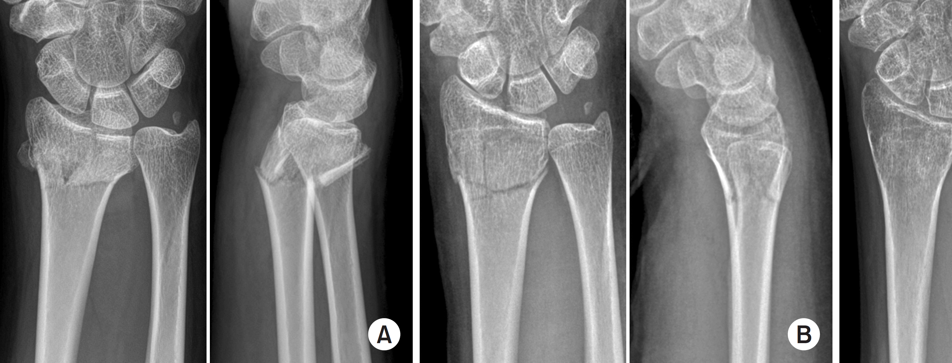
Fig. 2.
Result of conservative treatment of an elderly patient with a displaced distal radius fracture. X-rays of a seventy one-year-old woman. (A) Wrist anteroposterior and lateral X-ray after the initial reduction. Distal radius and ulnar styloid fracture with intra-articular comminution in the left wrist. The volar tilt and radial length were well restored after the reduction. (B) Wrist X-ray shows radial shortening and dorsal tilt of distal radius at 1 year after trauma. (C) Range of motion of the left wrist was decreased slightly compared to the right side, but she performed her daily activities without pain at 1 year after trauma.
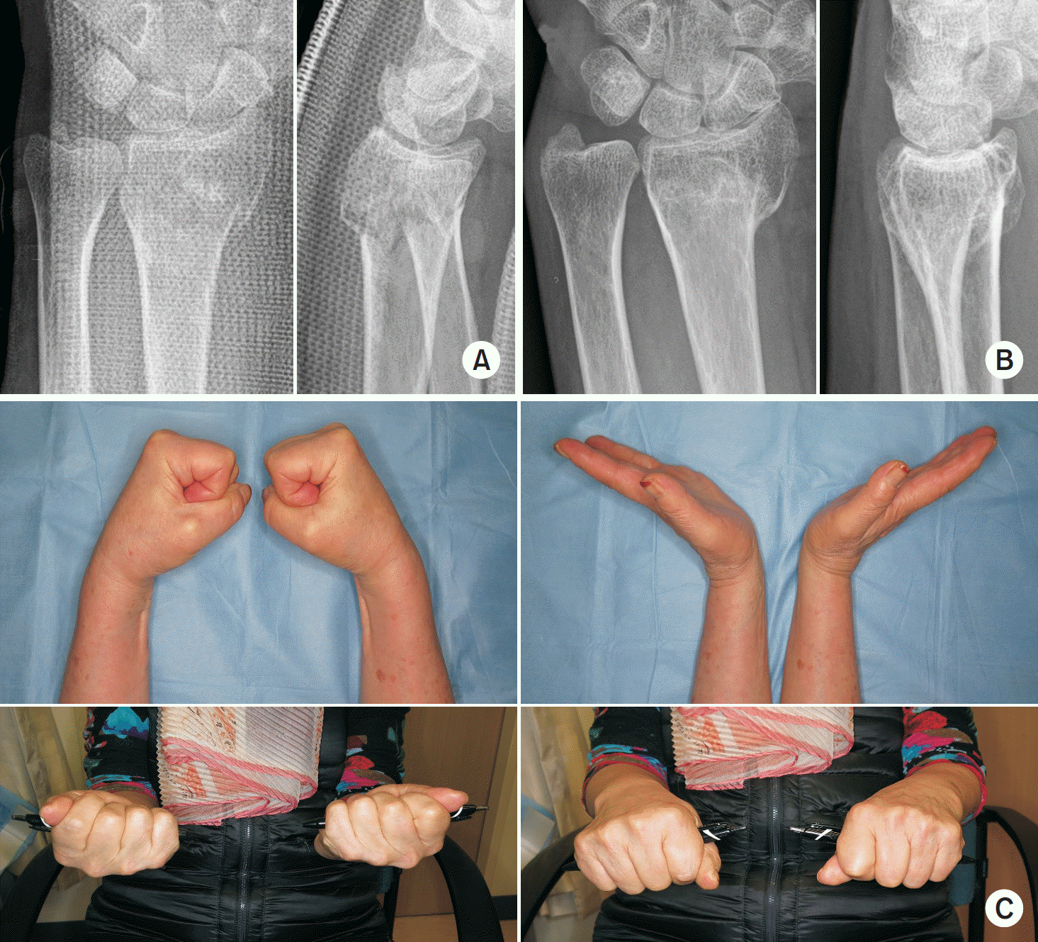
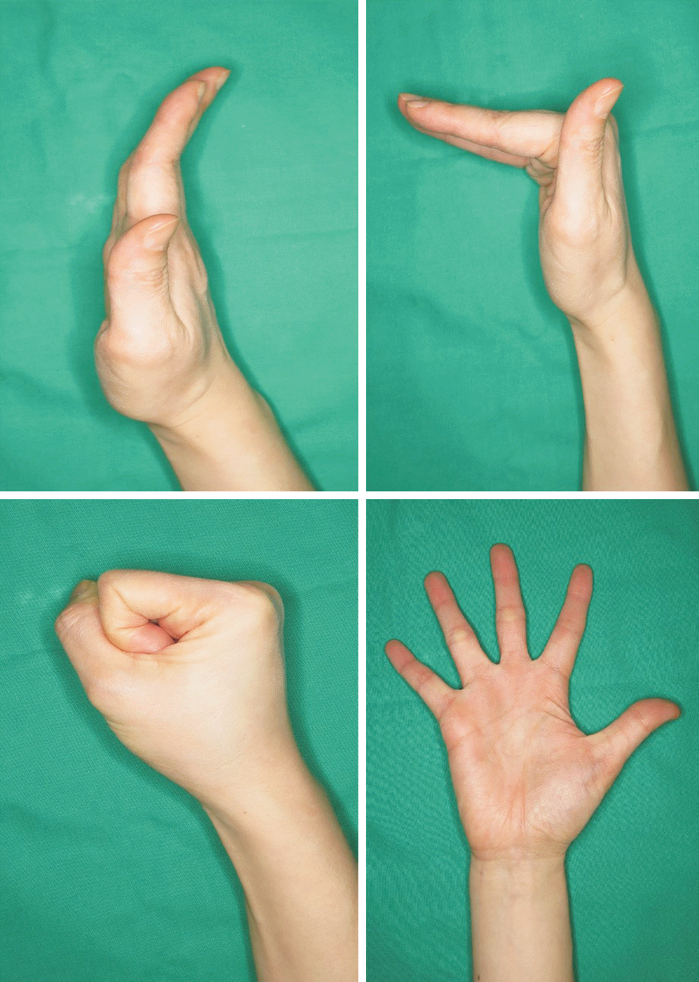
Table 1.
Summary of Randomized Controlled Trials for the Comparison between Non-Operative and Operative Treatment for Distal Radius Fractures
| Article/ candidate of study | Type of treatment | Number | Mean age (yr) | Sex (men: women) | Severity of fracture (AO type) (n) | Clinical outcomes at final F/U (mean) | Radiologic outcomes (mean±standard deviation) | ||||||||||
|---|---|---|---|---|---|---|---|---|---|---|---|---|---|---|---|---|---|
| Wrist flexion/ extension* | Grip strength* | Patient reported outcomes | Dorsal angulation (°)† | Radial inclination (°)† | Ulnar variance (mm)† | ||||||||||||
| Pre | Post | F/U | Pre | Post | F/U | Pre | Post | F/U | |||||||||
| Azzopardi et al.20) (2005)/ unstable, extraarticular fractures | Non-operative | 27 | 71 | 2:25 | A3 | 88.5% | 72% | SF-36 physical (38.2) | 29±16 | —5±7 | 4±8 | 18±6 | 21±3 | 19±6 | 3±3 | 0±1 | 3±2 |
| Percutaneous pinning | 27 | 72 | 4:23 | A3 | 90.5% | 77% | SF-36 physical (42.2) | 35±15 | —4±7 | —3±10‡ | 16±6 | 22±3 | 22±5 | 4±3 | 0±1 | 3±2 | |
| Wong et al.21) (2010)/ unstable, extraarticular fractures | Non-operative | 30 | 71 | 5:25 | Frykman classification I:II (18:12) | 143° | 9.0 kg | Mayo wrist scores (80.5±7.5) | 31±6 | —7.5±1 | 3±1 | 13±3 | 23±4 | 16±2 | 4.3±1.2 | 0.5±0.2 | 3.2±1.4 |
| Percutaneous pinning | 30 | 70 | 6:24 | Frykman classification I:II (17:13) | 145° | 8.5 kg | Mayo wrist scores (82.2±6.2) | 33±6 | —8±1 | —4±1‡ | 13±4 | 23±2 | 20±2‡ | 5.2±1.8 | 0.3±0.1 | 2.1±1.1 | |
| Arora et al.22) (2011)/ displaced, unstable fractures | Non-operative | 37 | 77.4 | 10:27 | A2 (3), A3 (9), C1 (11), C2 (8), C3 (6) | 103.7%‡ | 92.6% | DASH scores (8.0±9.3) | 3.6±11.2 | 10.4±19.1 | 20.3±3.3 | 15.9±9.0 | 0.8±1.7 | 3.2±2.9 | |||
| ORIF with volar locking plate | 36 | 75.9 | 8:28 | A2 (3), A3 (7), C1 (4), C2 (12), C3 (10) | 92.8% | 102.4%‡ | DASH scores (5.7±11.1) | —3.6±6.9‡ | —0.5±4.7‡ | 21.8±2.7‡ | 21.2±2.6‡ | 0.6±1.6 | 0.7±1.8‡ | ||||
| Sharma et al.23) (2014)/AO type B and C fractures | Non-operative | 32 | 48.1 | 10:22 | B (13), C (19) | 135.0° | 72.2% | DASH scores (14.0±10.2) | —8.4±0.4 | —5.2±0.5 | 18.1±0.9 | 15.2±0.8 | 0.2±0.1 | 0.3±0.1 | |||
| ORIF with volar locking plate | 32 | 52.4 | 9:23 | B (19), C (17) | 168.2°‡ | 89.1%‡ | DASH scores (5.0±9.4‡) | —10.1±1.5‡ | —8.4±1.0‡ | 20.5±1.3‡ | 17.9±0.8‡ | —0.3±0.3 | —0.3±0.2 | ||||
* Wrist flexion/extension arc and grip strength of the involved hand was presented as raw data or ratio compared to the contralateral side.
 XML Download