Journal List > J Korean Fract Soc > v.28(2) > 1038018

Chung, Choi, Yoon, Lee, and Park: Treatment of the Femoral Fracture Using Sirus® Nail: A Comparison of Complication according to the Entry Potal

Abstract

Purpose

The purpose of this study is to analyze the clinical results of fixation using Sirus® nail in patients with femoral subtrochanteric and shaft fracture and the difference in the frequency of complications according to the entry portal.

Materials and Methods

From July 2006 to August 2013, at least 1-year clinical follow-up, we retrospectively analyzed 36 cases with femoral subtrochanteric (15 cases) and shaft fracture (21 cases) who underwent surgery using Sirus® nail. We reviewed the records of operation time, intra-operative amounts of bleeding and complications. At last follow-up, we reviewed clinical results by Ray-Sanders criteria and analyzed the periods of bone union on the radiograph. We also measured changing of the femoral neck-shaft angle in the subtrochanteric fractures and angulation in the shaft fractures, respectively. Considering anatomical variation of the trochanter and fracture position of subtrochanteric and femoral shaft, entry points were divided into subgroups, and the clinical results were compared.

Results

The mean Ray-Sanders score was 27.4, 27 cases (75.0%) were good or excellent. The mean periods of bone union was 21.1 weeks in 31 cases. The mean neck-shaft angles were 135.7o preoperatively, 130.2o postoperatively. The mean angulation of the femur was 24.4o preoperatively, 2.4o postoperatively in patients of femoral shaft fractures. Despite no statistical significance, greater trochanter tip entry point and lateral entry point had a higher rate of frequency than medial entry point, with respect to the occurrence of iatrogenic fracture and malalignment.

Conclusion

Using Sirus® nail for femoral subtrochanteric and shaft fractures showed good clinical and radiographic results and a high rate of union. Medial entry point yielded slightly better results in the occurrence of iatrogenic fracture and malalignment, compared to greater trochanter tip entry point and lateral entry point.

Figures and Tables

Fig. 1

For estimation of proper entry portals, we assumed the length of Sirus® nail (s) to circular arc (EF). The radius (r) of the circle was informed by regular manufacturer's information. Finally we estimated the central angle (θ) from the circle and compared with contralateral neck-shaft angle.
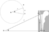
jkfs-28-103-g001
Fig. 2

Illustration of subdivision by entry points. ①: Lateral entry point, ②: greater trochanter tip entry point, ③: medial entry point.
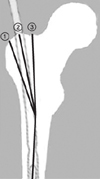
jkfs-28-103-g002
Fig. 3

Preoperative and postoperative antero-posterior view of iatrogenic fracture that occurred in Sirus® nail insertion using lateral entry point.
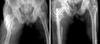
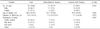
jkfs-28-103-g003
Table 1

Summary of Demographics

jkfs-28-103-i001

Values are presented as number (%) or median (range). *Two patients were injured with both subtrochanteric and segmental shaft fracture of femur.

Table 2

Comparison of Clinical Results and Complications between Subtrochanteric and Shaft Fracture of the Femur

jkfs-28-103-i002

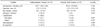
Values are presented as median (range) or number (%). *Includes screw irritations, heterotrophic ossification, device loosening & breakage and limping gait with pain. Mal-alignment was defined as more than or equal to 5 degrees of neck-shaft angle compared with contralateral of the subtrochanteric fractures in the coronal plane and 10 degrees of total angular deformity of the femoral shaft fractures in the coronal and sagittal planes, respectively.

Table 3

Comparison of Clinical Results between Entry Portals (n=36)

jkfs-28-103-i003

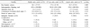
Values are presented as median (range) or number (%). *Includes screw irritations, heterotophic ossification, device loosening & breakage and limping gait with pain. Mal-alignment was defined as more than or equal to 5 degrees of neck-shaft angle compared with contralateral of the subtrochanteric fractures in the coronal plane and 10 degrees of total angular deformity of the femoral shaft fractures in the coronal and sagittal planes, respectively.

Notes

Financial support None.

Conflict of interest None.

References

1. Winquist RA, Hansen ST Jr, Clawson DK. Closed intramedullary nailing of femoral fractures. A report of five hundred and twenty cases. J Bone Joint Surg Am. 1984; 66:529–539.
crossref
2. Mileski RA, Garvin KL, Crosby LA. Avascular necrosis of the femoral head in an adolescent following intramedullary nailing of the femur. A case report. J Bone Joint Surg Am. 1994; 76:1706–1708.
crossref
3. Khan FA, Ikram MA, Badr AA, al-Khawashki H. Femoral neck fracture: a complication of femoral nailing. Injury. 1995; 26:319–321.
crossref
4. Dora C, Leunig M, Beck M, Rothenfluh D, Ganz R. Entry point soft tissue damage in antegrade femoral nailing: a cadaver study. J Orthop Trauma. 2001; 15:488–493.
crossref
5. Ansari Moein CM, Verhofstad MH, Bleys RL, van der Werken C. Soft tissue injury related to choice of entry point in antegrade femoral nailing: piriform fossa or greater trochanter tip. Injury. 2005; 36:1337–1342.
crossref
6. Yun HH, Oh CH, Yi JW. Subtrochanteric femoral fracture during trochanteric nailing for the treatment of femoral shaft fracture. Clin Orthop Surg. 2013; 5:230–234.
crossref
7. Afsari A, Liporace F, Lindvall E, Infante A Jr, Sagi HC, Haidukewych GJ. Clamp-assisted reduction of high subtrochanteric fractures of the femur. J Bone Joint Surg Am. 2009; 91:1913–1918.
crossref
8. Lakhwani OP. Correlation of trochanter-shaft angle in selection of entry site in antegrade intramedullary femoral nail. ISRN Orthop. 2012; 2012:1–5.
crossref
9. Wiss DA, Brien WW, Stetson WB. Interlocked nailing for treatment of segmental fractures of the femur. J Bone Joint Surg Am. 1990; 72:724–728.
crossref
10. Sanders R, Swiontkowski M, Rosen H, Helfet D. Double-plating of comminuted, unstable fractures of the distal part of the femur. J Bone Joint Surg Am. 1991; 73:341–346.
crossref
11. Ha SH, Kim WH, Lee GC. Results of intramedullary nailing of femoral shaft fracture: trochanteric entry portal (Sirus nail) versus piriformis entry portal (M/DN nail). J Korean Fract Soc. 2014; 27:50–57.
crossref
12. Ostrum RF, Marcantonio A, Marburger R. A critical analysis of the eccentric starting point for trochanteric intramedullary femoral nailing. J Orthop Trauma. 2005; 19:681–686.
crossref
13. Ricci WM, Bellabarba C, Lewis R, et al. Angular malalignment after intramedullary nailing of femoral shaft fractures. J Orthop Trauma. 2001; 15:90–95.
crossref
14. Linke B, Ansari Moein C, Bösl O, et al. Lateral insertion points in antegrade femoral nailing and their influence on femoral bone strains. J Orthop Trauma. 2008; 22:716–722.
crossref
TOOLS
Similar articles