

 PDF
PDF ePub
ePub Citation
 Print
Print


INTRODUCTION
The estrogen receptor (ER), progesterone receptor (PR), and human epidermal growth factor receptor 2 (HER2) are the most important therapeutic targets and are used to define breast cancer phenotypes. The clinical features and long-term outcomes of patients with breast cancer have been known to vary, based on the expression status of these receptors [12]. Triple-negative phenotype (TNP) is characterized by the lack of ER, PR, and HER2 overexpression. TNP breast cancer is known for its biological aggressive behavior and associated with poor clinical outcomes comparing with non-TNP breast cancer [3].
When the histopathologic confirmation and assessment of the ER, PR, and HER2 status of metastatic lesions was not performed, the treatment for metastatic disease was usually based on the ER, PR, and HER2 status of primary lesion [4]. However, recent studies have found that 14% to 42% of locoregional recurrences and distant metastases had different receptor status and tumor phenotype from the corresponding primary breast cancer [567]. The current clinical guidelines recommend determining the ER, PR, and HER2 status in recurrent lesion. To date, it is not clear how changes in the receptor status of distant metastasis affect the outcomes of breast cancer patients [8].
The purpose of this study was to evaluate the discordance of ER, PR, and HER2 status between primary breast cancer and the corresponding distant metastatic lesion. In addition, we examined the prognostic impact of discordant receptor status and phenotype after developing distant metastasis.
METHODS
Patients
The study included women with histologically confirmed breast cancer and subsequent distant metastasis. A prospectively maintained database (Seoul National University Hospital Breast Care Center Database) was used to identify 188 patients who underwent biopsy for distant metastases from 2000 to 2010. Among them, we excluded 44 patients with insufficient data for receptor status of metastatic lesions. Finally, our study included 144 breast cancer patients with distant metastasis. All patients provided written informed consent, and the study was approved by the Institutional Review Board of the Seoul National University Hospital (IRB number: 1304-041-479).
Pathology assessment
Immunohistochemical (IHC) analysis was performed to evaluate the expression of ER, PR, and HER2 of primary and metastatic lesions. The cutoff value for ER and PR positivity was ≥10% of tumor cells positive for nuclear staining [9]. HER2 were considered positive when either IHC score was 3+ or HER2 gene amplification was identified by fluorescence in situ hybridization (FISH) [10]. For information on metastatic lesions, needle biopsy or excisional biopsy was performed.
As described previously, TNP was considered to be negative expression of ER, PR, and HER2. Non-TNP was considered to be positive expression of at least one receptor. Concordant TNP was defined as both the primary tumor and metastatic lesion with TNP. Concordant non-TNP was defined as both the primary and metastatic breast cancer with non-TNP. A primary tumor and metastatic lesion with another phenotypic combination were considered to be discordant TNP including primary non-TNP with metastatic TNP and primary TNP with metastatic non-TNP breast cancer.
Statistical analysis
Patient characteristics and rates of discordance between the receptor status of primary and metastatic breast cancer lesions are presented descriptively as proportions. The κ-value was calculated to assess the agreement in receptor status between the primary and metastatic lesions. The κ-value was interpreted as follows: <0.20, slight or poor agreement; 0.21–0.40, fair agreement; 0.41–0.60, moderate agreement; 0.61–0.80, good agreement, and 0.81–1.00, very good agreement (perfect agreement=1.00) [1112].
The Kaplan-Meier analysis was used to estimate the followings: overall survival (OS), from the date of diagnosis of primary breast cancer to death; and postrecurrence survival (PRS), from the date of diagnosis of systemic recurrence to death. Cox proportional hazard regression model was used to calculate multivariate analysis. A two-sided test with p<0.05 was considered statistically significant. All statistical analyses were performed using SPSS version 17.0 software (SPSS Inc., Chicago, USA).
RESULTS
Patients and tumor characteristics
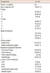
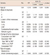
Table 1 summarizes the patient characteristics of the 144 patients. According to American Joint Committee on Cancer staging system, 23 patients (16.0%), 83 patients (57.6%), and 38 patients (26.4%) had stage I, II, and III disease at the diagnosis of primary breast cancer, respectively. Breast cancer surgery and adjuvant treatment, including endocrine therapy, chemotherapy, and radiotherapy, was performed according to clinical practice guidelines. However, only five patients received HER2-targeted therapy as adjuvant treatment, because until 2010, the Korean National Medical Insurance System permitted the use of trastuzumab only for patients with distant metastasis. Of 144 patients, lung was the most common metastatic lesion (39 patients, 27.1%), followed by bone (27 patients, 18.8%), contralateral lymph nodes (17 patients, 11.8%), and liver (12 patients, 8.3%). Of 49 patients, distant metastasis was found on multiple organs including lung, liver, bone, or brain and so on. Biopsy for histologic confirmation of distant metastasis was performed in all patients. Patients received chemotherapy, endocrine therapy, radiation therapy or HER2-targeted therapy after distant metastasis depending on tumor phenotype and physician's decision.
Discordant rates of ER, PR, HER2 expression and phenotype
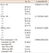
There was no change in the ER status of the primary tumor and corresponding metastasis in 118 of 144 patients (81.9%) (Table 2). A difference in ER status between the primary tumor and metastasis was observed in 26 patients (18.0%): 16 patients (11.1%) had an ER-positive primary and ER-negative metastatic lesion, and 10 patients (6.9%) had an ER-negative primary and ER-positive metastatic lesion. There was no difference in the PR status of the primary tumor and metastasis in 108 patients (75.0%). A difference in PR status between the primary tumor and metastasis was observed in 36 patients (25.0%): 25 patients (17.4%) had a PR-positive primary and PR-negative metastatic lesion, and 11 patients (7.6%) had a PR-negative primary and PR-positive metastatic lesion. A difference in HER2 status between the primary tumor and metastasis was observed in 11 patients (10.3%). There was no change in the HER2 status of the primary and metastatic lesion in 96 patients (89.7%).
Among 144 patients, 134 patients had available information on the tumor phenotype of both the primary and metastatic lesion. Concordant non-TNP and concordant TNP was found for 87 of 134 patients (65.0%) and 29 patients (21.6%), respectively. A difference in the phenotype between primary breast cancer and metastasis (discordant TNP) was found for 18 patients (13.4%).
The κ-values for ER, PR, and HER2 agreement were 0.639, 0.410, and 0.753, respectively. The ER and PR status of the primary and metastatic lesions showed moderate agreement. There was good agreement between the HER2 status of the primary and metastatic lesion. The κ-value of phenotypic agreement was 0.669 (good agreement).
Survival analysis
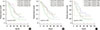
The PRS was estimated based on individual receptor status. The median PRS was 45.8 months (range, 30.5–61.1 months) for patients with concordance in ER positivity, 48.5 months (range, 39.6–57.4 months) for patients with an ER-positive primary and ER-negative metastatic lesion, 42.3 months (range, 34.3–50.2 months) for patients with and ER-negative primary and ER-positive metastatic lesion, and 37.7 months (range, 32.4–43.0 months) for patients with concordance in ER negativity. The median PRS of concordant ER-negative patients was worse than the PRS of concordant ER-positive or discordant ER patients (p=0.001) (Figure 1A).
The median PRS was 53.0 months (range, 19.3–86.6 moths) for patients with concordance in PR positivity, 41.8 months (range, 22.2–61.3 months) for patients with an PR-positive primary and PR-negative metastatic lesion, 62.4 months (range, 6.74–118.0 months) for patients with and PR-negative primary and PR-positive metastatic lesion, 25.4 months (range, 18.5–32.3 months) for patients with concordance in PR negativity. The median PRS of concordant PR-negative patients was worse than the PRS of concordant PR-positive or discordant PR patients (p=0.021) (Figure 1B).
The median PRS was 44.3 months (range, 14.4–74.1 months) for concordant HER2-positive patients, 36.2 months (range, 16.4–56.1 months) for patients with a HER2-negative primary and HER2-positive metastatic lesion, and 30.3 months (range, 19.4–41.2 months) for concordant HER2-negative patients. The median PRS of patients with a HER2-positive primary and HER2-negative metastatic lesion was 4.0 months (range, 3.4–4.6 months), significantly shorter than the PRS of the other patients (p=0.040) (Figure 1C).
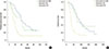
The 5-year PRS rates of concordant non-TNP patients (37.0%) and discordant TNP patients (25.8%) were not significantly different (p=0.280). The 5-year PRS rate of concordant TNP patients was significantly shorter than the 5-year PRS rates of discordant TNP and concordant non-TNP patients (p=0.03 and p<0.001, respectively) (Figure 2A). In patients with discordant TNP patients, the 5-year PRS rates of patients who changed from non-TNP to TNP and patients who changed from TNP to non-TNP were not significantly different (p=0.776). The 5-year OS rates of concordant non-TNP, discordant TNP, and concordant TNP patients were 77.4%, 70.6%, and 23.3%, respectively. The survival difference between concordant non-TNP and discordant TNP patients was not significant (p=0.799). The statistical difference in the 5-year OS between discordant TNP and concordant TNP patients and between concordant non-TNP and concordant TNP patients were significant (p=0.003 and p<0.001, respectively) (Figure 2B). In patients with discordant TNP, the 5-year OS rates of patients who changed from non-TNP to TNP and patients who changed from TNP to non-TNP were not significantly different (p=0.317). In multivariate analysis, stage at diagnosis, location of first metastasis and change of tumor phenotype were independent predictors for PRS (p=0.027, p=0.003, and p<0.001, respectively) (Table 3). Other factors including age at diagnosis, type of surgery were insignificant factors for PRS on multivariate analysis.
DISCUSSION
We reported the rates of discordance between the expression of ER, PR, and HER2 in primary breast cancer and meta-static lesions. The ER status changed in 18.1% of patients, with a κ-value of 0.639. The PR status changed in 25.0%, with a κ-value of 0.410. The rate of discordance between HER2 expression of the primary and metastatic lesion was 10.7%, with a κ-value of 0.753. Therefore, PR appeared to be most unstable receptor and HER2 appeared to be most stable receptor during development of a distant metastasis. In case with discordance between the receptor status of the primary lesion and distant metastasis, ER and PR discordance tended to be loss of expression; whereas HER2 discordance tended to be gain of expression. Previous studies have also reported on the discordance between the ER, PR, and HER2 status of primary and metastatic/recurrent breast cancer. Previous studies reported rates of discordance between primary and metastatic lesions of 10% to 32.4% for ER, 20% to 42% for PR, and 7% to 24% for HER2 [579131415].
Our results suggest that differences between the primary breast cancer and metastatic lesion in the receptor status had prognostic impact. According to our results, patients with concordant ER/PR-positivity and patients with discordant ER/PR status had longer PRS than patients with concordant ER/PR-negativity. Different from our results, Dieci et al. [16] reported that patients with discordant ER/PR status (ER/PR positive primary tumor and ER/PR negative metastatic lesion) had worse PRS than patients with concordant ER/PR positivity (p=0.001). Matsumoto et al. [14] found that patients with a gain in ER/PR status had a longer DFS compared with concordant ER/PR negative patients (p=0.011).
In our study, patients with concordance in HER2 positivity had the longest PRS, and those with HER2-positive primary cancer and HER2-negative metastasis had the shortest PRS. This result is consistent with the findings of a previous retrospective study that included 182 patients with HER-2 positive primary breast cancer. The patients with loss of HER2-positive status in their metastatic tumor had shorter OS and PRS than patients with concordant HER2-positive status [1314]. Another study has reported that the patients with HER2-negative primary cancer and HER-2 positive metastasis achieved the best survival [17].
With regard to tumor phenotype, previous studies reported that patients with concordant phenotype, either non-TNP or TNP, had longer PRS than patients with discordant phenotypes. In discordant phenotype, patients whose phenotype changed to TNP because of loss in ER, PR, and HER2 expression of distant metastasis had shorter OS and PRS [1618]. Liedtke et al. [18] hypothesized that the poor outcomes of patients with discordant receptor status was due to inaccurate assessments of receptor status, which could result in inadequate or ineffective therapy using targeted agents such as tamoxifen or trastuzumab for patients who would not benefit. However, our results showed that patients with concordance in non-TNP between primary and metastasis had better OS and PRS than those with concordance in TNP. Furthermore, concordant TNP was independent predictive factor for poorer PRS comparing with concordant non-TNP and discordant TNP in multivariate analysis. This result was consistent with our expectation that patients with TNP lesions had worse outcomes than patients with non-TNP lesions. Furthermore, an interesting finding in our study was that patients with discordant phenotype had longer OS and PRS than patients with concordance in TNP. Furthermore, distant metastasis developed earlier in patients with concordant TNP breast cancer than patients with concordant non-TNP and with discordant TNP breast cancer (data not shown). The characteristics of TNP breast cancer, which include aggressiveness and absence of therapeutic target, remain consistent throughout progression of the disease. Based on our results, we assume that the retention of triple-negativity in distant metastases adversely affects patient outcome.
A change in the receptor status of the primary breast cancer after neoadjuvant chemotherapy was associated with outcome in breast cancer patients. Chen et al. [19] reported that 15.2% of ER- or PR-positive breast cancer changed to ER- or PR-negative breast cancer after neoadjuvant chemotherapy. These patients had worse 5-year DFS and OS than patients whose receptor status remained positive after neoadjuvant chemotherapy (DFS, 43.2% vs. 67.9% and OS, 60.4% vs. 81.8%, respectively). Guarneri et al. [20] found that 27.5% of patients with HER2-positive primary breast cancer had a change to HER2-negativity after neoadjuvant chemotherapy. The study patients with loss of HER2 positivity tended to have a higher risk of recurrence than patients who retained HER2 positivity (hazard ratio, 2.41; p=0.063).
Although the mechanisms for changes in the expression of ER, PR and HER2 have not been completely elucidated, intratumoral heterogeneity has been proposed to account for the changes [21]. In general, a breast cancer lesion consists of ER, PR, and HER2-positive and ER, PR, and HER2-negative cancer cells. Breast cancer cells in the lesion that are sensitive to certain adjuvant treatments, including endocrine therapy, chemotherapy, and HER2-targeted therapy are eliminated by each treatment and cells that are relatively resistant to adjuvant treatment survive. Another hypothesis proposed that a change in receptor expression reflects a survival mechanism of tumor cells [22]. Change in individual genes and changes in tumor biology may occur after adjuvant treatment [23].
Changes in the expression of tumor receptors may also be a result of inconsistency in IHC staining, which is not actually a biological phenomenon. The methods used in sampling tissue and technical errors associated with IHC staining may result in inconsistent staining results. For instance, samples obtained by fine-needle aspiration performed to obtain a diagnosis of a metastatic lesion may provide less reliable IHC results on ER expression than core needle biopsies [24]. Different laboratories assessing the same tumor block by the same IHC staining method have shown highly discordant rates for the expression of ER, PR, and HER2 [18].
Our study has important strengths. All samples were obtained from primary breast cancer lesions and paired with corresponding metastatic lesion, which led to a complete histological assessment. All specimens were tested at the same laboratory, and evaluated by the same pathologist to minimize preanalytical and analytical error. However, there are several limitations to this study. First, this was a single-institution retrospective study based on medical records. Second, the cutoff value for ER and PR positivity was not 1% but 10%. In terms of current clinical guidelines, there were possibility of false-negative in assessing the ER and PR. Third, we included patients with distant metastasis with available IHC analysis but fine-needle aspiration was performed to obtain tissue from metastatic lesion in some cases. The IHC results from fine-needle aspiration might be inaccurate comparing with those from core-needle biopsy or excisional biopsy. Forth, adjuvant trastuzumab was not routinely administered for HER2-postive breast cancer patients because of the Korean National Medical Insurance System. Finally, the number of patients was small. Especially, the number of patients in discordant HER2 group was very small and there may be bias in the analysis of data.
In conclusion, this study confirmed that there are discordances between the receptor status and tumor phenotype of a primary breast cancer lesion and its metastasis. Patient with concordant TNP status have worse outcomes because of the persistently aggressive and treatment-refractory tumor characteristics of TNP breast cancers. It is important to identify the ER, PR, HER2 status and tumor phenotype of both primary breast cancer and metastatic lesion to estimate prognosis of breast cancer patients who developed distant metastases.
 XML Download