Abstract
Material and Method
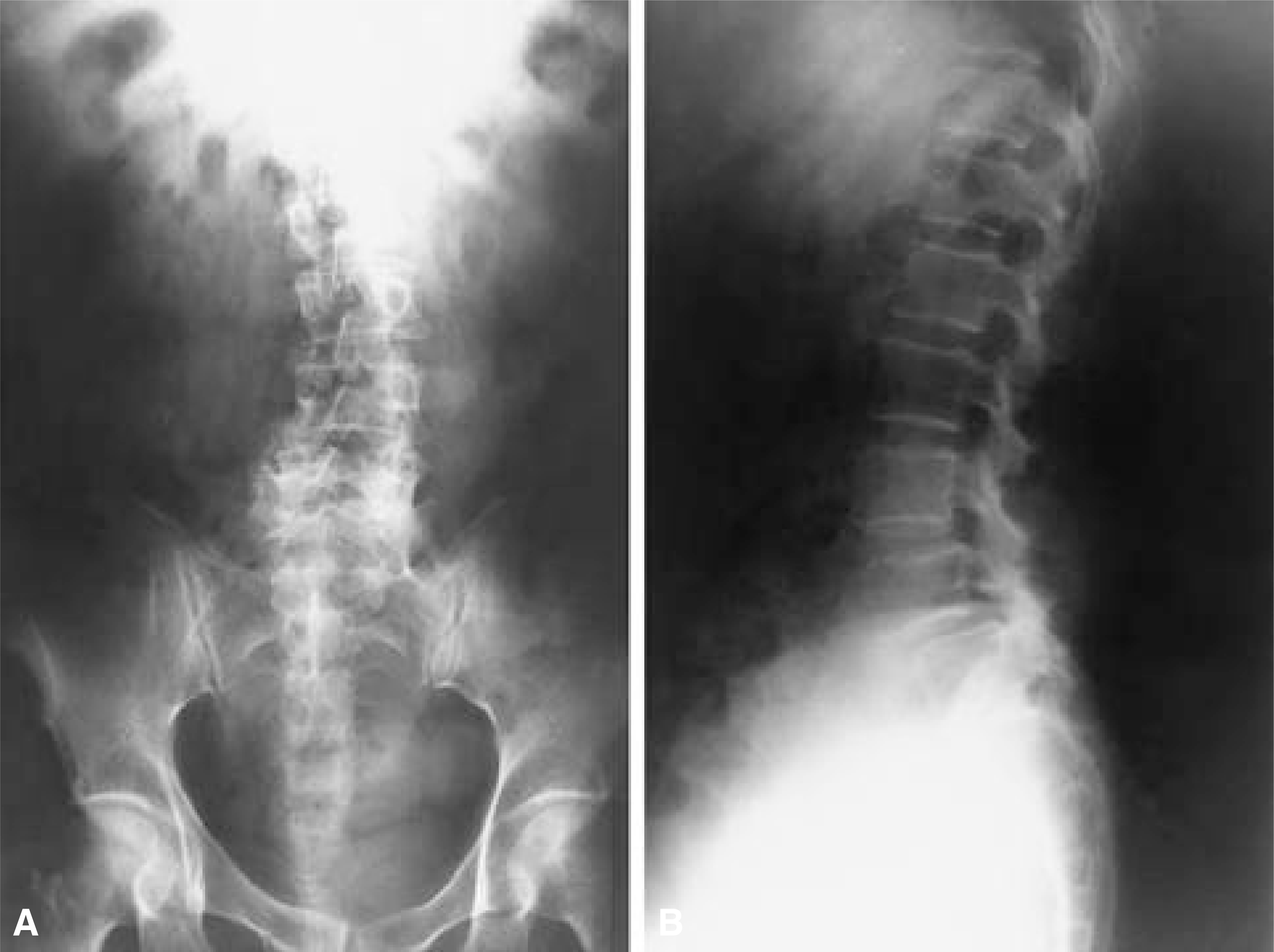
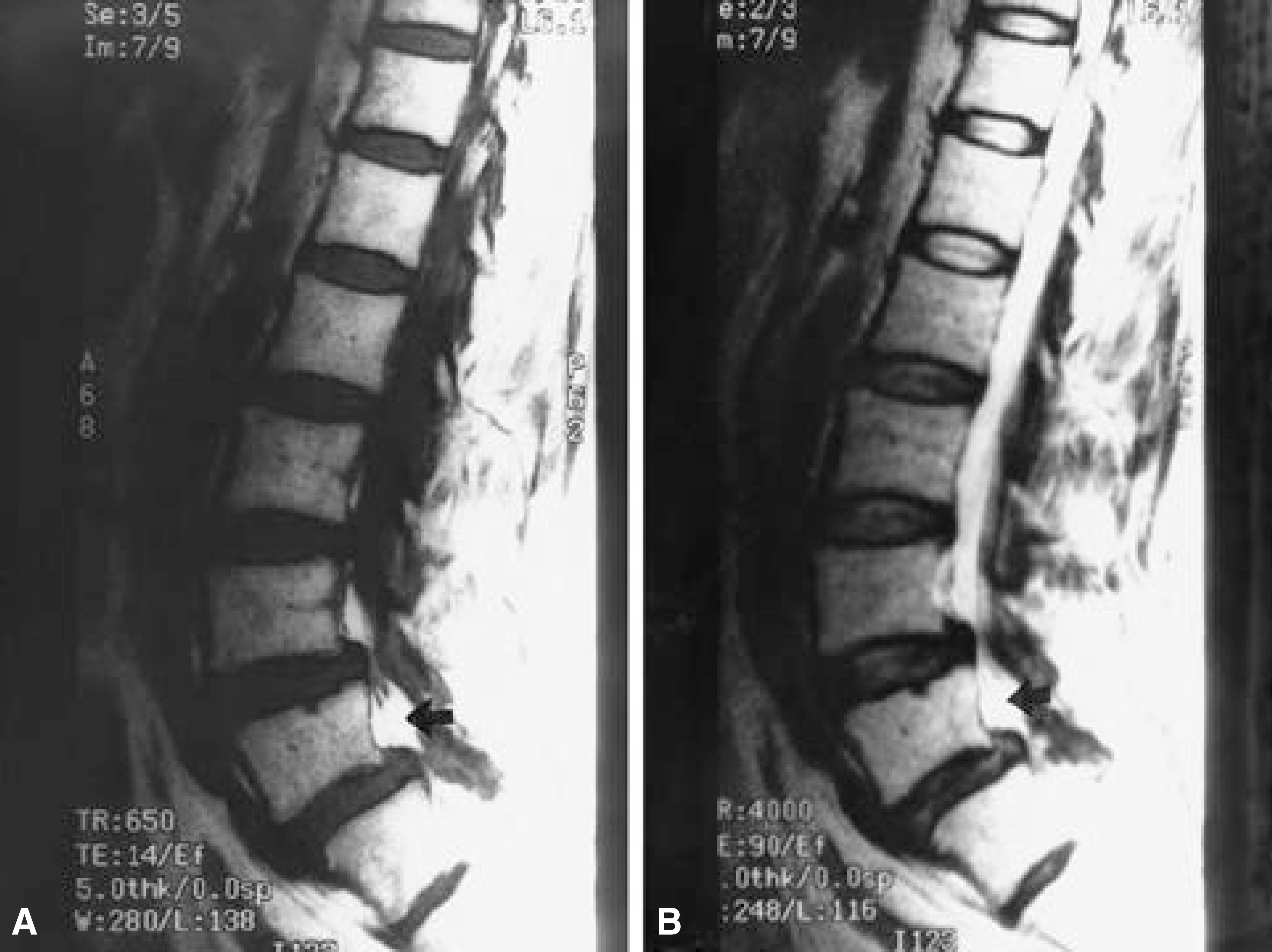
A 56- year- old woman had back pain and pain radiating to the left lower extremity that had gradually worse over 2 weeks. No definite history of trauma was disclosed. The straight leg raising test was positive at 60˚ on the left side. Sensation to pinprick was diminished in the L5, S1dermatome on her left leg. Examination of the left leg revealed weakness of the extensor hallucis longus(3/5 strength). Plain films of the lumbar spine showed degenerative scoliosis and degenerative L4- 5 spondylolisthesis with no bone involvement. In retrospective study, T1-, T2- weighted image showed a hyperintense signal mass, unlike an usual MR imaging of epidural hemangioma. A preoperative diagnosis of spondylolisthesis with spinal stenosis was made.
Result
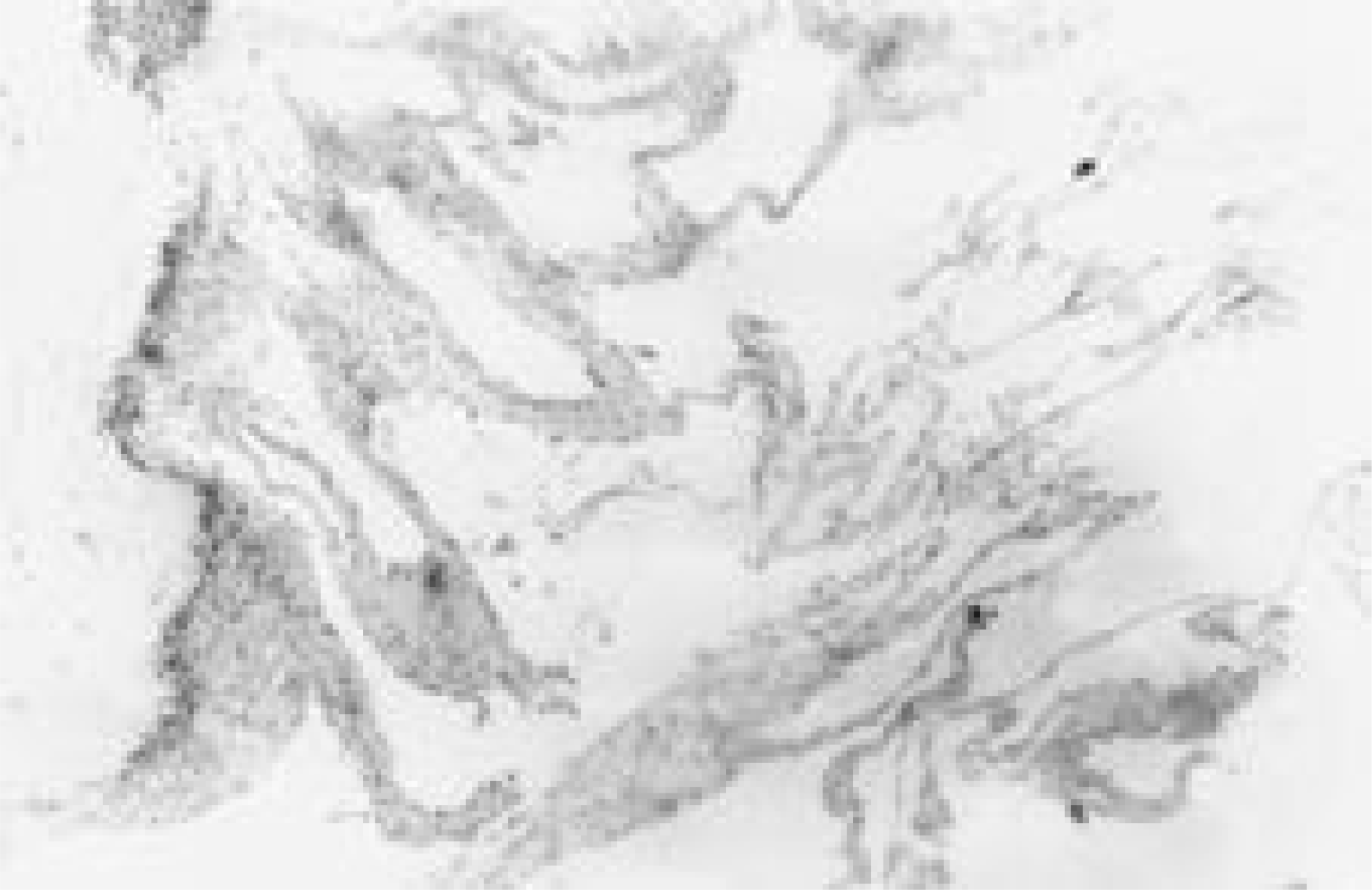
The mass compressing L5 root was excised through posterior approach and the fusion was performed from L4 to S1 with bone graft, instumentation. A purple encapsulated tumor, size 1.5× 1 × 0.8 cm, was found. Histopathologic examination revealed a thin walled sinusoidal vascular space of varying sizes, lined with a single layer of endothelial cells, consistent with typical hemangioma. The patient had a complete neurologic recovery and is doing well 5 months after surgery.
REFERENCES
1). Antoni R, Alex R, Jaume C, Martin Z, Rosa B, Mari-ana R. Lumbar extradural hemangiomas: Report of three cases, Am J Neuroadial. 20:27–31. 1999.
2). Dimitris Z, Andreas B, Serge W, Christoph H, Hans-Jrgen. Spinal epidural cavernous hemangiomas. J Neurosurg. 88:903–908. 1998.
3). Feider HK, Yuille DL. An epidural cavernous hemangioma of the spine. Am J Neuroradiol. 12:243–244. 1991.
4). Graziani N, Bouillot P, Figarella-Branger D. Carver-nous hemangioms and arteriovenous malformations of the spinal epidural space: report of 11 cases. Neurosurgery. 35:854–864. 1994.
5). Guthkelch AN. Haemangiomas involving the spinal epidural space. J Neurol Neurosurg Psychiatry. 11:199–210. 1948.

6). Harrington JF, Khan A, Grunet M. Spinal epidural an-gioma presenting as a lumbar radiculopathy with analysis of magnetic resonance imaging characteristics: case report. Neurosurgery. 36:581–584. 1995.
7). Kaplan A. Acute spinal cord compression following hemorrhage within extradural neoplasm. Am J Surg. 57:450–456. 1942.

8). Padovani R, Poppi M, Pozzali F, Tognetti F, Querzola C. Spinal epidural hemangiomas. Spine. 6:336–340. 1981.

9). Padovani R, Tognetti F, Proietti D, Pozzati E, Servadei F. Extrathecal cavernous hemangioma. Surg Neurol. 18:463–465. 1982; Epidural Hemangioma: case report. J of Korean Orthop Surgery,. 29:1026–1030. 1994.

11). Wyburn-Mason R. Vascular Abnormalities and Tumors of the Spinal Cord and its Membranes. London: Kimpton;p. 24–95. 1943.
Fig. 1.
Initial lumbar spine AP and lateral view show degenerative scoliosis, spondylolisthesis at L4-5 level, and degenerative change such as sclerosis, irregularity of posterior facet joint, but do not present any bony erosion of vertebral body.

Fig. 2.
Preoperative MR imaging show disk bulging in L3-4 level and spondylolisthesis of L4 on L5. But in retrospective study, T1-and T2- weighted MR images show a high signal intensity mass in the dorsal epidural space at the L5 level. Fig. 2-A. T1-weighted image B. T2-weighted image

Fig. 3-A.
Intraoperatively, a 1.5× 1.5 cm sized, well marginated, dark red, ovoid mass(arrow) was seen to posterolateral aspect of the root of L5, compressing a nerve root of L5 at left side. Fig. 3-B. The schematic drawing of intraoperative finding is an epidural hemangioma compressing a nerve root of L5(arrow: epidural hemangioma, L: L5 root).


 PDF
PDF ePub
ePub Citation
 Print
Print


 XML Download