

 PDF
PDF ePub
ePub Citation
 Print
Print


Abstract
Objectives
We wanted to verify the value of radiography and gait analysis to analyze the changes of the pelvic tilt before and after gait in the patients with LDK.
Summary of Literature Review
Patients with lumbar degenerative kyphosis show dynamic changes that are closely associated with the motion of pelvis.
Materials and Methods
We analyzed 18 lumbar degenerative kyphosis patients who didn't have multiple vertebral compressio fractures, a past history of spinal surgery or surgery for degenerative arthritis of the knee or hip, and obesity which causes marker errors on the gait analysis. Pelvic tilt was statistically evaluated by utilizing radiographs and dynamically utilizing the gait analysis. The linear parameters of the gait cycle and the kinematic data were obtained from the gait analysis.
Results
The LDK patients’ mean walking velocity was 80.7 cm/s, and it was largely decreased to 65% of the normal value. The cause of the decreased walking velocity was a decrease of stride length, and not a decrease of cadence. The mean static pelvic tilt in the gait analysis was -1.3±8.0。, and there were 8 cases of anterior tilt and 10 cases of posterior tilt. The mean pelvic tilt during gait was 12.5±8.2。, and there were 17 cases of anterior tilt and 1 case of posterior tilt. It was statistically significant difference (p<0.05) between the mean static pelvic tilt in gait analysis and the mean pelvic tilt during gait and the Pearson's correlation coefficient was -0.88.
Conclusions
Though there was no statistical significance, we observed anterior pelvic rotation after gait on the radiographs. As fatigue of the pelvic extensor muscles increases during gait, anterior pelvis tilt increases with statistical significance on the gait analysis. Therefore, we feel gait analysis is useful for evaluating the dynamic change of the pelvic tilt in patients with LDK.
REFERENCES
1). Takemitsu Y, Harada Y, Iwahara T, Miyamoto M, Miyatake Y. Lumbar Degenerative Kyphosis: Clinical, radiological and epidemiological studies. Spine. 1988; 13:1317–1326.
2). Takemitsu Y, Harada Y, Iwahara T. Low back pain and aging change of spine in Japanese farmers aged more than 40 years. Nippon Seikeigeka Gakkai. 1984; 58:551–552.
3). Andersson BJ, Ortengren R. Myoelectric back muscle activity during sitting. Scand J Rehab Med. 1974; 3:73–90.
4). Lee CS, Kim YT, Kim EG. Clinical study of Lumbar Degenerative Kyphosis. J Korean Spine Surg. 1997; 4:27–35.
5). Lee CS, Chung SS, Chung KH, Kim SR. Significance of Pelvic Incidence in the Development of Abnormal Sagittal Alignment. J Korean Orthop Assoc. 2006; 41:274–280.

6). Legaye J, Duval-Beaupe�re G, Hecquet J, Marty C. Pelvic incidence: a fundamental pelvic parameter for three-dimensional regulation of spinal sagittal curves. Eur Spine J. 1998; 7(2):99–103.
7). Murray MP, Drought AB, Kory RC. Walking patterns of normal men. J Bone Joint Surg. 1964; 46:335–360.
8). Sutherland DH, Olshen R, Cooper L, Woo SL. The development of mature gait. J Bone Joint Surg. 1980; 62:336–353.
9). Chung CY, Park MS, Choi IH, Cho TJ, Yoo WJ, Kim JY. Three dimensional gait analysis in normal Korean -A Preliminary Report. J Korean Orthop Assoc. 2005; 40:83–88.
10). Lee CS, Lee CK, Kim YT, Hong YM, Yoo JH. Dynamic sagittal imbalance of the spine in degenerative flat back: significance of pelvic tilt in surgical treatment. Spine. 2001; 26:2029–2035.
Figures and Tables%
Fig. 2.
Method of Pelvic Tilt Measurement. Pelvic tilt obtained in gait analysis means slope for horizontal line (A), but in standing lateral X-ray means slope for plumb line (B). So, β value is used for analysis of correlation between pelvic tilt in gait analysis and in standing lateral X-ray.
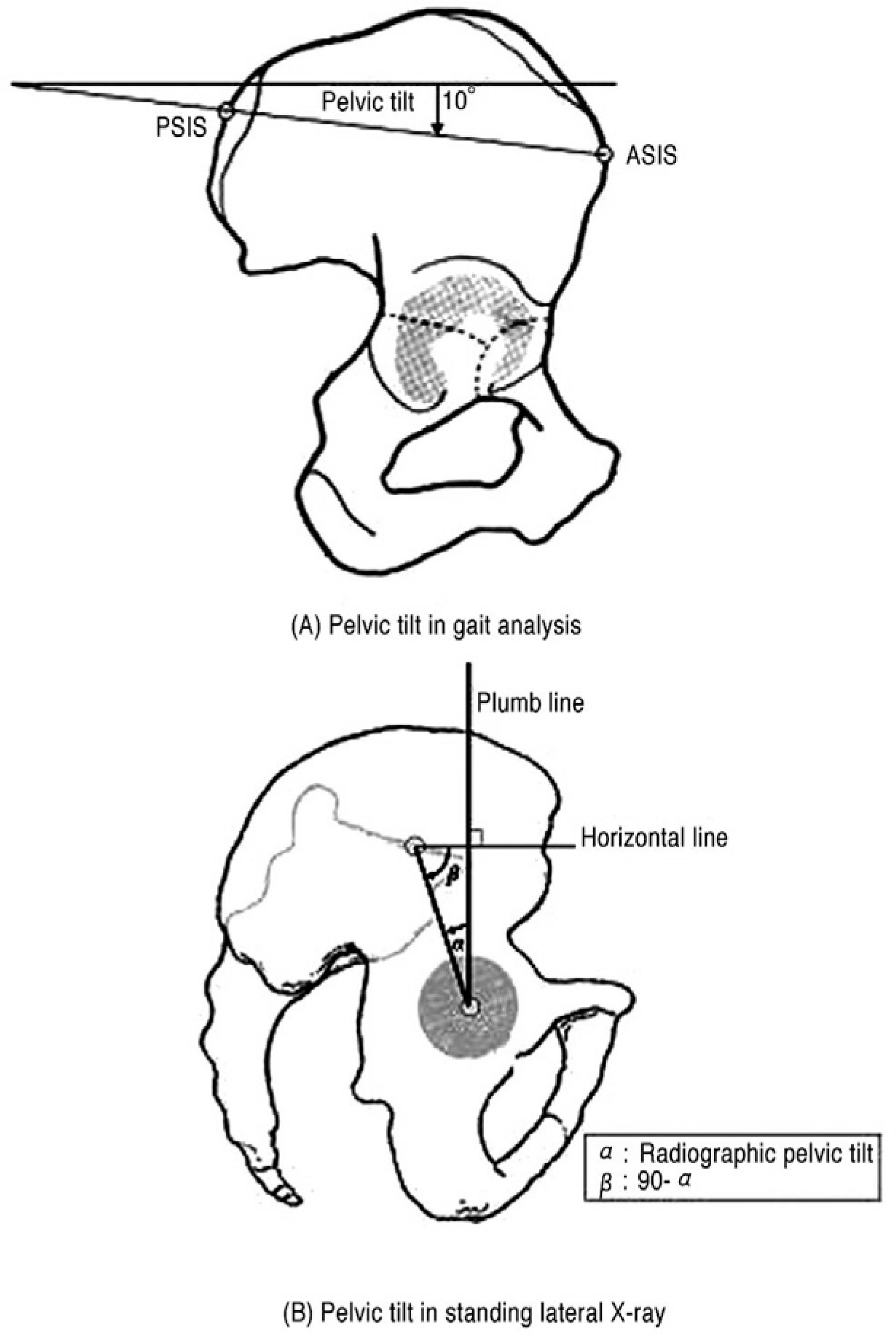
Fig. 3.
Classification of Lumbar Degenerative Kyphosis by Takemitsu. It is classified into 4 types by degree of lumbar kyphosis in radiographic findings.
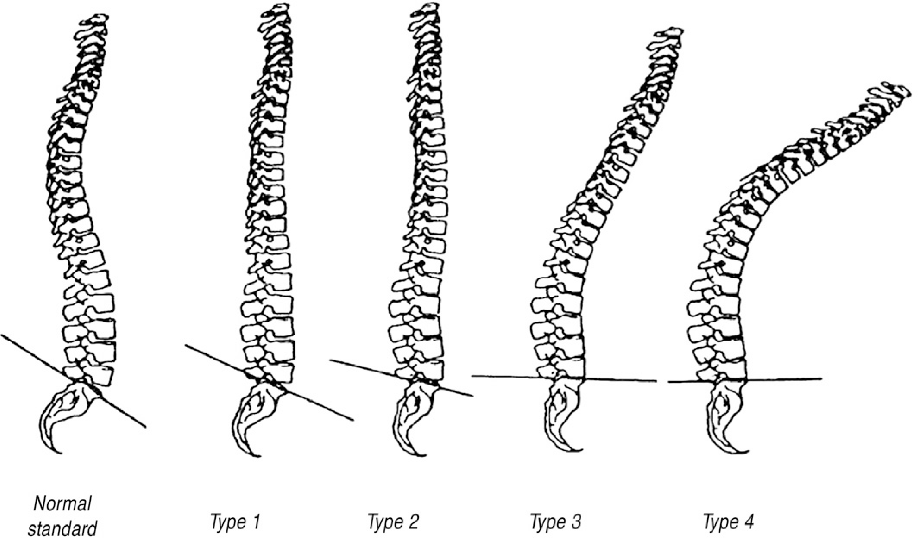
Fig. 4.
Pelvic Tilt in Gait Analysis. It shows that pelvis tilts anteriorly with statistical significance in gait analysis.
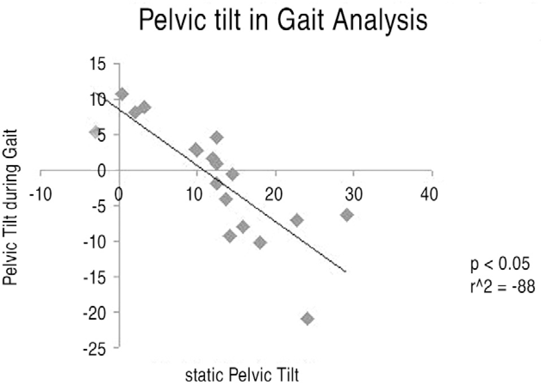
Fig. 5.
Radiographic Pelvic Tilt. There is no statistical significance in radiographic pelvic tilt before and after gait.
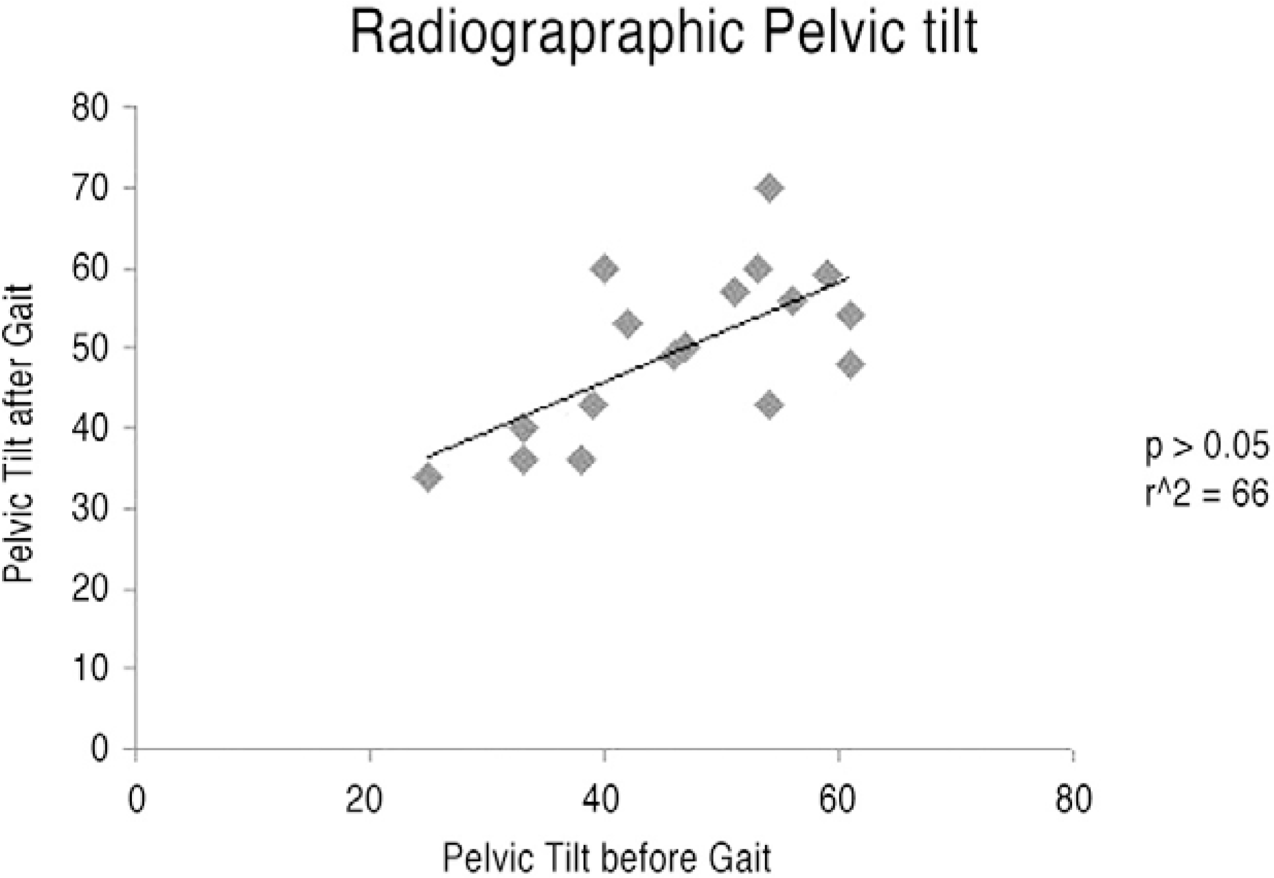
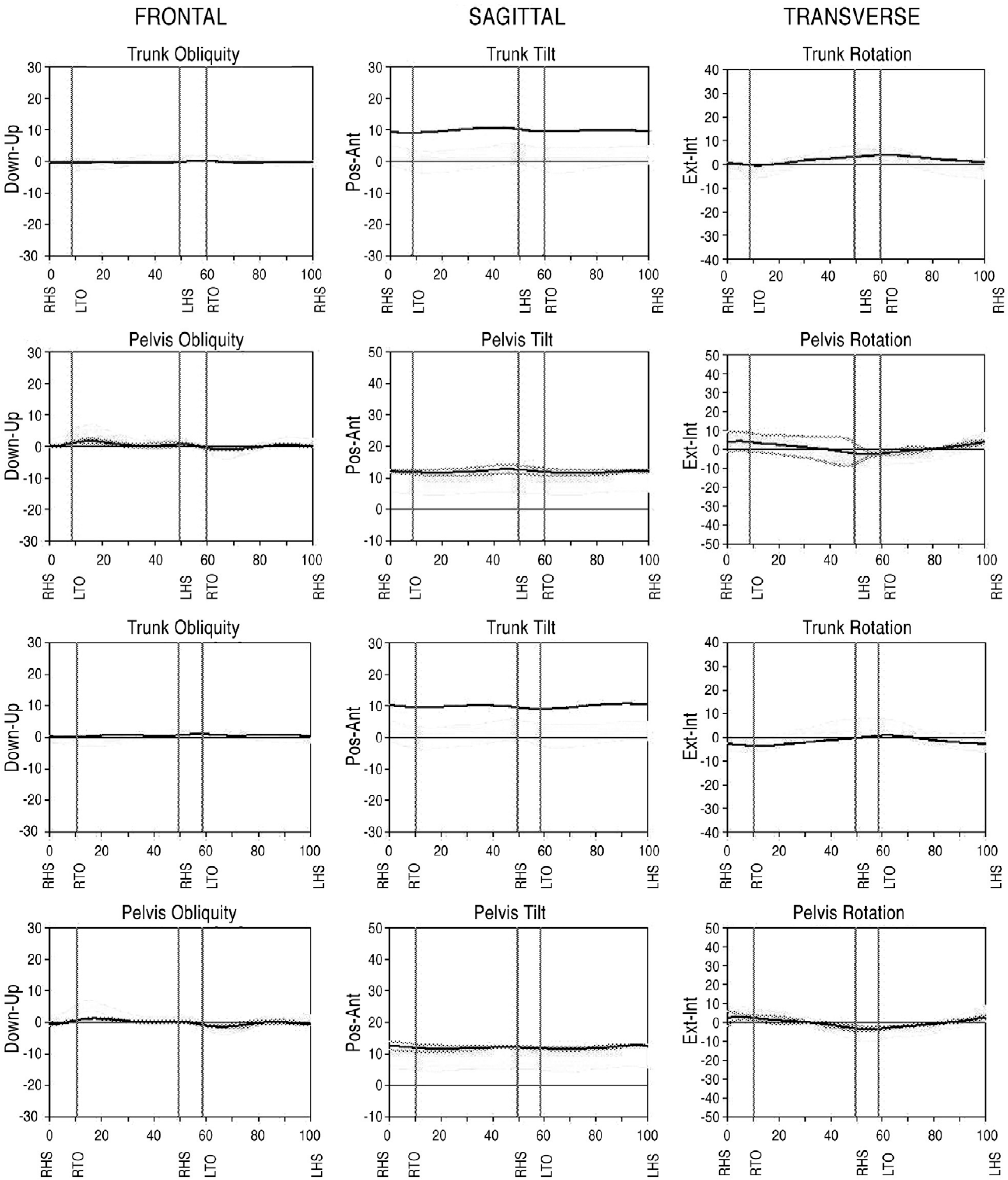
Fig. 7.
Difference between radiographic static posture and static posture in gait analysis. This difference may be one of causes that have no correlateion between radiographic examination and gait analysis.
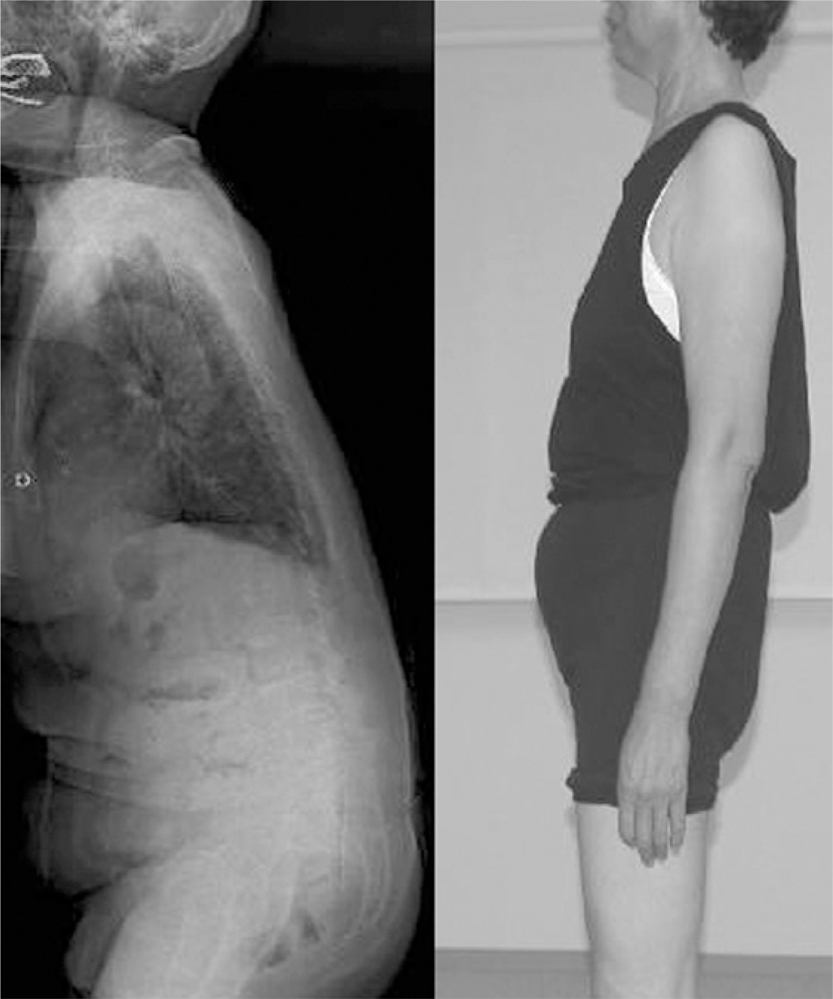
Table 1.
Demographics of the patients and radiographic and kinematic results
No, number; TT, Takemitsu type; RS, radiographic static pelvic tilt, RD, radiographic pelvic tilt after gait; GS, static pelvic tilt in gait analysis, GD, pelvic tilt during gait in gait analysis; LDK, Lumbar degenerative kyphosis, SS, spinal stenosis; SL, spondylolisthesis; DLS, Degenerative lumbar scoliosis; M, mean
Table 2.
Results of linear parameters during gait
 XML Download